Changes of Action Potential of Central Neuron by Maximal Volun- tary Isometric Contraction
Dal-Ju Moon, PT, MS; Tae-Youl Kim PT, PhD1; Kye-Yoep Kim, DVM, PhD1; Jin-Gyu Jeong, PT, PhD; Sue-hyun Kim, PT, MS
Department of Physical Therapy, Graduate School, Dongshin University; 1Department of Physical Therapy, Dongshin University
Purpose: This study analyzed changes in spinal neuron and action potential of motor unit depending on voluntary contraction on spinal neuron adaptation. Methods: It selected 80 university students in their twenties and divided into experimental groups of 25% MVIC (Ⅰ), 50% MVIC (Ⅱ), 75% MVIC (Ⅲ) and 100% MVIC (Ⅳ) depending on maximum voluntary isometric contraction (MVIC) and performed isometric exercise of plantar flexor muscle to each experimental group with given contraction for 20 times. It measured Mmax, H/Mmax, Hmax latency, V/Mmax, V wave latency before and after exercise, compared method and volume of contraction. Results: H/Mmax ratio showed significant difference in comparison among groups (p<0.01) and there was difference in I and IV groups. V/Mmax ratio showed significant difference in comparison among experimental groups (p<0.05) and there was difference in I and IV groups.
When voluntary contraction level was maximum, changes were greatest. However, no significantly difference was to Mmax, H latency and V wave latency. Conclusion: These results suggest that amplitude changes of voluntary contraction level, spinal neuron and supra-spinal neuron had a close connection that the more contraction level, the better central activation seem to decrease highly for a short time. (J Kor Soc Phys Ther 2006;18(3):37-45)
Key Words : Neural adaptation, V wave, H reflex
논문접수일: 2006년 3월 22일 수정접수일: 2006년 6월 8일 게재승인일: 2006년 6월 13일 교신저자: 김태열, [email protected]
최대 수의적 등척성 수축력에 의한 중추신경원의 활동전위 변 화
문달주, 김태열1, 김계엽1, 정진규, 김수현
동신대학교 대학원 물리치료학과, 동신대학교 물리치료학과1
Ⅰ. 서 론
신체활동이란 높은 에너지를 나타내는 여러 흥 분된 근육들의 조합이며 Aagaard 등(2002)은 신
체활동의 형태와 양은 인체 내 신경근육계의 수 행능력에 현저한 영향을 준다. 또한 신체활동의 수준에 의해 신경근계의 형태학적, 기능적 특징 이 바뀔 수 있는데, 이것은 신경순응(neural adaptation)의 생리학적 기전과 연결된다(Enoka, 1997).
Scaglioni 등(2002)은 운동신경원의 활성(motor neuron activity)이 척수 내 입력뿐만 아니라 상
척수신경원을 통해서도 얻어지며 근 수축에 의한 신경순응을 기대할 수 있다고 하였다. Enoka (1997)에 의하면 신체활동의 수준에 따른 인체 내 신경근계에 신경순응이 발생하고, 이러한 변화는 여러 가지 방법으로 측정될 수 있다. 근력강화를 위한 고강도 저항훈련은 근 수축력을 최대로 증 가시킬 수 있으며, 근 수축 에너지도 높일 수 있 다. 따라서 효과적인 근 수축은 척수수준(spinal nevel)의 통합된 반사반응에서 중추명령의 영구적 순응을 유도하고 근력향상을 가져온다(Scaglioni, 2002). 그러나 Mark 등(2004)은 근력강화에 따른 신경순응의 기전이 현재 명확하지 않다하였다.
상척수순응기전(supraspinal adaptation mechanism) 과 척수순응기전(spinal adaptation mechanism)은 중추운동주행(central motor drive)의 향상과 더불 어 운동신경원의 흥분성 증가와 연접 전 억제 (presynaptic inhibition)의 감소를 의미한다(Aa- gaard 등, 2002). McCrea (1996)는 수의적 운동시 상척수로부터 하행성 체계의 활동전위가 나타난 다 하였으며, 이러한 반응은 하행성 운동계의 활 성에 따른 연접 전 억제에 의해 항상 영향을 받 고 있다. 따라서 고강도 근력강화 운동은 상척수 신경원의 활성화가 반영되는 척수신경원의 활동 전위인 V wave(volitional wave)와 순수 척수신 경원의 활동전위인 H wave의 반응을 증가시킨다 (Aagaard 등, 2002). Pensini와 Pensini와 Martin (2004)은 근 수축에 의해 증가된 V wave 반응의 크기를 측정하고, 최대 수의적 수축 시 유발된 V wave와 휴식 시 기록 된 H wave 사이의 관계를 분석하였다.
V wave를 정의하면 근전도(electromyography) 에서 발생된 유발전위의 역방향 파가 수의적 근 수축 시 주행하는 정 방향성 신경원 흥분파와 충 돌하여 양쪽전위가 운동축삭들 내에 소실된다.
이때에 척수 전각세포에 남아있는 정방향성 α-운 동신경원의 활성전위가 나타나는 것을 의미한다 (McCrea, 1996; Pensini와 Martin, 2004). V wave 는 개재신경원(interneuron)인 Renshaw cell 억제 이기도 하며, 수의적 수축으로 얻어질 수 있고 (Hopkins 등, 1993), 전기자극을 최대 강도로 증
가시켜 H wave가 소멸되는 시점에서 최대 수의 적 근 수축 시 나타난다(Pensini와 Martin, 2004).
이는 중추인자가 변형된 수축력과 조합하여 운동 주행을 조절을 하는 것이다(Vollestad, 1997). 즉, V wave는 연접 전 억제에 의해서 영향을 받으 며, 근 수축 시 하행성 운동주행의 전위에 의하 여 나타난다(Scaglioni 등, 2002). 수축 강도에 따 라 V wave의 활동전위는 변하며 지속적인 저항 훈련이 V wave의 활동에 영향을 주어 상척수신 경원의 적응기전에 영향을 준다는 보고가 있다 (Aagaard 등, 2002; Pensini와 Martin, 2004).
수의적 수축 시 V wave가 대상자에 따라 다르 게 나타나는 이유는 각기 발달된 활성수준이 다 르기 때문이다. V wave 반응은 수의적 수축 시 하행성 운동주행과 구심성 Ia 척수회로의 흥분성 을 조절하는 상척수신경원의 흥분성 연구에 활용 할 수 있다.
H reflex는 1918년 Hoffmann에 의해 처음 기 술되었으며, 근육에 기원하는 Ia 구심성 감각 신 경을 자극하여 전달된 전위가 척수의 후각에서 단 연접을 통해 α-운동신경으로 전달되어 기록되 는 복합 활동전위이다(Scaglioni 등, 2002; Ho와 Waite, 2002). H wave는 후 경골신경에 전기적 자극을 하여 유발시키며(Sakamoto 등, 2004), 척 수를 통해 연결되는 단 연접반사와 빠른 건반사 의 전기적 신호이다(Stephen 등, 2002). 이 반사는 중추를 통한 단 연접반사로서 말초의 감각신경과 운동신경에 기능적으로 중요한 정보를 제공한다 (Kenneth 등, 1998). 즉, 단 연접으로 구성된 구심 성 Ia섬유로부터 척수의 α-운동신경원까지의 주행 경로를 측정하는 것이다(Simonsen, 1999). H wave는 최대 하 전기자극에 의해 나타나며, 소 연접과 연접 전 억제에 의해 반사반응이 조절된 다(Stephen 등, 2002). 이는 구심성 Ia 근 방추와 운동신경원사이의 중추연접의 흥분성 변화이기 때문이다(Mary와 Brian, 2001). 반사운동조절의 전달과 통합장소로서 척수의 개재신경원들은 운 동신경원 집단들의 수의적 운동명령을 보내고 받 는다(McCrea, 1996).
근 수축에 의한 신경순응과 관련된 지금까지의
Group Age(years) Height(㎝) Weight(㎏)
Ⅰ(N=10) MVIC 25% 22.70±2.98 174.50±4.19 67.00±8.65
Ⅱ(N=10) MVIC 50% 22.20±3.08 174.90±7.69 66.60±7.13
Ⅲ(N=10) MVIC 75% 19.40±1.34 170.20±5.76 68.10±14.20
Ⅳ(N=10) MVIC 100% 22.10±3.21 171.90±4.93 61.80±15.11
연구는 주로 표면 근전도와 H reflex의 변화를 분석하는 방법을 이용하였다(Lavoie 등, 1997;
Stephen 등, 2002). 또한 상척수신경원의 반응을 측정하기 위해서 체성감각유발전위(somatosen- sory evoked potential)와 운동유발전위(motor evoked potential) 등을 이용하였다(Nozaki 등, 2003; Lentz와 Nielsen, 2002; Siemionow 등, 2004; Vrana 등, 2005). 최근 들어 상척수신경원 의 영향이 반영되는 척수신경원의 활동전위인 V wave 측정방법이 소개되어 점차 관심이 높아지 고 있다. V wave는 전기자극과 함께 운동피질의 활성에 의한 자발적 활동전위가 반영된 복합반응 이기 때문에 상척수신경원의 신경순응에 대한 수 의적 반영으로 이해할 수 있는데 착안하여 수의 적 수축에 의한 저항운동의 신경순응에 대한 연 구가 주를 이루고 있다. 따라서 본 연구에서는 이러한 전기생리학적 측정방법을 이용하여 수의 적 수축에 의한 수축 전·후의 중추신경원의 활동
전위의 변화를 분석하여 수축력에 따른 신경순응 의 전·후 차이를 알아보고자 한다.
Ⅱ. 연구방법
1. 연구대상
본 연구는 수의적 근 수축에 의한 중추신경원 의 활동전위 변화에 따른 순응현상을 알아보기 위한 것으로서 20대 건강한 남자 대학생 40명을 대상으로 하였다. 대상자들은 실험에 영향을 줄 만한 신경계 및 근골격계 병력과 기능장애가 없 는 자로 본 연구에 동의하여 자발적으로 참여 하 였다. 실험군은 최대 수의적 수축(maximum voluntary isometric contraction; MVIC) 수준에 따라 구분하였다(표 1).
표 1. 대상자의 일반적인 특성.
모든 값은 평균±표준편차
2. 근수축유발
대상자는 실험대에 슬관절을 신전하고 앉은 자 세에서 등받이를 고정하여 운동 시 밀려나지 않 도록 하였으며, 최대 등척성 수축력 측정은 좌측 족관절이 중립위가 유지되게 하고 dynamometer (JLW instruments Inc., CS200 Dynamometer, USA)는 applicator를 족저면에 밀착시킨 후 실험
대의 고정장치에 고정시켰다.
대상자는 좌측 족관절의 저측굴곡근에서 최대 등척성 수축이 일어나도록 applicator를 최대한 밀도록 하여 최대 수의적 등척성 수축력을 측정 하였다. 최대 수의적 등척성 수축력(MVIC)을 산 출하여 이를 기준으로 25% MVIC, 50% MVIC, 75% MVIC, 100% MVIC 군으로 구분하고, 각 실 험군에 설정된 MVIC로 등척성 운동을 실시하였
다.
운동방법은 MVIC를 측정할 때와 같은 조건에 서 좌측 족관절의 저측굴곡근에 대한 등척성 운 동을 수축시간 5초, 휴식시간 2초로 하여 20회 반복하였다(Stylianou 등, 2005; Mcloda 등, 2000).
3. 중추신경원활동전위측정
중추신경원 활동전위로 H wave와 V wave를 측정하기 위해 근전도기(Cadwell Laboratories Inc., CADWELL II Wedge, USA)를 사용하였다.
1) H reflex 측정
H wave의 측정을 위해 먼저 알코올(70%)로 피부를 깨끗이 닦고 습기가 없도록 건조시킨 후 전극을 부착하였다.
기록전극으로 활동전극은 슬와부 주름 중앙과 족관절 내과의 가장 근위부를 연결하는 선상을 양분하는 중심점, 기준전극은 아킬레스건, 접지전 극은 활동전극에서 3 ㎝ 위의 외측 비복근 위에 배치하였고, 기록전극은 1×1 ㎝ 크기의 자가 부 착식 일회용 전극(Medicotest A/S, Neuroline Disposable Neurology Elecrodes 700 10-K, Malaysia)을 사용하였다.
기록조건은 주파수 여과범위(filter setting)가 10 ㎐~10,000 ㎐, 소인속도(sweep speed)는 15 msec/division, 감응도(gain)는 10 ㎷/division이 었다. 대상자는 엎드려 누운 자세에서 슬관절을 약간 굴곡 시킨 후 족관절 밑에 받침대를 놓아 완전히 이완되도록 하고 족관절이 중립에 놓이게 하였다.
먼저 H wave를 확인하고 나서 강도를 서서히 조절하면서 Hmax와 Mmax를 구하고 두 개의 활 동전위에 대한 최대 진폭비율로 H/Mmax비를 산 출하였다. 전기자극은 후경골신경의 정중 슬와근 주름에서 이극전극을 사용하여 강도를 최대 하 자극(submaximal stimulation) 수준으로 하여 2초 간격으로 자극하였다.
2) V wave 측정
V wave의 측정을 위해 H wave 측정과 마찬 가지로 먼저 피부를 알코올로 깨끗이 닦고 피부 에 습기가 없도록 건조시킨 후 전극을 부착하였 다. 기록전극의 배치와 기록조건은 H wave와 동 일하며, 대상자는 엎드려 누운 자세에서 슬관절 을 약간 굴곡 시킨 후 족관절 밑에 받침대를 놓 아 완전히 이완되도록 한 후 족관절이 중립위치 에 놓이게 하였다. 먼저 H reflex를 확인하고 나 서 자극강도를 서서히 높여 H wave의 진폭이 최 소화되고 M wave의 진폭이 최대하가 되는 지점 에서 대상자로 하여금 족관절 저측굴곡근을 최대 한 수축하도록 하여 V wave를 얻었다(Pensini와 Mar- tin, 2004).
4. 자료분석
모든 통계자료는 Window SPSS 10.0 프로그램 으로 분석하였다. 각 측정항목의 군 간 비교는 공분산분석(ANCOVA)을 이용하였으며, 사후분석 으로 본페로니(Bonferroni) 검정을 실시하였다. 통 계학적인 유의성을 검증하기 위해서 유의수준은 α=0.05로 하였다.
Ⅲ. 결 과
1. H reflex의 변화
1) Mmax의 변화
Mmax의 변화로 모든 군에서 수축 전에 비하여 수 축 후에 약간 증가되었으나 군 간에 차이는 없었다 (그림 1).
2) H/Mmax ratio의 변화
척수신경원 활동전위의 진폭으로 H/Mmax ratio의 변화는 100% MVIC로 수축한 Ⅳ군에 수 축 전․후의 차이가 가장 컸고, 다음으로 75%
MVIC로 수축한 Ⅲ군순으로 나타났다. 실험군 간
0 5 10 15 20 25
1 2 3 4
Mmax(㎷)
pre post
Ⅰ Ⅱ Ⅲ Ⅳ
Ⅰ; 25%MVIC, Ⅱ; 50%MVIC, Ⅲ; 75%MVIC, Ⅳ; 100%MVIC
그림 1. 각 집단의 전·후의 Mmax 진폭 변화.
0 0.5 1 1.5 2 2.5 3 3.5 4
1 2 3 4
H/Mmax ratio(%)
pre post
Ⅰ Ⅱ Ⅲ Ⅳ
*
Ⅰ; 25%MVIC, Ⅱ; 50%MVIC, Ⅲ; 75%MVIC, Ⅳ; 100%MVIC.
* 집단 사이의 현저한 차이
그림 2. 각 집단에서 H/Mmax의 전·후 변화.
0 0.05 0.1 0.15 0.2 0.25 0.3 0.35
1 2 3 4
V/Mmax ratio(%)
pre post
Ⅰ Ⅱ Ⅲ Ⅳ
*
Ⅰ; 25%MVIC, Ⅱ; 50%MVIC, Ⅲ; 75%MVIC, Ⅳ; 100%MVIC * 집단 사이의 현저한 차이
의 차이를 알아보기 위하여 공분산분석을 한 결 과에서 유의한 차이가 나타났으며(p<.01), 사후검 정에서 25% MVIC 수축군인Ⅰ군과 Ⅳ군이 차이 가 있었다(그림 2).
3) H wave 잠복시의 변화
H wave 잠복시의 변화로 모든 군에서 수축 전에 비하여 수축 후에 약간 증가되었으나 군 간 에 차이는 없었다(그림 3).
0 1 2 3 4 5 6 7 8
1 2 3 4
Hmax latency(㎳)
pre post
Ⅰ Ⅱ Ⅲ Ⅳ
Ⅰ; 25%MVIC, Ⅱ; 50%MVIC, Ⅲ; 75%MVIC, Ⅳ; 100%MVIC
그림 3. 각 집단에서 H wave 잠복시의 전·후 변화.
2. V wave의 변화
1) V/Mmax ratio의 변화
상척수 신경원의 영향에 의한 척수신경원 활동 전위의 진폭으로 V/Mmax ratio의 변화는 Ⅳ군에 서 수축 전․후의 차이가 가장 컸으며, Ⅲ군, 25% MVIC로 수축한 Ⅱ군 순으로 나타났다. 실 험군 간의 차이를 알아보기 위하여 공분산분석을 한 결과에서 군 간 유의한 차이가 나타났으며 (p<.05), 사후검정에서 Ⅰ군과 Ⅳ군이 차이가 있 었다(그림 4).
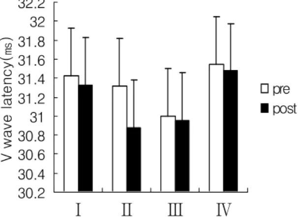
2) V wave 잠복시의 변화
V wave 잠복시의 변화로 모든 군에서 수축 전 에 비하여 수축 후에 약간 감소되었으나 군 간에 차이는 없었다(그림 5).
30.2 30.4 30.6 30.8 31 31.2 31.4 31.6 31.8 32 32.2
1 2 3 4
V wave latency(㎳)
pre post
Ⅰ Ⅱ Ⅲ Ⅳ
Ⅰ; 25%MVIC, Ⅱ; 50%MVIC, Ⅲ; 75%MVIC, Ⅳ; 100%MVIC
그림 5. 각 집단에서 V잠복기의 전·후 변화.
Ⅳ. 고 찰
본 연구는 수의적 근수축력에 따라 중추신경원 의 활동전위의 변화를 비교하여 척수 및 상척수 수준에서 신경순응에 어떠한 영향을 주는지를 알 아보고자 하였다. 대상자는 건강한 20대 성인 남 성 80명이었으며, 실험군은 수의적 수축력에 따 라 4개 실험군으로 구분하였다. 좌측 족관절 저 측굴곡근에 대한 등척성 운동 전·후에 중추신경 원의 활동전위를 측정하여 Mmax, H/Mmax, H wave 잠복시, V/Mmax, V wave 잠복시를 분석 하였다.
척수신경원 흥분성의 변화는 H wave의 진폭 변화를 측정하면 가능하며, H/Mmax비를 산출하 면 가장 정확하다(Palmieri 등, 2004). Hmax는 척 수신경원의 수를 간접적으로 표현하며 척수운동 신경원의 전체적인 흥분성을 나타내고 지구력 훈 련의 적합한 지표가 된다(Nicola 등, 2001).
Palmier 등(2004)은 전류강도의 점진적인 증가로 H wave를 구하고 6번에 걸쳐 측정한 H wave 중에서 가장 진폭값이 큰 것으로 선택하여 Hmax 라 하였다. 본 연구에서 말초신경-근 구간의 흥분 성을 반영하는 Mmax의 전․후 변화가 모든 실험 군에서 크지 않아 수의적 수축력에 따른 신경근 연접부의 흥분성 변화는 크지 않은 것으로 나타 났다. 그러나 운동 전․후 H/Mmax ratio의 변화 는 100% MVIC에서 가장 크게 나타났으며, 25%
MVIC와 유의한 차이가 있어 수축력에 따라 척수 신경원의 흥분성이 차이가 있다는 것을 알 수 있 었다. Tsuboi 등(1995)도 수의적 운동이나 전기자 극에 의한 근수축이 모두 H wave의 진폭을 감소 시키며, 수축력과 상관성이 있다고 하여 본 연구 비슷한 결과를 보고하였다.
이러한 결과는 상척수신경원으로부터 개재신경 원의 Ia억제가 수의적 움직임에 의해 영향을 받 기 때문으로(Perez 등, 2003; Romaniello 등, 2002; Stein, 1995; Tsuboi 등, 1995), 수축력에 따 라 차이가 나타난 것도 수의적 운동이 개재신경 원 Ia억제에 영향을 더 주기 때문으로 볼 수 있 는데, 근육에 기원하는 Ia 구심성 감각 신경이 척 수의 후각에서 단 연접을 통해 운동신경원으로 전달되는 과정에서 감소되는 것으로 본다. 그러 나 신경전도와 관련된 H wave 잠복시는 전․후 의 변화에 큰 차이가 없으며, 수축력에 다른 차 이도 유의하지 않아 수의적 수축에 의한 척수신 경원의 흥분성 변화는 활동전위의 진폭변화와 밀 접한 관계가 있는 것으로 생각된다.
상척수신경원의 흥분성변화 연구는 V wave의 진폭 변화를 측정하면 가능하며, V/Mmax ratio 를 산출하여 분석하는 방법이 가장 정확하다.
V/Mmax ratio는 하행성 운동주행로의 활성도에 따른 α-운동신경원의 흥분빈도와 양을 나타내는 정확한 지표라 할 수 있다(Aagaard 등, 2002;
Pensini와 Martin, 2004). 이는 전기자극 강도가 증가하여 H wave가 소멸되고 Mmax가 출현할 때 근육을 최대로 수축시켜 얻어내는 상척수신경 원의 활성도가 반영된 활동전위로 몇 차례에 걸 쳐 Vmax를 측정하고 그중에서 가장 진폭값이 큰
것으로 선택하여 기록한다. 본 연구에서 운동 전․후 V/Mmax ratio의 변화는 수의적 수축력을 최대로 하였을 때 가장 크게 나타났으며, 수의적 수축은 수축력이 증가할 수 록 상척수신경원의 활동전위의 변화에 크게 영향을 주는 것으로 나 타났다. 이러한 결과는 V wave 반응이 하행성 운동주행로의 활성도에 의존하기 때문에 수의적 수축이 연접 전 억제에 의한 영향을 더 받는다 (Mary와 Brian, 2001; Masaki 등, 2002; Stephen 등, 2002). 따라서 수의적 수축력이 증가할수록 척수신경원의 순응과정에 상척수신경원의 영향을 더 크게 받는 것으로 나타났다. 그러나 상척수신 경원의 영향력이 반영된 상태에서의 신경전도를 의미하는 V wave 잠복시도 H wave 잠복시와 마찬가지로 크게 차이가 없는 것으로 나타나 중 추운동주행에 의한 척수신경원의 흥분성 변화에 서도 활동전위의 진폭변화와 밀접한 관계가 있는 것으로 생각된다.
Ⅴ. 결 론
본 연구는 수의적 수축력에 따라 중추신경원의 활동전위 정도와 차이를 비교하여 근 수축력이 신경순응에 어떠한 영향을 주는지 알아보고자하 였다.
H/Mmax ratio는 최대 수의적 수축력에서 변화 가 가장 컸으며, 실험군 간의 비교에서 유의한 차이가 있었다(p<0.01). 최소 수의적 등척성 수축 군인 Ⅰ군과 최대 수의적 등척성 수축군이 Ⅳ군 사이에서 차이가 있었다. V/Mmax ratio도 수의 적 수축력을 최대로 하였을 때 변화가 가장 컸으 며, 실험군 간의 비교에서 유의한 차이가 이었다 (p<0.05). 최소 수의적 등척성 수축군인 Ⅰ군과 최대 수의적 등척성 수축군인 Ⅳ군 사이에서 차 이가 있었다.
결론적으로 수의적 수축력과 척수신경원 및 상척수신경원의 활동전위 진폭의 변화는 밀접한 관계를 가지며, 짧은 시간동안의 운동에서는 수 축력이 증가할수록 중추신경원의 흥분성 크게 감
소하는 것으로 나타났다.
참고문헌
Aagaard P, Simonsen EB, Andersen JL et al. Neural adaptation to resistance training: changes in evoked V-wave and H-reflex responses. J Appl Physiol. 2002;92(6):2309-18.
Aagaard P, Andersen JL, Dyhre-Poulsen P et al. A me- chanism for increased contractile strength of human pennate muscle in response to strength training: changes in muscle architecture. J Physiol. 2001;534(Pt. 2):613-23.
Avela J, Kyrolainen H, Komi PV. Altered reflex sensi- tivity after repeated and prolonged passive muscle stretching. J Appl Physiol. 1999;86(4):
1283-91.
Bishop MD, N pathare. Consideration for the Use of Surface Electromyography. J Korean Acad Univ Trained Phys Ther 2004, 11(4):61-70.
Callaghan MJ, Oldham JA. Electric muscle stimulation of the quadriceps in the treatment of patellofemoral pain. Arch Phys Med Rehabil. 2004;85(6):956-62.
Cauraugh JH, Summers JJ. Neural plasticity and bilateral movements: A rehabilitation approach for chro- nic stroke. Prog Neurobiol. 2005;75(5):309-20.
Cliffer KD, Tonra JR, Carson SR et al. Consistent repeated M-and H-wave recording in the hind limb of rats. Muscle Nerve. 1998;21(11):1405-13.
Courtney C, Rine RM, Kroll P. Central somatosensory changes and altered muscle synergies in subjects with anterior cruciate ligament deficiency. Gait Posture. 2005;22(1):69-74.
Dongren Y, Tao L, Fengsheng H. Electroneurophysiolo- gical studies in rats of acute dimethoate. Toxicol Lett. 1999;107(1-3):249-54.
Duchateau J, Balestra C, Carpentier A et al. Reflex re- gulation during sustained and intermittent submaximal contractions in humans. J physiol.
2002;541(Pt 3):959-67.
Enoka RM. Neural adaptations with chronic physical activity. J Biomech. 1997;30(5):447-55.
Ferrante S, Pedrocchi A, Ianno M et al. Functional elec- trical stimulation controlled by artificial neural networks: pilot experiments with simple move-
ments are promising for rehabilitation applica- tions. Funct Neurol. 2004;19(4):243-52.
Fumoto M, Komiyama T, Nishihira Y. Soleus H-reflex dynamics during fast plantarflexion in humans. J Electromyogr Kinesiol. 2002;12(5):367-74.
Garrett M, Caulfield B. Increased H(max):M(max) ratio in community walkers poststroke without in- crease in ankle plantarflexion during walking.
Arch Phys Med Rehabil. 2001;82(8):1066-72.
Gazzoni M, Farina D, Merletti R. A new method for the extraction and classification of single motor unit action potentials from surface EMG signals. J Neurosci Methods. 2004;136(2):165-77.
Glanz M, Klawansky S, Stason W et al. Functional elec- trostimulation in poststroke rehabilitation: A meta-analysis of the randomized controlled trials. Arch Phys Med Rehabil. 1996;77(6):549-53.
Holtermann A, Roeleveld K, Karlsson JS. Inhomoge- neities in muscle activation reveal motor unit recruitment. J Electromyogr Kinesiol. 2005;15(2):
131-7.
Hopkins JT, Ingersoll CD, Edwards JE et al. Changes in soleus motoneuron pool excitability after arti- ficial knee joint effusion. Arch Phys Med Rehabil. 2000;81(9):1199-203.
Ho SM, Waite PM. Effects of different anesthetics on the paired-pulse depression of the H reflex in adult rat. Exp Neurol. 2002;177(2):494-502.
Kenneth D. Cliffer, James R. Tonra, Susan R. Carson, Ba et al. Consistent Repeated M-and H-wave recording in the hind limb of Rats. Muscle Nerve 1998;21: 1405-1413.
Lentz M, Nielsen JF. Post-exercise facilitation and depression of M wave and motor evoked potentials in healthy subjects. Clinical Neuro- physiology 2002;113:1092-1098.
Lambertz D, Goubel F, Kaspranski R et al. Influence of long-term spaceflight on neuromechanical pro- perties of muscles in humans. J Appl Physiol.
2003;94(2):490-8.
Lavoie BA, Devanne H, Capaday C. Differential control of reciprocal inhibition during walking versus postural and voluntary motor tasks in humans.
J Neurophysiol. 1997;78(1):429-38.
Lentz M, Nielsen JF. Post-exercise facilitation and depre- ssion of M wave and motor evoked potentials in healthy subjects. Clin Neurophysiol. 2002;
113(7):1092-8.
Levin O, Mizrahi J, Gornish M et al. Muscle strength and geometrical changes in a paralysed muscle following FES. Hong Kong Physiotheraphy Journal 2000;18(1):3-11.
Maffiuletti NA, Martin A, Babault N et al. Electrical and mechanical H(max)-to-M(max) ratio in power- and endurance-trained athletes. J Appl Physiol. 2001;90(1):3-9.
Maria B, Elena P. Increase in the releasable pool of synaptic vesicle underlies facilitation. Neurocom- puting. 2004;58-60:496-76.
Mark DB, Neeti P. Consideration for the Use of Surface Electromyography. KAUTPT 2004, 11(4):61-70.
Mary Garrett and Brian Caulfield : Increased Hmax:
Mmax ratio in community walkers poststroke without increase in ankle plantarflexion during walking. Arch Phys Med Rehabil 2001;82:1066- 72.
Masaki F, Tomoyoshi K, Yoshiaki N. Soleus H-reflex dynamics during fast plantarflextion in human. J Electomyogr Kinesiol. 2002;12:367-74.
McCrea DA. Supraspinal and segmental interactions.
Can J Physiol Pharmacol. 1996;74(4):513-7.
McLoda TA, Carmack JA. Optimal burst duration during a facilitated quadriceps femoris contrac- tion. J Athl Train. 2000;35(2):145-150.
Naomi CC, William KD. Surface EMG as a fatigue indi- cator during FES-induced isometric muscle con- tractions. J Electromyogr Kinesiol. 1997;7:27-37.
Nicola AM, Alain M, Nicolas B et al. Electrical and mechanical Hmax-to-Mmax ratio in power- and eduranced-trained athletes. J appl Physiol. 2001;
90:3-9.
Nozaki D, Kawashima N, Aramaki Y et al. Sustained muscle contractions maintained by autono- mous neuronal activity within the human spinal cord. J Neurophysiol. 2003;90(4):2090-7.
Ollivier K, Portero P, Maisetti O et al. Repeatability of surface EMG parameters at various isometric contraction levels and during fatigue using bipolar and laplacian elecrode configurations. J Electromyogr Kinesiol. 2005;15(5):466-73.
Palmieri RM, Ingersoll CD, Hoffman MA. The Hoff- mann reflex: Methodologic considerations and applications for use in sports medicine and athletic training research. J Athl Train. 2004;
39(3):268-77.
Pavesi G, Cattaneo L, Tinchelli S. Masseteric repetitive nerve stimulation in the diagnosis of myasthenia gravis. Clin Neurophysiol. 2001;112(6):1064-9.
Pensini M and Martin A. Effect of voluntary contraction intensity on the H-reflex and V-wave response.
Neurosci Lett. 2004;367(3):369-74.
Perez MA, Field-Fote EC, Floeter MK. Patterned sensory stimulation induces plasticity in reciprocal Ia inhibition in humans. J Neurosci. 2003;23(6):
2014-8.
Palmieri RM, Ingersoll CD, Hoffman MA. The hoffmann reflex: Methodologic considerations and applica- tions for use in sports medicine and athletic training research. Journal of Atheletic Taniners 2004;39(3):248-77.
Romaniello A, Valls-Sole J, Iannetti GD et al. Nocice- ptive quality of the laser-evoked blink reflex in humans. J Neurophysiol. 2002;87(3):1386-94.
Sakamoto M, Nakajima T, Wasaka T et al. Load- and cadence-dependent modulation of somatosen- sory evoked potentials and soleus H-reflexes during active leg pedaling in humans. Brain Res. 2004;1029(2):272-85.
Scaglioni. G, Ferri. A, Minetti. A E et al. Plantar flexor activation capacity and H reflex in older adults: adaptations to strength training. J Appl Physiol 2002;92:2292-302.
Stein RB. Presynaptic inhibition in humans. Prog Neuro- biol. 1995;47(6):533-44.
Stephen G, Ferri A, Minetti AE et al. Plantar flexor activation capacity and H reflex in older adults:
adaptations to strength training. J Appl Physiol.
2002;92(6):2292-302.
Schillings ML, Kalkman JS, van der Werf SP et al.
Diminished central activation during maximal voluntary contraction in chronic fatigue synd- rome. Clin Neurophysiol. 2004;115(11):2518- 24.
Siemionow V, Fang Y, Calabrese L et al. Alter central nervous system signal during motor perfor- mance in chronic fatigue syndrome. Clin Neuro- physiol. 2004;115(10):2372-81.
Simonsen EB, Dyhre-Poulsen P. Amplitude of the human soleus H reflex during walking and running. J Physiol. 1999;515(Pt 3):929-39.
Stylianou AP, Luchies CW, Lerner DE et al. The use of correlation integrals in the study of localized
muscle fatigue of elbow flexors during maximal efforts. J Electromyogr Kinesiol. 2005;15(5):437-43.
Thomas CK, del Valle A. The role of motor unit rate modulation versus recruitment in repeated sub- maximal voluntary contractions performed by control and spinal cord injured subjects. J Electromyogr Kinesiol. 2001;11(3):217-29.
Tsuboi T, Sato T, Egawa K et al. The effect of fatigue caused by electrical induction or voluntary contraction on Ia inhibition in human soleus muscle. Neurosci Lett. 1995;197(1):72-4.
Vollestad NK. Measurement of human muscle fatigue. J Neurosci Methods. 1997;74(2):219-27.
Vrana J, Polacek H, Stancak A. Somatosensory-evoked potentials are influenced differently by isometric muscle contraction of stimulated and non-sti- mulated hand in humans. Neurosci Lett. 2005;
386(3):170-5.
Ward AR, Shkuratova N. Russian electrical stimulation:
The early experiments. Phys Ther. 2002;82(10):
1019-30.