• 검색 결과가 없습니다.

Effects of Spinal Stabilization Exercises on the Cross

N/A
N/A
Protected

Academic year: 2021

Share "Effects of Spinal Stabilization Exercises on the Cross"

Copied!
7
0
0

로드 중.... (전체 텍스트 보기)

전체 글

(1)

Seong-Ho Kim, PT, MS; Wan-Hee Lee, PT, PhD

1

Department of Physical Therapy, Shingu University;

1

Department of Physical Therapy, Sahmyook University

Purpose: To evaluate, in patients with degenerative disc disease (DDD), the efficacy of using spinal stabilizing exercises for the reversal? of atrophy of the multifidus and psoas major, reductions in pain and disability, and for increases in paraspinal muscle strength.

Methods: Nineteen patients diagnosed with DDD participated for 10 weeks in a spinal stabilization exercise program. Pain and disability were measured before and after exercise using, respectively, a visual analogue scale (VAS) and the Oswestry Disability Index (ODI). Paraspinal muscular strength in four directions was evaluated using CENTAUR. Both before and after exercise we used computed tomography (CT) too measure cross-sectional areas (CSAs) of both the left and right multifidus and the psoas major at the upper & lower endplate of L4.

Results: After 10 weeks of a spinal stabilization exercise program, pain was significantly decreased from 5.7±0.9 to 2.5±0.9 (p<0.01); the ODI score decreased from 16.7±4.9 to 7.3±3.1. Paraspinal muscle strength was significantly increased (p<0.01) and the CSAs of the left and right multifidus and psoas major muscles were significantly increased (p<0.01).

Conclusion: Spinal stabilization exercise is effective in reversing atrophy in DDD patients, in reducing pain and disability, and in increasing paraspinal muscle strength. It is an effective treatment foro aiding rehabilitation in these cases.

Keywords: Degenerative disc disease (DDD), Spinal stabilization, Multifidus, Psoas major, Cross section areas (CSA) Received: April 22, 2010

Revised: June 1, 2010 Accepted: June 14, 2010

Corresponding author: Wan-Hee Lee, [email protected]

Areas of the Lumbar Multifidus and Psoas Major Muscles of Patients with Degenerative Disc Disease

The Journal Korean Society of Physical Therapy

I. Introduction

Low back pain (LBP) is one of the most common diseases in modern society, occurring in nearly 80% of the population.

There is a high potential for recurrence, so successful rehabili- tation is important in preventing the return of LBP.

1

Though there are various causes of this pain, the primary factors for developing LBP are damage to the trunk’s soft tissues or weakening of the muscles, which causes pain, decreased muscle endurance and flexibility and restriction of spine movement.

2

In particular, it has been shown that LBP patients’ deep layer lumbar spine muscles experience more atrophy when compared with healthy controls, and the contraction speed of their muscles decreases.

3-5

Such damage and muscle weakening gives rise to disc degeneration and causes pain and instability of the spine.

6,7

Another study reported that excessive mechanical load applied

to the spine destroys its tissues, leads to disc degeneration, and

worse, can cause irreversible alterations of cellular tissues.

8

Once

disc degeneration has progressed, elimination of cartilage is

quickly increased, thus, the height of disc is narrowed, instability

of the spine is developed, and the deep intrinsic muscles undergo

atrophy, taking on a major role in stabilizing the spine.

3,4,9,10

Recent studies have shown that spinal stabilization exercise for

acute and subacute including chronic lower back pain (CLBP)

patients has produced a positive effect in terms of relieving pain,

improving spinal function and reducing limitations in daily

life.

6,8,11-14

The basic concept is that these spinal stabilization

exercise programs enhance musculoskeletal capacity, which main-

tains the neutral posture of the spine by preventing excessive

movement.

15

Some researchers have focused on activating the

transversus abdominis, multifidus and psoas major through

various exercise implements

6

because strengthening these deep

(2)

Table 1. Patient Characteristics (N=19)

Mean Range Standard Deviation

Age (yr) 47.31 29∼66 19.51

Height (cm) 164.78 153∼178 6.94

Weight (kg) 61.31 50∼80 8.99

Duration of symptoms (weeks) 22.26 5∼84 19.50

muscles stabilizes the spine quickly.

1

Other researchers insist that in the first occurrence of LBP, atrophy of the multifidus occurs quickly; such a dysfunction of this muscle in CLBP patients may be related to relapsed LBP, which means that its structure may return to its former state after appropriate surgical and therapeutic intervention.

16

In a recent study, it was found that not only the multifidus of CLBP patients atrophied, but their paraspinal muscles and psoas major were reduced as well,

3,5,17

reduction of the CSA of paraspinal muscles is caused by spinal instability and progressive spine dysfunction; paraspinal muscle atrophy is caused by LBP.

4,5,9,17-19

Comparisons of the atrophy of the para- spinal muscles between healthy controls and CLBP patients, or investigations of the atrophy of the left and right muscles for LBP patients have been focused on in previous studies.

3,4,9,20,21

Also, the use of spinal stabilization exercise for CLBP patients was primarily concerned with relieving pain and decreasing disability,

6,11-14,22

studies related to clinical symptoms such as the increase of the CSA of the paraspinal muscles using image devices, and related to pain and lumbar disability or improve- ment of atrophy of the multifidus muscle account for the greater part of the research on this topic.

18,19,23

Therefore, the purpose of this study is to determine the effect of conducting spinal stabili- zation exercise on the improvement of atrophy in the multifidus and psoas major, and on pain, disability and paraspinal muscle strength in DDD patients by using CT technology.

II. Methods

1. Subjects

19 persons (8 males, 11 females) ranging in age from 29∼66 who were diagnosed with DDD, and were listed as outpatients of W hospital in Seoul from May 2009 through Feb 2010, participated in this study. Only patients who had symptom duration of over 5 weeks and who had LBP without radiating leg pain were included. The study was approved by the ethics committee of W hospital. All the included patients were given

the clinical explanation of the study, and each signed an informed consent form. Their averages were determined: age 47.3 years (29∼66), height 164.7 cm (153∼178 cm), weight 61.3 kg (50

∼80 kg) and duration of symptoms 22.2 weeks (5∼84 months) (Table 1).

2. Measures and Procedures

19 patients were given a questionnaire (self-reported form) to determine their pain score using the Visual analogue scale (VAS)

6

and their lumbar function using the Oswestry disability index (ODI).

12,24

After the experiment, the same questionnaire was given.

Paraspinal muscular strength was measured using the Cen- taur 3D Spatial Rotation Device (BFMC, Germany); it is possible to test muscular strength in 4 directions, as the device gradually increases resistance. The patient placed both hands over his/her navel in a standing posture after the pelvis and femur were fixed into position with a pad. The patient was required to put his/her feet on the device pedal while it was slanted up to 90 degrees using gravity toward the surface by each angle.

25

The test was immediately suspended if the patient either complained of pain or was unable to stand. In the above manner, the paraspinal muscular strength test was conducted at 4 angles (0, 90, ‑90, 180). A “+” indicates the clockwise direction, while a

“‑” indicates the counterclockwise direction. Muscle strength was automatically calculated by the computer with the maximal torque at the time of suspending the test. Patients were instructed to give their best effort; repose was approximately 10 sec after each angle.

26

The CSA of the deep muscles were measured by CT (Simens,

Germany). Patients were instructed to distribute their weight

evenly on both sides in the supine position, with a cushion

supporting their knees. In this posture, the L4 upper & lower

endplate was photographed, and the CSA of the muscles of the

left and right (Lt & Rt) psoas major and the Ltlt & Rtrt

multifidus were measured. The CSA was measured twice by a

clinically-experienced radiologist from an axial image before and

(3)

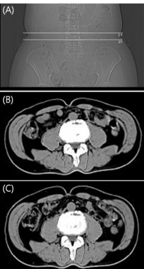
Figure 1. View of computer tomography of L4 upper and lower plate (A), before exercise (B), after exercise (C).

Table 2. Outcome of clinical variables (N=19)

Pre-test Post-test t p

VAS of LBP 5.77±0.93 2.51±0.95 14.11 0.00

ODI 16.73±4.96 7.36±3.04 16.79 0.00

VAS: Visual analogue scale, ODI: Oswestry disability index

after the experiment, the program was exploited by PACS (Medi- face, Seoul, Korea). The psoas major and multifidus muscles were drawn along the outline of the muscles to avoid fat, skeletal structures and other flexile tissues; the images were uniformly enlarged to 150.04% in order to better visualize the circum- ference of the psoas major and multifidus.

4

Various numbers of points (55∼60) were selected depending on the size and shape of each muscle; the CSA of the Lt & Rt muscles of the psoas major and multifidus were calculated automatically in mm

2

by the computer (Figure 1).

Spinal stabilization exercise began with riding the stationary bicycle for 10minutes as a warm-up, followed by 45∼50 minutes of using CENTAUR, ball maneuvers, or mats to do psoas major muscle strengthening exercises. The intensity test of exercise was performed at 30∼40% from weeks 1∼5 and at 40∼50% from weeks 6∼10. Stretching was conducted for 10 minutes as a cool-down. The exercise sessions lasted for 65∼70 minutes each, twice a week, for 10 weeks. All the exercise was conducted under the direction of a supervisor.

3. Reliability of Measurement

All of the measurements were taken by the same individual. Intra- tester reliability was checked by repetitive measurement with the same protocol, and the intraclass correlation coefficient (ICC) of the CSA was 0.95, which is high because both tests were conducted by the same researcher.

4. Statistical Analysis

Statistical analyses of the findings were performed with the SPSS for Window v. 12.0 software program. A paired-t test was used to assess the differences between paired measurements of CSA, VAS, ODI and paraspinal muscle strength in individual patients.

A significance level of p<0.015 was set.

III. Results

1. Clinical Outcome Assessment

The clinical symptom changes are shown in Table 2. After spinal stabilization exercise for 10 weeks, pain was significantly de- creased from 5.7 to 2.5 (p<0.01), and the limitation of daily life was also significantly decreased from 16.7 to 7.3 (p<0.01).

2. CSA of the Multifidus & Psoas major muscle in patients with DDD

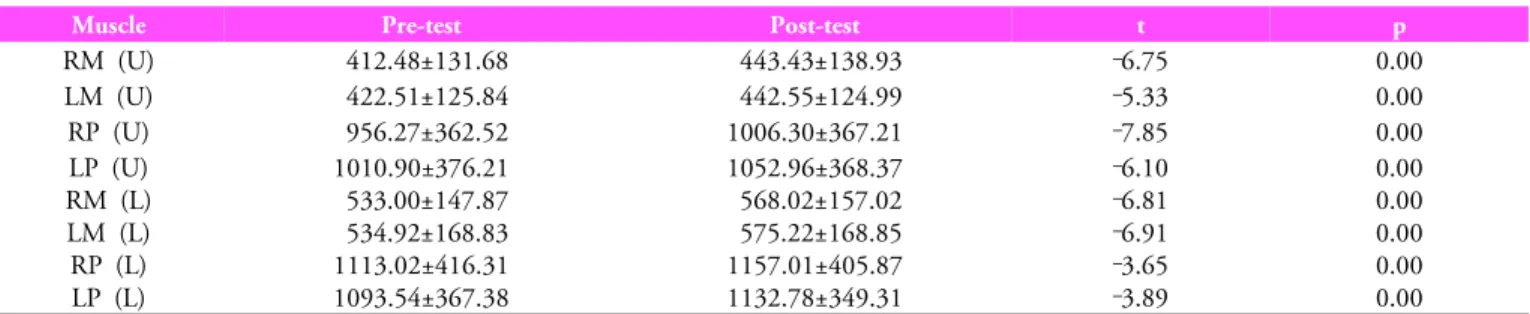
The CSA deep muscle changes after 10 weeks of spinal stabili-

zation exercise for 19 DDD patients are shown in Table 3. L4

upper end plate size of the right multifidus was significantly

increased from 412.4 mm

2

before exercise to 443.4 mm

2

after

exercise (p<0.01); the left one was significantly increased from

422.5 mm

2

before exercise to 442.5 mm

2

after exercise (p<0.01),

(4)

Table 3. CSA of the multifidus & psoas muscle in DDD patients (N=19)

Muscle Pre-test Post-test t p

RM (U) 412.48±131.68 443.43±138.93 ‑6.75 0.00

LM (U) 422.51±125.84 442.55±124.99 ‑5.33 0.00

RP (U) 956.27±362.52 1006.30±367.21 ‑7.85 0.00

LP (U) RM (L) LM (L) RP (L) LP (L)

1010.90±376.21 533.00±147.87 534.92±168.83 1113.02±416.31 1093.54±367.38

1052.96±368.37 568.02±157.02 575.22±168.85 1157.01±405.87 1132.78±349.31

‑6.10

‑6.81

‑6.91

‑3.65

‑3.89

0.00 0.00 0.00 0.00 0.00 RM: Right multifidus, LM: Left multifidus, RP: Right psoas, LP: Left psoas

(U): L4 upper end plate, (L): L4 lower end plate Unit: mm

2

Table 4. Outcome of paraspinal muscle strength testing (N=19)

angle Pre-test (NkmkNm) Post-test (NkmkNm) t p

0° 72.69±20.01 79.29±19.15 ‑4.52 0.01

90° 64.53±22.03 71.05±20.88 ‑8.17 0.00

180° 38.85±13.83 75.43±19.67 ‑11.51 0.00

‑90° 62.84±69.90 69.90±21.26 ‑4.11 0.00

0°: erector spinae, multifidus, 180°: rectus abdominis, inter/exter oblique, 90°: Rt internal obilique, ‑90°: Lt intrenal oblique

and the right psoas was significantly increase form 956.2 mm

2

before exercise 1006.3 mm

2

after exercise (p<0.01); the lt psoas was significantly increased from 1010.9 mm

2

before exercise 1052.9 mm

2

after exercise (p<0.01). L4 lower end plate size of right

& left psoas, multifidus were significantly increased (p<0.01).

3. Paraspinal Muscle Strength

Paraspinal muscle strength values pre- and post-experiment are shown in Table 4.

Muscular strength in all 4 angles increased after exercise (p<0.01). Muscular strength in the 180˚ angle showed the greatest increase from 38.8 kNm before exercise to 75.4 kNm after exercise (p<0.01). Strength in the other angles, 0˚, 90˚ and

‑90˚ was significantly increased by as much as 6.6 kNm, 6.5 kNm and 7.0 kNm, respectively (p<0.01).

IV. Discussion

The results of this study show that spinal stabilization exercise has the positive effects of reducing pain and lumbar disability, improving atrophy of the multifidus and psoas major muscles and improving paraspinal muscle strength in DDD patients.

A recent study reported that spinal stabilization exercise is a very effective method for relieving pain and improving function

in LBP patients; it is focused on strengthening the deep intrinsic muscles such as the multifidus, psoas major, transversus abdo- minis, and oblique internus abdominis

16

because these muscles limit excessive rotation and dislocation at the intervertebral level.

These muscles improve intervertebral disc stability; in particular,

strengthening the multifidus in order to stabilize lumbar seg-

ments was found to reduce pain and lower the recurrence rate of

LBP.

1,15,27,28

Improvement of function is emphasized in treating

CLBP patients because if pain is related to a dysfunction of local

stabilization which occurs after onset, it can be eliminated. How-

ever, if dysfunction continues, it causes the individual to be more

susceptible to the recurrence of LBP. Both the multifidus and

psoas major provide stabilization for the spine and are sensitive

to pathological alteration.

17

In particular, multifidus provides

stabilization biomechanically to the spine by segmental adhe-

rence and nerve control;

10

image technologies have shown

atrophy of the multifidus in LBP patients.

9

Several previous

imaging studies have reported evidence of multifidus, psoas and

paraspinal muscle atrophy in patients with LBP.

3,4,9,20,21

In an

MRI study concerning atrophy of the multifidus and psoas major

of unilateral LBP patients, atrophy of the multifidus was found

in 80% of the patients. It was shown that the CSA of the psoas

major, multifidus, paraspinal muscles and quadratus lumborum

are smaller in CLBP patients than in healthy subjects,

5

(using

CT) in special, atrophy of multifidus is serious (L4 upper end

(5)

plate).

4

Even in a study using ultrasound, unilateral atrophy of acute and subacute LBP patients was discovered;

1

the CSA of the multifidus at L4-L5 in chronic LBP patients was reduced, and there was the greatest difference at the L5 level,

21

which suggests that muscle atrophy of chronic LBP patients is localized rather than general.

4

Accordingly, it was reported that it is necessary for CLBP patients to perform special exercises to recover from this local damage. In a study of the CSA forthe psoas major using MRI, the unilateral CSA in 25 patients suffering from unilateral radiation pain due to single level disc was reduced.

3

I was also reported that atrophy exists in LBP, and that atrophy of the multifidus and psoas major is positively correlated with pain.

17

Evidence that the CSA of the paraspinal muscle of CLBP patients is being reduced gets gradually increased.

20,29

Therefore, the present study focused on the effect that spinal stabilization exercise has on clinical symptoms such as size of the CSA of the multifidus and psoas major and pain in DDD patients.

Studies concerning the effect of spinal lesions on the size of the psoas major have reported contradictory results. It was shown that the CSA of the multifidus in LBP patients was reduced without a change in the psoas major;

20,29

Conversely, the CSA of the psoas major in herniated nucleus pulpous patients and LBP patients was significantly reduced.

3,5,9,17

They reported that psoas major plays an important role in providing spinal stabilization;

together with multifidus, it may coactivate or facilitate contraction of the transversus abdominis and multifidus,

5,17

and atrophy of the psoas major has been related with pain.

17

It was suggested that stretching of the psoas major to treat lumbar spine disorders, may be able to increase spine mobility.

3

However, they concen- trated only on stretching, so the effect of selective muscle training of the psoas major on the symptoms of LBP has not yet been established. It appears that excessive contraction of the psoas major may increase intervertebral disc pressure. As a result of the combination of strengthening and stretching of the psoas major in the exercise program in this study, its CSA showed a lesser increase than that of multifidus however, its CSA was increased and pain was reduced (43.5%). Thus, spinal stabilization exercise including appropriate strengthening exercises of the psoas major may be effective for rehabilitation of DDD patients; the difference in the CSA increase between the two muscles can be attributed to the fact that an implement was used to strengthen the multi- fidus, while simple isomeric exercise was used for the psoas

major. It was reported that spinal stabilization exercise may be more effective than any other conventional therapy in terms of pain relief and improvement of body function, while special exercise is effective for spondylolysis, spondylolisthesis and re- duction of pain and improvement of dysfunction in DDD patients,

6,7

and rate of recurrence of LBP patients was lowered.

15

Also, even in studies for nonspecific LBP patients, spinal stabi- lization exercise is more effective than general physical therapy and manual therapy in relieving pain, improving dysfunction and preventing recurrence of LBP for subacute or chronic LBP patients.

11-13

In the previous study, patients were divided into the spinal stabilization exercise group and another therapeutic group.

The main focus was pain relief and improvement of dys- function, not improvement of atrophy of the multifidus and psoas major, which plays a major role in spine stabilization. After comparing the CSA of the multifidus through CT imaging of the L3, L4 upper endplate and the L4 lower endplate after conducting 10 weeks of exercise patients were divided into spinal stabilization exercise groups: the spinal stabilization+dynamic resistance group and the spinal stabilization+dynamic-stabtic resistance group. It was reported that the CSA of spinal stabi- lization+dynamic-static resistance group were increased at every level, but there was no difference between the two groups. Thus, it appeared that an intensive dynamic-static spinal stabilization exercise program is the most proper for reversing atrophy of the multifidus.

19

from the comparison of the CSA of the paraspinal muscles, it could be seen that the CSA was significantly increased in the spinal stabilization+dynamic resistance group and the spinal stabilization+dynamic-static resistance group.

18

However, as the spinal stabilization exercise group, unlike the other experimental group, did not use an implement, it appeared that this limited the increase of CSA. In contrast, as a result of having conducted spinal stabilization exercise program using an implement in this study, the CSA of the multifidus was signifi- cantly increased, even paraspinal strength was increased. Young elite cricketers increased the CSA of the multifidus (L5 level) and relieved pain by conducting spinal stabilization exercises, ceasing to lift heavy implements;

23

this hypertrophic effect on the deep intrinsic muscles only occurred with low-intensity exercise and accurate contraction of the multifidus. In this study, the supervisor instructed the patient to maintain contraction of the multifidus during exercise.

However, there have been some objections in recent studies

(6)

to using spinal stabilization exercise for chronic LBP patients because it is argued that using these exercises for nonspecific LBP patients has no advantage over other therapeutic approaches in terms of pain relief and recurrence of LBP.

14,30

In particular, it is claimed that previous studies of spinal stabilization exercise were conducted for nonspecific LBP patients, not on LBP patients diagnosed through accurate radiation checks. As the duration of symptoms was not clearly classified, there exists a limitation in the generalization of the results.

11-13,15,23

Spinal stabilization exer- cise should be used with the LBP patients whose deep intrinsic muscles are weakened, not simply any LBP patients; selection of patients should be clearly limited depending on the symptoms, the duration of the symptoms and the diagnosis. Thus, this study was clearly limited to patients who were diagnosed as DDD with symptom duration of over 5 months. Further, debate remains regarding whether special spinal stabilization exercise is more effective for rehabilitation of the spine than general phy- sical exercise. Undoubtedly, there is a body of current work showing that spinal stabilization is important,

28,31

and there is much evidence supporting that spinal stabilization exercise is more effective than a general physical exercise program; thus, spine rehabilitation exercise programs must be prescribed.

31

This study has a number of limitations. It may not able to reflect actual tissue relationships in provoking symptoms. It also may not be able to accurately represent the relationships of muscles while conducting functional activities, as all the CT scans are done in the supine position.

21

The study was not able to represent relationships between the increase of paraspinal musclestrength and change of the size of CSA of the multifidus and psoas major muscles. Also, the sample size is small

3,9

and devoid of a control group. It was not able to show if spinal stabilization exercise for the DDD patients is more effective than other therapeutic approaches.

V. Conclusion

Multifidus and psoas major play a significant role in stabilizing the spine and in its functional movements. As a result of having conducted spinal stabilization exercise, including the psoas major strengthening exercise, pain was relieved, disability was reduced and paraspinal muscle strength was increased in DDD patients.

In addition, the CSA of the multifidus muscles and psoas major

muscle were also significantly increased. Accordingly, it can be said that spinal stabilization exercise is an effective treatment method for rehabilitation of DDD patients.

Author Contributions Research design: Kim SH Acquisition of data: Kim SH

Analysis and interpretation of data: Kim SH Drafting of the manuscript: Kim SH Research supervision: Lee WH

References

1. Hides JA, Richardson CA, Jull GA. Multifidus muscle recovery is not automatic after resolution of acute, first- episode low back pain. Spine (Phila Pa 1976). 1996;21(23):

2763-9.

2. Koumantakis GA, Watson PJ, Oldham JA. Trunk muscle stabilization training plus general exercise versus general exercise only: randomized controlled trial of patients with recurrent low back pain. Phys Ther. 2005;85(3):209-25.

3. Dangaria TR, Naesh O. Changes in cross-sectional area of psoas major muscle in unilateral sciatica caused by disc herniation. Spine (Phila Pa 1976). 1998;23(8):928-31.

4. Danneels LA, Vanderstraeten GG, Cambier DC et al. CT imaging of trunk muscles in chronic low back pain patients and healthy control subjects. Eur Spine J. 2000;9(4):266-72.

5. Kamaz M, Kiresi D, Oguz H et al. CT measurement of trunk muscle areas in patients with chronic low back pain. Diagn Interv Radiol. 2007;13(3):144-8.

6. Lie H, Frey S. Mobilizing or stabilizing exercise in degenera- tive disk disease in the lumbar region? Tidsskr Nor Laegeforen.

1999;119(14):2051-3.

7. O’Sullivan PB, Phyty GD, Twomey LT et al. Evaluation of specific stabilizing exercise in the treatment of chronic low back pain with radiologic diagnosis of spondylolysis or spon- dylolisthesis. Spine (Phila Pa 1976). 1997;22(24):2959-67.

8. Adams MA, Roughley PJ. What is intervertebral disc de- generation, and what causes it? Spine (Phila Pa 1976). 2006;

31(18):2151-61.

9. Cooper RG, St Clair Forbes W, Jayson MI. Radiographic demonstration of paraspinal muscle wasting in patients with chronic low back pain. Br J Rheumatol. 1992;31(6):389-94.

10. Kader DF, Wardlaw D, Smith FW. Correlation between the

(7)

MRI changes in the lumbar multifidus muscles and leg pain.

Clin Radiol. 2000;55(2):145-9.

11. Moseley L. Combined physiotherapy and education is efficacious for chronic low back pain. Aust J Physiother. 2002;

48(4):297-302.

12. Niemisto L, Lahtinen-Suopanki T, Rissanen P et al. A rando- mized trial of combined manipulation, stabilizing exercises, and physician consultation compared to physician consul- tation alone for chronic low back pain. Spine (Phila Pa 1976).

2003;28(19):2185-91.

13. Rasmussen-Barr E, Nilsson-Wikmar L, Arvidsson I. Stabilizing training compared with manual treatment in sub-acute and chronic low-back pain. Man Ther. 2003;8(4):233-41.

14. Arokoski JP, Valta T, Kankaanpaa M et al. Activation of lumbar paraspinal and abdominal muscles during therapeutic exercises in chronic low back pain patients. Arch Phys Med Rehabil. 2004;85(5):823-32.

15. Hides JA, Jull GA, Richardson CA. Long-term effects of specific stabilizing exercises for first-episode low back pain.

Spine (Phila Pa 1976). 2001;26(11):E243-8.

16. Hides JA, Stokes MJ, Saide M et al. Evidence of lumbar multifidus muscle wasting ipsilateral to symptoms in patients with acute/subacute low back pain. Spine (Phila Pa 1976).

1994;19(2):165-72.

17. Barker KL, Shamley DR, Jackson D. Changes in the cross- sectional area of multifidus and psoas in patients with unilateral back pain: the relationship to pain and disability. Spine (Phila Pa 1976). 2004;29(22):E515-9.

18. Danneels LA, Cools AM, Vanderstraeten GG et al. The effects of three different training modalities on the cross- sectional area of the paravertebral muscles. Scand J Med Sci Sports. 2001;11(6):335-41.

19. Danneels LA, Vanderstraeten GG, Cambier DC et al. Effects of three different training modalities on the cross sectional area of the lumbar multifidus muscle in patients with chronic low back pain. Br J Sports Med. 2001;35(3):186-91.

20. Gibbons LE, Latikka P, Videman T et al. The association of trunk muscle cross-sectional area and magnetic resonance image parameters with isokinetic and psychophysical lifting strength and static back muscle endurance in men. J Spinal

Disord. 1997;10(5):398-403.

21. Hides J, Gilmore C, Stanton W et al. Multifidus size and symmetry among chronic LBP and healthy asymptomatic subjects. Man Ther. 2008;13(1):43-9.

22. O’Sullivan PB. Lumbar segmental ‘instability’: clinical pre- sentation and specific stabilizing exercise management. Man Ther. 2000;5(1):2-12.

23. Hides JA, Stanton WR, McMahon S et al. Effect of stabi- lization training on multifidus muscle cross-sectional area among young elite cricketers with low back pain. J Orthop Sports Phys Ther. 2008;38(3):101-8.

24. Yi S-J. Oswestry low back pain disability index and related factors in patients with low back pain. J Kor Soc Phys Ther.

2008;20(4):21-8.

25. Anders C, Brose G, Hofmann GO et al. Evaluation of the EMG-force relationship of trunk muscles during whole body tilt. J Biomech. 2008;41(2):333-9.

26. Kim HR, Kim YS. The effects of spinal stabilization exercise using gravity on patients with degenerative disc disease. J Kor Soc Phys Ther. 2008;20(1):23-31.

27. Comerford MJ, Mottram SL. Movement and stability dysfunction--contemporary developments. Man Ther. 2001;

6(1):15-26.

28. Foster NE, Thompson KA, Baxter GD et al. Management of nonspecific low back pain by physiotherapists in Britain and Ireland. A descriptive questionnaire of current clinical practice.

Spine (Phila Pa 1976). 1999;24(13):1332-42.

29. Parkkola R, Rytokoski U, Kormano M. Magnetic resonance imaging of the discs and trunk muscles in patients with chronic low back pain and healthy control subjects. Spine (Phila Pa 1976). 1993;18(7):830-6.

30. Cairns MC, Foster NE, Wright C. Randomized controlled trial of specific spinal stabilization exercises and conventional physiotherapy for recurrent low back pain. Spine (Phila Pa 1976).

2006;31(19):E670-81.

31. Lindstrom I, Ohlund C, Eek C et al. Mobility, strength, and fitnessafter a graded activity program for patients with sub- acute low back pain. A randomized prospective clinical study with a behavioral therapy approach. Spine (Phila Pa 1976).

1992;17(6):641-52.

수치

Table 1. Patient Characteristics (N=19)
Figure 1. View of computer tomography of L4 upper and  lower plate (A), before exercise (B), after exercise (C).
Table 3. CSA of the multifidus &amp; psoas muscle in DDD patients (N=19)

참조

관련 문서

Purpose: The purpose of this study was finding out the effects of spinal stabilization exercise using Centaur which is a 3D spinal stabilization sports implement on Chronic

Such results are believed to be due to the fact that the postoperative stabilization exercise showed decreased cervical pain and cervical disability index caused

The Effect of Lumbar Stabilization Exercises with Neurodynamic Techniques on Lumbar Muscular Strength and Oswestry Disability Index in Lumbar Disc Herniation Patient's..

Objective: The purpose of this study was to examine the effects lumbar stabilization exercise with kinesio taping on pain, muscle strength, and oswestry disability index

Objective: This study was aimed at investigating the effects of lumbar stabilization exercise according to correct verbal instructions in pain and

The purpose of this study was to evaluate the effects of using cervical spinal stabilization exercise for the improvement of pain intensity, cervical range of

Effects of Active Release Technique and Lumbar Stabilization Exercise on Pelvic Asymmetry, Muscle Activation and Pain in Chronic Low Back Pain Patients.. Nam Seungmin,

Purpose : The purpose of this study was to identify the effect of lumbar stabilization exercise (LSE) and isometric lumbar strengthening exercise (ILSE) on lumbar strength