pISSN 2288-9272 eISSN 2383-8493 J Oral Med Pain 2016;41(3):91-98 http://dx.doi.org/10.14476/jomp.2016.41.3.91
Oral Lichen Planus and Oral Lichenoid Lesion:
Diagnosis and Assessment of Direct Immunofluorescence
Kyung-Eun Lee
Department of Oral Medicine, School of Dentistry, Chonbuk National University, Institute of Oral Bioscience, Chonbuk National University, Jeonju, Korea
Received August 22, 2016 Revised September 17, 2016 Accepted September 19, 2016
Purpose: Oral lichen planus (OLP) has generated many discussions and been associated with much controversy for a long time. A reliable diagnosis of OLP has proven challenging and sig- nificant disagreements concerning its diagnosis has continued. Therefore, the aim of this study was to apprehend newly proposed diagnostic criteria of OLP and oral lichenoid lesion (OLL) and to evaluate difference of final diagnosis of OLP and OLL in accordance with type of diag- nostic criteria. Also, direct immunofluorescence (DIF) was compared to evaluate the value of DIF between two groups.
Methods: Fifty-two patients with DIF result were retrospectively reviewed. The selected patients were classified by the modified World Health Organization (WHO) diagnostic criteria of OLP and OLL and by criteria proposed by American Academy of Oral and Maxillofacial Pathology (AAOMP). Results of DIF in OLP and OLL were classified by deposition intensity or pattern of fibrinogen. The classification of fluorescence pattern in each specimen was graded as positive, possibly positive or negative.
Results: Patients diagnosed as OLP were a few more when the modified WHO diagnostic cri- teria were used than when criteria proposed by AAOMP were used. There was no statistical difference of DIF between OLP and OLL by applying the WHO modification criteria or criteria proposed by AAOMP.
Conclusions: The final diagnosis of OLP could be changed in accordance with type of diagnos- tic criteria and difference of DIF between OLP and OLL was not found.
Key Words: Criteria; Direct immunofluorescence; Fibrinogen; Fluorescent antibody technique;
Lichen planus, oral; Oral lichenoid lesion
Correspondence to:
Kyung-Eun Lee
Department of Oral Medicine, School of Dentistry, Chonbuk National University, 567 Baekje-daero, Deokjin-gu, Jeonju 54896, Korea Tel: +82-63-250-2044 Fax: +82-63-250-2058 E-mail: [email protected]
This paper was supported by Clinical Fund of Chonbuk National University Hospital.
JOMP
Journal of Oral Medicine and PainCopyright Ⓒ 2016 Korean Academy of Orofacial Pain and Oral Medicine. All rights reserved.
CC This is an open-access article distributed under the terms of the Creative Commons Attribution Non-Commercial License (http://creativecommons.org/licenses/by-nc/4.0/), which permits unrestricted non-commercial use, distribution, and reproduction in any medium, provided the original work is properly cited.
INTRODUCTION
Lichen planus (LP) is a chronic immune-mediated muco- cutaneous disease which can affect oral mucosa, skin, geni- tal mucosa, scalp and nail.
1)When LP is found in the oral cavity without associated dermatologic lesions, it is referred to as oral lichen planus (OLP).
2)The set of clinical and his- tologic criteria for diagnosis of OLP was published by the World Health Organization (WHO) collaborating centre for diagnosis of oral precancerous lesion in 1978. Diagnostic criteria of this seminal article are frequently referred to in
the literature on OLP although this article was focused for differential diagnosis of leukoplakia and related precancer- ous lesions.
3)However, no disease in the field of oral medicine and pa-
thology has generated more discussion and been associated
with more controversy than OLP for a long time. Although
much effort has been investigated in clinical, pathologic,
and basic science research studies, inconsistent results and
diverse opinions still leave many questions. A reliable diag-
nosis of OLP has proven challenging for a few reasons and
significant disagreements concerning its diagnosis continue
to be found among pathologists and clinicians.
4)van der Meij et al.
5,6)showed variability in both interob- server and intraobserver reliability in the clinical and his- tologic assessment of OLP by the WHO criteria. The authors proposed the set of strict diagnostic criteria based on the 1978 clinical and histologic definition of OLP by the WHO in 2003.
7)The criteria were referred to as the modification WHO diagnostic criteria of OLP and oral lichenoid lesion (OLL). Rad et al.
8)compared the correlation between clini- cal and histologic diagnosis of OLP for both the WHO cri- teria and the 2003 modified WHO diagnostic criteria. They found increased agreement between clinicians and patholo- gist in the diagnosis of OLP when the modified WHO diag- nostic criteria were used compared with the WHO criteria.
8)However, Cheng et al.
4)proposed a new stricter set of diag- nostic criteria by adding additional elements to the existing modified WHO diagnostic criteria through a position paper of American Academy of Oral and Maxillofacial Pathology (AAOMP).
Above these criteria, several studies suggested that im- munopathological finding may occasionally be additional markers in the diagnosis of OLP as it showed fibrinogen de- position at the mucosal-submucosal interface.
9-12)Author wondered whether final diagnosis of oral lesions similar to OLP in accordance with types of criteria could be changed and immunopathology between OLP and OLL had differences. In a previously published study, author investi- gated direct immunofluorescence (DIF) in only clinically di- agnosed OLP, based by the modified WHO diagnostic crite- ria. In previous study, difference of fluorescence pattern in OLP and OLL was not found when oral lesions suspected to OLP were classified by only clinical features.
13)In this study, author included both clinical and histologic features for diagnosis of OLP and OLL by using both the modified WHO diagnostic criteria and the criteria proposed by AAOMP. The aim of this study was to evaluate differ- ence of final diagnosis of OLP and OLL in accordance with type of diagnostic criteria to be complied and to compare the prevalence and intensity or pattern of immunofluores- cence in OLP and OLL.
MATERIALS AND METHODS
1. Patients Selection
This study was conducted on outpatients who visited at the Department of Oral Medicine in Chonbuk National University Hospital (Jeonju, Korea) from January 2007 to June 2016. Patients who had lesions suspected to OLP clini- cally and took a biopsy on the buccal mucosa for DIF were retrospectively reviewed. Patients who diagnosed as vesi- culo-mucosal disease in DIF were excluded and those hav- ing either fibrinogen or negative deposition in DIF were selected.
2. Classification by the Modified WHO Diagnostic Criteria of OLP and OLL
The modified WHO diagnostic criteria were used for di- agnosis of OLP and OLL.
7)In criteria, the clinical criteria in- cluded the following conditions: 1) the presence of bilateral, more or less symmetrical lesions, 2) presence of lace-like network of slightly raised white lines (reticular pattern), and 3) erosive, atrophic, bullous, or plaque type lesions accepted as a subtype only in presence of reticular lesion. The his- tologic criteria included the followings: 1) the presence of well-defined band-like zone of cellular infiltration confined to the superficial part of the connective tissue, consisting mainly lymphocytes, 2) signs of ‘liquefaction degeneration’
in the basal cell layer, and 3) absence of epithelial dysplasia (Fig. 1A).
Clinical features of the patients were evaluated on basis of medical record and histologic feature were evaluated on basis of microscopic slides obtained from Department of Pathology in Chonbuk National Hospital. When the clinical features and histologic features included all of the afore- mentioned criteria, oral lesion was classified as OLP. Lesions that did not complete either clinical or histologic criteria were diagnosed as OLL.
3. Classification by Criteria Proposed by AAOMP
The criteria by prosed by AAOMP were used for diagno-
sis of OLP and OLL.
3)The clinical criteria included the pres-
ence of multifocal symmetrical lesions with white or mixed
white and red lesions exhibiting one or more of the follow-
ing forms: reticular/papular, erosive, atrophic, bullous, and
plaque type. The histologic criteria included the followings:
1) the presence of well-defined band-like or patchy lym- phocytic infiltrate in the lamina propria confined to the epi- thelium lamina propria interface, 2) basal cell liquefactive degeneration, 3) absence of epithelial dysplasia, 4) lympho- cytic exocytosis (Fig. 1B), and 5) absence of verrucous epi- thelial architectural change.
Clinical and histologic features of patients were evaluated on basis of medical record and microscopic slides. When the clinical features and histologic features included all of
the aforementioned criteria, oral lesion was classified as OLP. Lesions that did not complete either clinical or histo- logic criteria were diagnosed as OLL.
4. Examination and Diagnostic Classification of Direct Immunofluorescence
The results of DIF were reviewed through the pictures taken under a fluorescent microscope and obtained from Department of Pathology in Chonbuk National Hospital.
Results of DIF were classified by intensity or pattern of
Fig. 1. (A) Histologic features of oral lichen planus (OLP). A dense predominantly lymphocytic infiltrate is situated in the laminal propria abutting oral mucosa stratified squamous epithelium. Liquefaction degeneration in basal cells is apparent (H&E staining, ×100). (B) Histologic features of OLP. The presence of lymphocytic infiltrate in the lamina propria and basal cell liquefactive degeneration is shown. Lymphocytic exocytosis also is shown (arrow; H&E staining, ×200).
A B
Fig. 2. (A) Positive pattern: direct immunofluorescence stained section showing deposition of fibrinogen along the basement membrane in linear-to-fibrillary shaggy pattern and clearly and consistently greater intensity than the background. (B) Possibly positive pattern: direct immunofluorescence stained section showing deposition of fibrinogen along the basement membrane in homogenous linear pattern and slightly greater intensity than the background.
A B
fibrinogen deposition. The classification of fluorescence de- position in each specimen was graded as positive, possibly positive or negative. Positive pattern (Fig. 2A) was defined as intensity that was clearly and consistently greater than the background intensity or as pattern presented linear-to- fibrillary shaggy deposition of fibrinogen along the base- ment membrane (BMZ). In the possibly positive pattern (Fig.
2B), the observable intensity was only slightly greater than the background or homogenous linear pattern of fibrino- gen was along the BMZ. Negative fluorescence was defined when no observable intensity greater than the background fluorescence was noted.
9-12)5. Statistic Methods
The chi-square test was used to compare DIF in OLP and OLL. Statistical test was done at the 5% significant lev- el. Statistical calculations were performed with the SPSS Statistics version 12.0 (SPSS Inc., Chicago, IL, USA).
RESULTS
A total of 52 outpatients were selected. Of 52 patients, 18 patients were males and 34 patients were females. The
overall mean age was 55.40 years. The youngest patient was 17 years and the oldest was 73 years (Table 1).
The most commonly affected site was buccal mucosa fol- lowed by buccal fold, buccal gingiva. All patients did not have the cause relationship between onset of medication and of oral lesions. All lesions are not localized adjacent to and in contact with dental restoration. Also onset of all le- sions did not correlate with the start of restoration treat- ment and use of allergenic products.
1. Diagnosis of OLP and OLL by the Modification WHO Criteria and DIF Assessment
Thirty-one patients were diagnosed as having OLP and 21 patients were diagnosed as having OLL by the modification WHO criteria (Table 2). Twenty-two of 31 patients diag- nosed as OLP had medical history and some of those were taking medicine associated with general disease. Ten of 31 patients did not medical history. The most commonly medi- cal history was hypertension (7 of 22 patients) followed by thyroid disease, gastric cancer operation, osteoporosis, hep- atitis B, asthma.
Fibrinogen deposition was found in 37 of 52 patients.
Both OLP and OLL had positive and possibly positive pat- tern. Prevalence of positive pattern was 51.6% in OLP and 33.3% in OLL and that of possibly positive pattern was 22.6% in OLP and 28.6% in OLL. Prevalence of nega- tive was 25.8% in OLP and 38.1% in OLL (Table 3). There was no statistical difference of DIF between OLP and OLL (p=0.419).
2. Diagnosis of OLP and OLL by the Criteria Proposed by AAOMP and DIF Assessment
According to criteria proposed by AAOMP, 28 of 52 pa- tients were diagnosed as OLP. The others were diagnosed as
Table 1. Age and sex distribution of 52 patients
Age group (y) Male
(n=18)
Female (n=34)
11-20 1 0
21-30 0 0
31-40 1 3
41-50 7 10
51-60 2 6
61-70 5 13
70-80 2 2
Values are presented as number only.
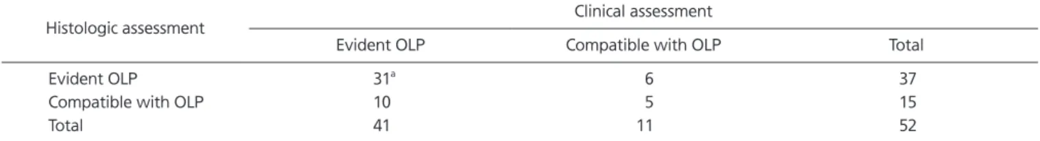
Table 2. Final diagnosis of 52 patients according to the modified WHO diagnostic criteria
Histologic assessment Clinical assessment
Evident OLP Compatible with OLP Total
Evident OLP 31
a6 37
Compatible with OLP 10 5 15
Total 41 11 52
WHO, World Health Organization; OLP, oral lichen planus; OLL, oral lichenoid lesion.
Values are presented as number only.
a
Thirty-one patients were finally diagnosed as having OLP; the remaining 21 were diagnosed as having OLL.
OLL (Table 4). Seventeen of 28 patients had medical history and 11 patients had no medical history.
Fibrinogen deposition was found in 37 of 52 patients.
Both OLP and OLL had positive and possibly positive pat- tern similarly to result by the modification WHO criteria.
Prevalence of positive pattern was 46.4% in OLP and 41.6%
in OLL and that of possibly positive pattern was 25.0% in OLP and 29.2% in OLL. Prevalence of negative was 28.6%
in OLP and 29.2% in OLL (Table 5). There was no statistical difference of DIF between OLP and OLL (p=0.972).
DISCUSSION
OLP is a chronic inflammatory disorder having un- known etiology and affecting stratified squamous epithe- lia.
14)In general, it was known that mucosal lesions in OLP
are usually multiple and often have a symmetrical distri- bution.
3,7)In newly criteria proposed by AAOMP, multifo- cal symmetric distribution as clinical feature of OLP was proposed.
4)It is divided into six types: reticular, papular, plaque-like, erosive, atrophic, and bullus.
3,4,7)A characteris- tic feature is the presence as slender white lines (Wickham’s striae) radiating from the papules. In the reticular form there is lacelike network of slightly raised gray-white lines, often interspersed with papules or rings.
3)Whereas reticular pattern occurs as isolated lesions, erythematous lesions are accompanied by reticular lesions, and erosive lesions are accompanied by reticular and erythematous lesions in al- most all cases.
15)These features help clinically differentiate OLP from other vesiculo-erosive disease such as pemphigus and pemphigoid.
14)Histological features included presence of keratinized layer with either ortho- or parakeratinzed and
Table 3. Comparison of the outcome of diagnosis by the modified WHO diagnostic criteria and direct immunofluorescence assessment
Deposition of fibrinogen OLP OLL Total p-value
Positive 16 (51.6) 7 (33.3) 23 (44.2)
Possibly positive 7 (22.6) 6 (28.6) 14 (26.9) 0.419
Negative 8 (25.8) 8 (38.1) 15 (28.8)
Total 31 (100) 21 (100) 52 (100)
WHO, World Health Organization; OLP, oral lichen planus; OLL, oral lichenoid lesion.
Values are presented as number (%).
p-value was obtained from chi-square test.
Table 4. Final diagnosis of 52 patients according to criteria proposed by AAOMP
Histologic assessment Clinical assessment
Evident OLP Compatible with OLP Total
Evident OLP 28
a8 36
Compatible with OLP 9 7 16
Total 37 15 52
AAOMP, American Academy of Oral and Maxillofacial Pathology; OLP, oral lichen planus; OLL, oral lichenoid lesion.
Values are presented as number only.
a