www.i-mri.org 192
Giant Arachnoid Granulations in Headache Mimicking Migraine with Aura
INTRODUCTION
Arachnoid granulations are projections of arachnoid membrane into dural sinuses, frequently located parasagitally in the transverse and superior sagittal sinuses. When arachnoid granulations are enlarged, they are termed “giant”. Previous reports have found these structures to be sometimes associated with headaches, and in some patients, idiopathic intracranial hypertension (IIH) may occur (1-3). The pathophysiology of these enlarged structures seen as filling defects on imaging is not clearly defined, although in some cases they are presumed to cause symptoms such as headache via pressure resulting from secondary venous sinus obstruction. We present a case of headache mimicking migraine with aura, not previously described in the setting of giant arachnoid granulations in a 39-year-old, previously healthy man.
CASE REPORT
A 39-year-old previously healthy man presented to the neurology clinic with the chief complaint of headaches described as a dull, pressure-like sensation associated with lightheadedness that began approximately three weeks prior to presentation.
He reported the headaches to occur three to four days weekly on average and experienced worsening of his headaches with sleep deprivation or alcohol consumption.
The headaches were not precipitated by changes in position. He reported no other associated symptoms such as visual obscurations, tinnitus or diplopia. He reported no
This is an Open Access article distributed under the terms of the Creative Commons Attribution Non-Commercial License (http://creativecommons.org/licenses/
by-nc/3.0/) which permits unrestricted non-commercial use, distribution, and reproduction in any medium, provided the original work is properly cited.
Received: June 1, 2017 Revised: June 30, 2017 Accepted: July 3, 2017 There was no financial support for this work.
Correspondence to:
Jung E Park, M.D.
Department of Neurology, Dongguk University Ilsan Hospital, 27, Dongguk-ro, Ilsandong-gu, Goyang-si 10326, Korea.
Tel. +82-31-961-7271 Fax. +82-31-961-7216 E-mail: [email protected]
Copyright © 2017 Korean Society of Magnetic Resonance in Medicine (KSMRM)
iMRI 2017;21:192-194 https://doi.org/10.13104/imri.2017.21.3.192
Case Report
Giant arachnoid granulations have been reported to be associated with headaches, which can be acute or chronic in presentation. In some cases, idiopathic intracranial hypertension, previously called pseudotumor cerebri, may occur. The pathophysiology of these enlarged structures seen as filling defects on imaging is not clearly defined, although they are presumed to cause symptoms such as headache via pressure resulting from secondary venous sinus obstruction. We present a unique presentation of secondary headache in a 39-year-old man with no prior history of headaches found to have giant arachnoid granulations, presenting as migraine with aura.
Keywords: Arachnoid; Granulation; Headache; Migraine; Aura
pISSN 2384-1095 eISSN 2384-1109
Jung E Park1, Eun-ja Lee2
1Department of Neurology, Dongguk University Ilsan Hospital, Goyang, Korea
2Department of Neuroradiology, Dongguk University Ilsan Hospital, Goyang, Korea Magnetic resonance imaging
www.i-mri.org 193
https://doi.org/10.13104/imri.2017.21.3.192
a b
c
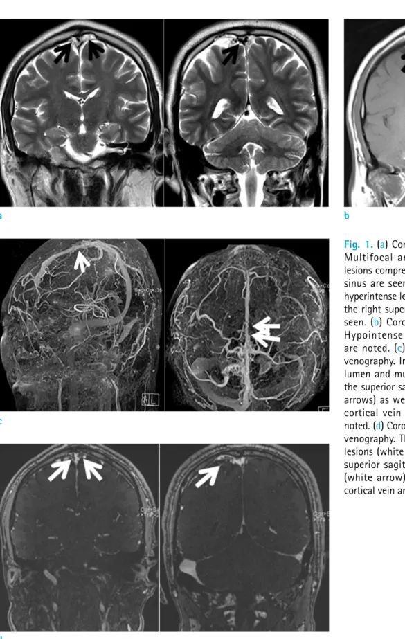
Fig. 1. (a) Coronal T2-weighted image.
Multifocal amorphous hyperintense lesions compressing the superior sagittal sinus are seen (black arrows). Tubular hyperintense lesion (short black arrow) in the right superficial cortical vein is also seen. (b) Coronal T1-weighted image.
Hypointense lesions (black arrows) are noted. (c) Contrast-enhanced MR venography. Irregular narrowing or the lumen and multifocal filling defects in the superior sagittal sinuses (long white arrows) as well as the right superficial cortical vein (short white arrow) are noted. (d) Coronal source imaging of MR venography. There are poorly opacified lesions (white arrows) compressing the superior sagittal sinus. Filling defects (white arrow) in the right superficial cortical vein are seen.
d
www.i-mri.org 194
Giant Arachnoid Granulations in Headache Mimicking Migraine | Jung E Park, et al.
history of previous headaches. Family history was negative for headache or other neurological disorders. Past medical history was unremarkable and he did not take any daily medications.
Magnetic resonance imaging (MRI) of the brain was conducted, which revealed an amorphous lesion compressing the superior sagittal sinus, shown as high signal on T2-weighted image and low signal on T1- weighted image (Fig. 1a, b). MR venography of the brain was subsequently obtained, which revealed irregular narrowing of the lumen and multifocal filling defects of the superior sagittal sinus suggestive of arachnoid granulations (Fig. 1c, d). His symptoms improved with the use of symptomatic medications including naproxen for headache and dimenhydrinate for dizziness, but he was eventually started on propranolol for headache prophylaxis. The patient stated that his headache frequency was dramatically reduced approximately three weeks following the daily use of propranolol.
DISCUSSION
Giant arachnoid granulations, thought to represent extension of endothelium-lined venous sinuses, are seen as focal ovoid masses with surrounding blood flow on neuroimaging. There structures are termed “giant” when they are sufficiently large enough to cause local dilation or filling defects. Giant arachnoid granulations are commonly seen as nonenhancing, hypointense lesions on T1-weighted MR images (4). Headaches have been reported in patients with arachnoid granulations, and imaging findings, as well as clinical presentation can lead to the differential diagnosis of venous sinus thrombosis or IIH. Although IIH is a reported manifestation, venous sinus pressure has commonly been found to be normal despite the presence of headaches, suggesting that increased pressure may not be the sole explanation for patient symptomatology (5, 6). The headache characteristics of our patient were not indicative
of venous sinus thrombosis or IIH and therefore he did not undergo further relevant evaluation. His headaches which were associated with vertigo met the International Classification of Headache Disorders (ICHD-3) criteria for migraine with aura, defined as recurrent attacks of central nervous system symptoms usually followed by headache.
However, the patient did not report history of previous headaches, which is therefore suggestive of secondary headache that is migraine-like. The patient’s headaches were responsive to propranolol, a widely-used medication used for prophylaxis in migraine patients. Our case is an example of headache mimicking migraine with aura in a patient with giant arachnoid granulations, a clinical manifestation not previously described in the literature.
Therefore, it may be useful to consider such structural etiology in a patient presenting with headaches suggestive of migraine who reports no previous history of migraines in one’s younger years.
REFERENCES
1. Arjona A, Delgado F, Fernandez-Romero E. Intracranial hypertension secondary to giant arachnoid granulations. J Neurol Neurosurg Psychiatry 2003;74:418
2. Kiroglu Y, Yaqci B, Cirak B, Karabulut N. Giant arachnoid granulation in a patient with benign intracranial hypertension. Eur Radiol 2008;18:2329-2332
3. Zheng H, Zhou M, Zhao B, Zhou D, He L. Pseudotumor cerebri syndrome and giant arachnoid granulation:
treatment with venous sinus stenting. J Vasc Interv Radiol 2010;21:927-929
4. Peters SA, Frombach E, Heyer CM. Giant arachnoid granulation: differential diagnosis of acute headache.
Australas Radiol 2007;51 Spec No.:B18-20
5. Choi HJ, Cho CW, Kim YS, Cha JH. Giant arachnoid granulation misdiagnosed as transverse sinus thrombosis. J Korean Neurosurg Soc 2008;43:48-50
6. Kan P, Stevens EA, Couldwell WT. Incidental giant arachnoid granulation. AJNR Am J Neuroradiol 2006;27:1491-1492