지난 십 수년동안 자기공명을 이용한 영상기법은 가시적 형 태의 해부학적 묘사 뿐만 아니라 생명체내의 분자상태를 영상 화 하고자 하는 노력으로 발전해왔다. 그 결과 확산 현상이 영 상화 되고 급성 뇌 경색 진단에서 확산영상의 탁월한 가치가 입증되어 현재 임상적으로 널리 이용되고 있다. 근래에 조직 내에서 확산현상의 텐서(tensor) 성질을 자기공명영상으로 구 현하여 뇌 백질내에 얽혀있는 신경다발을 그 역할과 주행경로 에 따라 볼 수 있게 되었다. 확산텐서영상(diffusion tensor imaging; DTI)은 신경 해부 및 생리학적 연구, 여러 질병의 병 태생리학적 연구와 그 임상적용에 새로운 장을 열고 있다. 이 에 저자들은 뇌 확산텐서영상의 원리와 현재 진행되고 있는 임상적용의 예를 살펴보고자 한다.
확산텐서영상의 원리
확산과 확산영상
확산은 브라운 운동(Brownian motion)이라 불리는 열에너 지에 의한 분자의 불규칙한 운동을 의미하며 확산에 의해 분 자는 공간상에서 위치가 변한다(translational motion). 이러한 위치변화는 자기공명 시 스핀의 T1 또는 T2 붕괴(decay)와 는 또 다른 탈위상을 조장하여 자기공명신호를 감소시킨다(1).
물 분자는 뇌조직속에서 50 msec 동안 평균 10 μm의 거리를
세포막, 신경섬유, 거대분자와 같은 조직구성 인자들 사이를 지 나기도 하고 부딪히기도 하며 확산한다. 이는 mm단위의 자기 공명영상의 해상력으로는 가시화 할 수 없는 미세 운동이다.
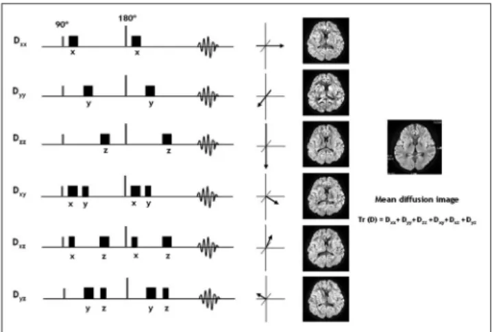
1965년 Stejskal과 Tanner 등(2)은 90°와 180°펄스를 이용 한 스핀에코 펄스대열의 180°펄스 전후에 각각 강한 경사자 장을 추가해서 다양한 조직들이 가지는 확산력의 차이를 증폭 시킴으로 확산현상을 가시세계로 이끌었다(Fig. 1). 이 때 걸 어주는 경사자장의 특성을 b 값(factor)이라 칭하는데 이는 경 사자장의 세기(G), 각 경사자장의 시간(δ), 경사자장간 시간 (Δ), 자기공명기기의 자기회전율(gyromagnetic ratio; γ)에 의 해 결정된다.
b=γ2G2δ2 (Δ - δ/3)
확산영상(diffusion imaging)의 신호강도는 확산 강조를 위 한 경사자장 후의 신호강도의 감소 정도로 결정되며 이는 조 직의 확산계수(diffusion coefficient; D)와 경사자장의 b 값에 따라 달라진다.
In(S / S0)=- bD
, S0는 경사자장의 b 값이 0일 때의 신호강도 이다.
즉, 확산이 큰 조직(예, 뇌척수액)은 경사자장에 의한 위상 차가 확산이 작은 조직(예, 뇌 백질)보다 크므로 신호강도가 더 감소되고 같은 조건이다 하더라도 경사자장의 b 값이 커질
뇌 확산텐서자기공명영상
1김현정・최충곤2・이정현2・양보성・강시원・이연수・김지창・황보설
자기공명영상 기술의 발전은 이미 해부학적 형태 묘사를 뛰어넘어 확산이라는 생리 상태를 보여주기에 이르렀고 이제 확산 현상을 이용하여 비침습적으로 뇌의 미세 구조인 신경 섬유를 그려 낼 수 있는 확산텐서영상이 가능해졌다. 더불어 신경 섬유의 발달과 퇴행 과정이 밝혀지 고 있으며 구조에 따른 뇌기능의 연구에도 새로운 장이 열려 신비로운 뇌로 인류는 한발 더 접근하게 되었다. 신경섬유추적지도를 실제와 같이 구현하고자 하는 많은 자기공명 물리학자 들의 기술적인 노력에서 뇌의 구조와 기능을 밝히고자 하는 뇌 과학자들의 연구, 그리고 확산 텐서영상을 이용하여 뇌 질환의 병태생리를 밝히고 유용한 임상적 쓰임새를 알아내려는 의사 들의 연구에 이르기까지 확산텐서영상은 뇌 과학 분야의 한 축을 이루고 있다. 이에 저자들은 본 종설에서 확산텐서영상의 원리와 현시점에서의 문제점들을 알아보고 지금까지 진행되어왔 고 진행되고 있는 임상응용을 위한 여러 연구들을 살펴 보고자 한다.
1가톨릭대학교 의과대학 대전성모병원 영상의학과
2울산대학교 의과대학 서울아산병원 방사선과
이 논문은 2005년 5월 22일 접수하여 2005년 7월 15일에 채택되었음.
수록 분자의 위상차가 커지므로 확산정도를 더 민감하게 반영 한다.
이제 확산영상을 얻음으로 조직의 확산정도를 알 수 있고 확산계수를 계산할 수 있는데 이렇게 얻어진 조직의 확산계수 (D)는 무제한적이고 무작위적인 본래의 확산형태가 아닌 제한 적이고 덜 무작위적인 조직 내에서의 확산을 반영하는 것으로 현성확산계수(apparent diffusion coefficient; ADC)라 한다(3, 4).
ADC = In(S / S0) / -b
텐서(Tensor)
텐서란 물질이 가지는 운동 또는 힘의 원리에 대한 다소 추 상적인 수학적 개념이다. 물질세계를 수리화 할 때 쓰이는 스 칼라(scalar)나 벡터(vector)와 같은 물리량은 우리에게 익숙
하다. 스칼라는 좌표축에 관계없이 일정한 양을 가지는 값, 예 를 들어 온도, 무게 등의 물리량을 일컬으며 벡터는 배의 위 치정보와 같이 지구의 반지름 외에 힘의 크기와 방향이라는 두개의 요소를 포함하며 하나의 화살표로 표시될 수 있는 물 리량으로 좌표축의 변환과 같은 방식으로 변한다. 3차원 공간 에서 물질의 변화는 흔히 하나의 화살표로 표시할 수 없는데 예를 들어 콜라 캔이나 타이어를 누를 때 누르는 방향 뿐 만 아니라 옆으로도 힘을 받는 것을 볼 수 있다. 자동차의 차체 를 설계할 때도 자동차에 수직으로 다른 자동차가 부딪힐 때 충격을 최소화하기 위해 힘이 옆으로 분산되게 하는데 이런 물리량을 텐서라 한다.
확산텐서영상
뇌 백질에서 물 분자는 모든 방향으로 자유로이 확산하지 않고 세포구조물과 세포막과 같은 장벽에 의하여 특정한 방향
Fig. 1. Principles of diffusion MR imaging . To measure signal loss by diffusion, a pair of pulsed diffusion sensitizing gradi- ents (αand β) is arranged on each side of the 180°radiofre- quency (RF) pulse in spin-echo sequence. G is the strength of the gradients that can be applied to any direction, δis the dura- tion of each gradient pulse, and ∆ refers to the time interval between their onsets. Suppose a and b in static molecules and c and d in diffusion molecules are positions of each molecules when the first gradient pulse αis applied. Because of this pulse, molecules in time will acquire a phase shift that is a function of their position with respect to the gradient. After a second pulse β, for “static”molecules, the second phase shift is identical to that produced by the first pulse, so that pulses cal- cel each other out (initial phase shift is reversed by 180°RF pulse). For “diffusion”molecules, position changes between the two pulses (c and d). A net phase shift is then observable, a function of displacement of the molecules during diffusion time. Phase shifts acquired by all molecules interfere with each other, resulting in imperfect refocusing of echo (i.e., at- tenuation of an echo depends directly on molecular mobility rate [diffusion coefficient]).
A
B
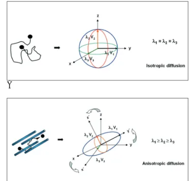
Fig. 2. A. Isotropic diffusion model. Path (tortuous line) of wa- ter molecule (black dots) under no spatial constrain (left). The molecule moves randomly by Brownian motion, resulting in a sphere displacement profile (right). This condition is termed isotropic.
B. Anisotropic diffusion model. Motion (tortuous arrow) of wa- ter molecule (black dots). Under ordered structure (rods) (e.g., myelin sheath, axon in white matter) (left). The probable loca- tion of the molecule after unit time will be within an ellipsoid (right). This condition is termed anisotropy. The ellipsoid can be characterized by three variables (eigenvalues: λ1, λ2, λ3) to describe the shape and relationship between the principal co- ordinate axes (X , Y , and Z ) and the laboratory coordinate axes (X, Y, and Z). Each of the principal axes (X , Y , and Z ) are represented by three eigenvectors (V1, V2, and V3) of the diffusion tensor. Diagonalization means rotating the laboratory coordinate system and adjusting it to the principal coordinate system of ellipsoid (curved arrows in B).
성을 띠게 되며 이러한 특성을 확산비등방성(diffusion ani- sotropy)이라고 한다(5, 6). 예를 들어 뇌 백질에서 물 분자 는 축삭과 신경섬유다발에 의해, 축삭에 대하여 직각인 경우 보다 축삭에 평행한 경우 확산이 더 잘되는 비등방성을 보인 다. 조직 속에서 물 분자의 비등방성 확산은 본질적으로 텐서 의 물리적 성상을 갖는다. 이러한 뇌 백질 내의 비등방성 확 산텐서는 타원체(ellipsoid)로 도식화할 수 있다(Fig. 2). 자기 공명영상의 기본 단위인 화소(voxel) 내의 확산텐서 타원체 모양은 타원체의 주축(principal coordinate axes: x , y , and z )을 이루는 세 축상의 확산력(고유값[eigenvalue]: λ1, λ2, λ
3)과 확산방향(고유벡터[eigenvector]: v1, v2, and v3)에 의 해 결정된다. 하나의 3차원 화소 내의 비등방성 확산텐서량 (D)은 수학적으로 9개 요소의 3×3행렬조합으로 계산할 수 있 다(7, 8).
Dxx Dxy Dxz
D = Dyx Dyy Dyz Dzx Dzy Dzz
확산의 정규분포적인 통계적 성격을 고려하면 Dxy = Dyx, Dxz= Dzx, Dyz= Dzy이므로 실제 Dxx, Dyy, Dzz, Dxy, Dxz, Dyz의 여섯 가지 요소를 알면 계산할 수 있다. 이는 자기공명 영상 획득 시 xx, yy, zz, xy, xz, yz축(laboratory coordinate axes)에 경사자장을 추가한 영상과 경사자장 없이 획득한 영 상(non-diffusion-weighted images)의 신호강도 차이로 알 수 있다(Fig. 3). 이렇게 얻어진 Dxx, Dyy, Dzz, Dxy, Dxz, Dyz 를 타원체 주축(principal coordinate axes)으로 대각화(diag- onalization)하면 화소 내 확산텐서 타원체 주축(x , y , z )의 고유값(eigenvalues: λ1, λ2, λ3)을 구할 수 있다(7-10). 이 세 개의 고유값은 타원체의 모양을 결정하며 이들의 합인 trace
= λ1 + λ2 + λ3 은 타원체의 방향과는 관계없는 타원체 크 기를 반영한다. 이렇게 형성된 타원체의 주축 방향(eigenvec- tors: v1, v2, and v3) 중 가장 큰 고유값을 가진 축 방향은 화 소 내 신경섬유의 주행방향을 반영한다. 즉, 확산텐서영상으로 자기공명영상 화소의 확산텐서 타원체를 구현할 수 있고 뇌 백질의 생리적 그리고 구조적인 정보를 알 수 있다.
확산텐서영상의 정량적 지표와 그래픽 지도
정량적 지표
확산텐서 영상에서 조직의 비등방성 확산을 나타낼 수 있는 몇 가지 정량적 지표를 계산할 수 있고 이 지표들로 정량적 뇌 영상(quantitative brain map)을 만들 수 있다. 가장 기본적인 정량적 값은 비등방성 확산텐서 형태를 결정하는 고유값 (eigenvalue: λ1, λ2, λ3)이다. 이 고유값을 이용하여 확산의 비 등방성 정도를 나타내는 몇 가지 지표들이 제안되어왔다. 그 중 가장 직관적이고 간단한 방법은 주확산력(principal diffu- sion)으로 고유값 λ1, λ2, λ3을 λh(highest diffusivity), λ m(intermediate diffusivity), λl(lowest diffusivity)의 확산력 크
기 순으로 구분한 뒤 λh / λl 또는 2λh / (λm + λl) 방식으로 계산한 값이다(11). 식에서도 알 수 있듯이 주확산력은 확산 타원체의 가장 큰 축의 확산력을 다른 축의 확산력에 대하여 상대적으로 표시한 값이다. 고유값의 크기에 따른 구분에 의 존한 주확산력 값은 잡음에 의한 통계적 오차를 유발하기 때 문에 그런 구분에 의존하지 않는 방법으로 volume ratio(VR), relative anisotropy(RA), fractional anisotropy(FA) 지표들이 쓰이고 있다(9, 11-14). VR은 반지름이 곧 평균 확산력이 되 는 구형 확산 형태의 부피에 대한 타원체 확산 형태의 부피 비 이고 0에서 1사이의 값을 가진다. 0에 가까울 수록 확산의 비 등방성이 큰 것을 의미한다.
VR = λ1 λ2λ3 / [(λ1 + λ2 + λ3)/3]3
RA는 고유값의 평균과 그 평균값에서 벗어난 각 고유값의 변이의 비이며 FA는 전체 확산텐서와 비등방성을 가지는 확 산텐서의 비이다. 이 둘은 0에서 1사이 값을 가지며 0은 등방 성 확산은 1은 무한 비등방성 확산을 의미한다. 즉, 확산의 비 등방성 정도가 커질수록 RA와 FA는 1에 가까운 값을 갖는다.
RA = [((λ1- Dave)2+ (λ2- Dave)2+ (λ3- Dave)2)] 1/2 / 31/2Dave
FA = 3/2/2((λ1 - Dave)2 + (λ2 - Dave)2 + (λ3 - Dave)2)1/2 / (λ12+ λ22 + λ32)1/2
, Dave는 평균확산값(mean diffusivity) 이다.
이러한 지표들은 가지공명영상에서 획득된 신호강도에 기반
Fig. 3. Scheme of diffusion tensor imaging. The six elements (Dxx, Dyy, Dzz, Dxy, Dxz, Dyz) for each voxel are calculated from six images obtained by applying diffusion-sensitizing gra- dients (squares) in at least six directions (for example: xx, yy, zz, xy, xz, and yz) in addition to a non-diffusion-weighted im- age. From the difference in attenuation of signal intensity be- tween non-diffusion-weighted and diffusion-weighted images, six voxel by voxel maps of ADC are generated. The image ob- tained by applying diffusion-sensitizing gradients in xx direc- tion (top law) shows that corpus callosum among the white matter tract, transverse to the x direction, has maximum diffu- sivity as a hypointense area than the other areas of the white matter.
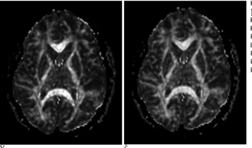
을 둔 고유값을 근거로 하는 바 영상의 신호대잡음비(signal to noise ratio)가 감소할수록 고유값과 지표들이 조직확산의 참값에서 벗어난다. 이를 극복하기 위해 알고자 하는 화소의 확산텐서 고유벡터정보와 인접한 화소의 고유벡터정보와의 상 관관계를 고려한“Lattice”비등방성 지표(LIN)나 평균확산 값 에 대한 확산텐서 6요소(xx, yy, zz, xy, xz, yz)의 확산텐서 변이를 고려한 total anisotropy(Aσ)지표가 제안되기도 하였다 (11, 15, 16). 각 화소의 비등방성 지표로 RA 지도, FA 지도 등을 만들 수 있으며 영상을 통해 육안으로 조직간의 비등방 성 차이를 구분할 수 있다(Fig. 4A, B).
그래픽 지도
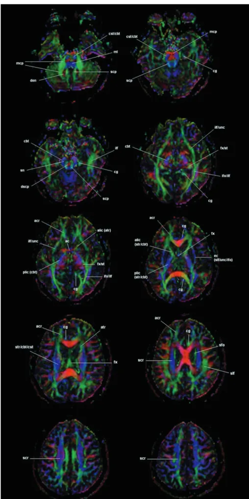
앞에서 구현된 지표영상 위에 화소 마다의 확산텐서 고유벡 터에 색깔을 결합시키거나 벡터방향을 선으로 나타내어 확산 텐서의 방향, 즉 뇌 백질 신경섬유의 방향을 표시하는 색지도 (color map) 나 벡터지도(vector map) 등을 만들 수 있다(Fig.
5)(17). 이는 뇌조직의 확산이 신경섬유나 세포에 의해 제한 을 받는 비등방성을 띄며 비등방성의 역학 모형인 타원체내에 서 가장 큰 확산력을 갖는 벡터방향이 신경섬유의 방향이라는 전제 하에 자기공명으로 원하는 조직의 확산텐서를 신호화해 서 각 화소의 고유벡터와 고유값을 정량화 하여 역으로 뇌조 직 신경섬유의 구조를 밝히는 일련의 과정으로 얻어지는 결과 물이다.
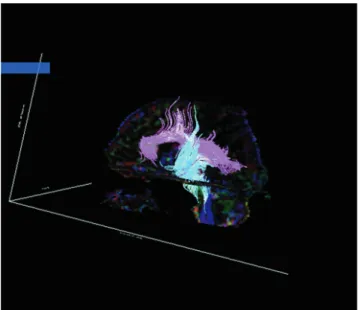
한 화소 안의 조직 정보 뿐만 아니라 화소와 화소 사이의 확 산텐서의 역학적 연결고리를 이용하여 뇌 전장에 걸친 삼차원 신경섬유지도를 그릴 수 있는데 이를 신경섬유추적지도(trac- tography)라 한다(Fig. 6). 좀 더 실제에 가까운 심경섬유추적 지도를 구현하기 위한 여러 가지 방법들이 제안되어 왔는데 크게 두 가지 범주로 나누어 볼 수 있다. 첫번 째는 line prop- agation 기법(18-22)이고 두번 째는 energy minimization 기 법(22-24)이다. Line propagation 기법은 시작점이 되는 화 소의 가장 큰 고유벡터를 기준선으로 하여 그 전후 방향으로 인접한 화소의 가장 큰 고유벡터를 따라 하나의 선을 이어나
가는 방식으로 대개 추적을 끝내는 한계점을 FA 값으로 정하 게 된다(Fig. 7). 예를 들어 뇌 회백질의 FA 값인 0.2를 한계 점으로 한다면 신경섬유추적 프로그램은 도착한 화소의 FA 값 이 0.2에 이르면 추적을 멈추게 된다. 또한 추적 각도에도 한 계점을 두는데 이는 추적 시 신경섬유의 주행방향이 급격히 꺾이는 경우는 다른 경로의 섬유의 가능성이 크기 때문이다.
Energy minimization 기법은 옷감이나 화장지에 잉크가 떨어 져 조직의 결에 따라 번져나갈 때 그 속도와 방향을 예측할 수 있는 원리를 이용하여 신경섬유를 추적하는 방식이다(Fig. 8).
임상 응용
앞에서 살펴본 바와 같이 확산텐서 자기공명 영상은 조직에 서 물 분자 확산의 비등방성정도를 수학적으로 계산하여 얻는 정량적 지도라고 할 수 있다. 즉, 사람이 태어나서 뇌의 수초 화가 진행됨에 따라 비등방성은 커질 것이고 노화에 의해 뇌 백질의 구조가 엉성하게 되면 비등방성은 다시 감소될 것이다.
이러한 변화를 객관적으로 분석하고 일반화 시키고자 하는 연 구가 확산텐서 영상이 가능해지면서 선행되었다(25-29). 신 생아, 아동, 청소년 들을 대상으로 한 연구에서 나이에 따라 ADC는 감소하고 FA는 증가됨을 보이고 있으며 특히 운동영 역을 담당하는 피질척수로(corticospinal), 시상피질로(thala- mocotical tract)과 언어영역 중추인 좌측 궁상속(left arcuate fasciculus)의 FA 변화가 유의한 증가를 보여 뇌 발달의 양상 을 밝혀내는데 확산텐서 영상의 유용성을 입증하고 있다(25- 27). 또한 노화에 따라 뇌량과 내포, 전두엽, 두정엽, 후두엽 등의 뇌 백질에서 유의한 FA 감소가 보고 되었고 이는 신경 축삭과 수초의 감소 등에 의한 세포 외 공간의 확장을 반영하 는 소견으로 확산텐서 영상이 기존의 자기공명영상에서는 알 수 없었던 미세한 뇌의 구조적인 변화를 보여줌을 시사하고 있다(28, 29). 나아가 확산텐서의 정량적 지표들과 행동이나 인지 능력과의 관계, 알츠하이머 병에서 증상과의 관계에 대 한 연구에서 전두엽의 FA 감소와 행동, 인지능력의 감소정도
A B
Fig. 4. Diffusion tensor MR images in 12-year-old health boy. Relative anisotropy map (A) and Fractional anisotropy map (B) describe degree of diffusion anisotropy in each voxel. In white matter, where anisotropy is high, bright end of gray scale is assigned; in gray matter, where anisotropy is low, dark end of gray scale is assigned.
Fig. 5. Diffusion tensor MR images in 12-year-old health boy. Color-coded white matter fiber maps are generated on basis of the three vector elements of the eigenvector v1for each voxel. The absolute values of the vector elements are assigned to red (x element), green (y element), blue (z element). If the princi- pal eigenvector is aligned along the x- axis, pure red is assigned to the corre- sponding voxel, whereas if the eigen- vector is 45°between the x- and y-axes, yellow (red plus green) is assigned to the voxel. The intensity of color in each voxel is gauged by the degree of frac- tional anisotropy. Ac = anterior com- missure, acr = anterior region of coro- na radiata, alic = anterior limb of inter- nal capsule, atr = anterior thalamic ra- diation, cbt = corticobulbar tract, cg = cingulum, cst = corticospinal tract, dscp = decussation of superior cerebel- lar peduncle, ec = external capsule, fx
= fornix, ifo = inferior fronto-occipital fasciculus, ilf = inferior longitudinal fasciculus, mcp = middle cerebellar peduncle, ml = medial lemniscus, ot = optic tract, plic = posterior limb of in- ternal capsule, scp = superior cerebel- lar peduncle, scr = superior region of corona radiata, sfo = superior fronto- occipital fasciculus, slf = superior lon- gitudinal fasciculus, sn = substantia ni- gra, st = stria terminalis, str = superior thalamic radiation, unc = uncinate fas- ciculus.
가 유의한 관계가 있음을 보고하는 등 확산텐서영상에 의한 퇴행성 질환의 조기 발견과 그에 따른 적절한 치료의 가능성 을 보여주고 있다(30-33).
확산영상은 허혈성 뇌졸중에서 물의 확산이 느려지는 초기 변화를 진단할 수 있는 전기를 마련했고 이로 말미암아 약물 이나 중재적 시술로 혈관을 재개통 시키는 허혈성 뇌졸중의 적극적인 치료가 가능해져 임상적으로 널리 쓰이고 있다(34- 38). 허혈성 뇌 질환의 확산텐서영상 연구는 뇌의 허혈성 영 역에서 중요한 시간적, 공간적인 변화양상에 대한 정보를 밝 히고 있다. 허혈 급성기와 아급성기에 허혈 부위 물 분자의 확 산 비등방성이 증가되며 아급성기에서 만성으로 진행하면서 확산 비등방성은 지속적으로 감소된다는 보고가 있으며 ADC 감소영역에서 비등방성이 증가되는 원인은 허혈에 의한 세포 부종으로 세포간질이 좁아지고 세포 내 물 확산의 제한이 허
Fig. 6. Diffusion tensor MR image in 44-year-old health woman. Three dimensional fiber tracts are generated on basis of fractional anisotropy by line propagation technique.
Fig. 7. Schematic shows white matter tracking used by fiber assignment by continuous tracking (FACT) as a kind of line propagation techniques. Diagram was provided by courtesy of Dr Mori, Johns Hopkins University. Degree of diffuse anisotropy is indicated by gray scale (white is highest), and di- rection of principal eigenvector in each image voxel is indicat- ed by an arrow. In this algorithm, the lines (long curved ar- rows) represent trace paths starting from the center of the user-defined seed voxels (asterisks in A area) bidirectionally (forward and backward) by following a direction of the eigen- vector with the largest eigenvalue (arrow in each voxel). The continuous line can be kept by memorizing information on in- tercept when the tracking line leaves a border of the voxel.
Algorithm can distinguish between tract A and B because they are separated by voxels with low anisotropy, and between tracts A and C because of differences in direction of principal eigenvectors. Asterisks indicate starting points of tracking.
Fig. 8. The principle of fiber tracking is fast marching method as a kind of energy minimizing techniques. Diagram was pro- vided by courtesy of Dr Parker, University of Manchester.
Vectors used in the calculation of the speed function, F, of the spreading ink. The relationship between the positions of the front (arrowheads), ε1(r), ε1(r′), grid points passed by front (black circles) defining the set S, grid points in the narrow band (gray circles), and grid points not yet reached by front (white circles). The speed for the spreading front propagation is de- fined by F(r) = A| ε1(r′)・n (r) | (A is the anisotropy, ε1is the eigenvector and n the orientation normal to the front. This equation reflects that the spreading speed is largest when the propagating front line is co-linear with the eigenvector and minimal when it is perpendicular. Using this equation, the shape of the stain can be calculated from the vector field, which is equivalent to a contour line showing the distance from the origin traveled by the ink within the same amount of time.
Multiple contour lines can be calculated, each representing the stain shape at a different time point. These multiple contours represent a likelihood-of-connection map. The most likely path between an arbitrary point to the seed voxel (origin the stain) can be found by following the gradient of steepest path.
혈 변화로 가중되기 때문으로 설명하고 시간이 지남에 따라 비등방성이 감소되는 것은 뇌 조직 내 신경세포 및 신경줄기 의 파괴에 따른 구조적인 느슨함을 반영한다고 밝히고 있다 (39, 40). 또한 이들 연구 결과는 조직의 구조적 보존 여부를 민감하게 반영하는 확산 비등방성의 정량적 지표들이 조직의 가역적 또는 비가역적 변화를 조기에 나타낼 수 있을 것이라 는 가능성을 제시하고 있다. 뇌의 허혈성 손상에 대한 또 다 른 연구에서 허혈에 따른 뇌 회백질과 뇌 백질의 확산 비등방 성 변화 양상이 다름을 밝혀 조직에 따른 허혈 변화의 이질성 을 설명하고 있다(40-42). 이는 허혈에 의한 뇌 손상의 치료 에 있어서 조직에 따라 다른 접근이 필요함을 보여주고 있어 이후 neuroprotective 치료나 연구에 새로운 계기를 제공할 것 으로 보인다.
확산텐서영상이 뇌 백질의 구조적 상태를 민감하게 반영하 는 영상인 만큼 다발성 경화증(43-48), CADASIL(cerebral autosomal dominant arteriopathy with subcortical infarcts and leukoencephalopathy) (49,50), 근위축성측삭경화증 (Amyotrophic lateral sclerosis)(51-54), Krabbe dis- ease(55), X-linked cerebral adrenoleukodystrophy (Fig. 9) (56, 57), HIV 감염(58,59) 등의 뇌 백질 질환에서의 연구가 활발히 이루어졌다. 이들 연구에서 확산텐서의 정량적 지표가 기존의 자기공명영상에서 정상으로 보이는 뇌 백질에의 병변 침범을 알게 하고 지표의 변화정도가 병변의 병리적인 변화 및 임상소견에 상관관계가 있음을 밝히고 있다. 이로서 확산 텐서영상이 질환의 진행상태와 치료에 대한 반응을 평가하는 데 유용한 도구가 될 수 있음을 언급하고 있다.
사후 부검 등의 병리적인 검사, 자기공명 분광법(MR spec- troscopy), DNA 분석 등을 이용하여 정신과 질환의 병태 생 리를 밝히고자 하는 여러 연구에서 질환에 따라 뇌의 일정부 분에서 구조적인 이상이 관계되어 있음을 증명해 왔다(60-
69). 알코올 중독(70, 71), 우울증(72, 73), 정신분열증(74- 76) 등에서 확산텐서영상은 해당 영역에서 확산 비등방성이 감소함을 보여 상기 질환들의 병태 생리 연구에 그 유용성을 보이고 있다.
신경섬유추적지도(tractography)를 이용하여 뇌허혈이나 종 양성 질환에서 병변 부위와 침범된 신경섬유와의 관계, 침범 된 정도와 증상과의 관계를 살펴본 연구들이 진행되고 있으며 이러한 노력들이 해부학적 구조에 바탕을 둔 뇌 기능 연구에 한 축을 이루고 있고 실제 환자들의 수술 전 계획이나 수술 후 또는 허혈 시점 이후의 예후를 예측하고 대처하는데 실질적인 도움을 줄 수 있을 것으로 보인다(77, 78).
뇌확산텐서영상과 신경섬유추적지도의 문제점
알고자 하는 영역의 정확한 확산텐서 정보를 얻고 그를 바 탕으로 한 실제와 같은 신경섬유추적지도를 만들기 위해서는 무엇보다 충분한 신호대잡음비와 높은 해상력의 영상획득과 운동이나 회오리 전류(eddy current) 등에 의한 인공물을 최 소화 하여 영상 왜곡을 막아야 한다. 우선은 확산을 가시화 할 만큼의 강한 경사자장을 적용할 수 있는 자기공명영상 기기의 발달이 있어야 했고 확산현상을 따라잡기 위한 빠르고도 충분 한 신호를 획득할 수 있는 영상기법이 뒷받침 되어야 했다. 자 기공명영상 기기는 이제 3 Tesla 시대가 시장화 될 만큼 눈부 신 발전을 거듭하고 있고 단발 또는 다발평면에코(single shot- or multi-shot echo planar) 기법과 parallel 영상기법 의 발달로 영상획득시간을 줄이고 양질의 영상을 획득할 수 있게 되었다(79-82). 또한 운동 인공물을 최소화하기 위하여 호흡 및 심장운동에 대한 동기화(gaiting)와 회오류 전류에 의 한 영상 왜곡의 보정을 위한 여러 가지 기술적인 기법의 적용 등 정확한 정보 획득을 위한 노력이 이루어지고 있다(83, 84).
A B
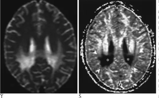
Fig. 9. 9-year-old boy with cerebral X- linked adrenoleukodystrophy (ALD) who presented with progressive motor and visual deficits. Pictures were pro- vided by courtesy of Dr Mori, Johns Hopkins University.
A. T2-weighted MR image (TR/TE, 5000/92) shows abnormal signal inten- sity involving peritrigonal white mat- ter.
B. Fractional anisotropy (FA) map shows central zone (asterisks) of marked hypointensity (marked de- crease in anisotropy) and peripheral zone (arrowheads) of mild hypointensi- ty (mild decrease in anisotropy). The gradation of signal intensity on FA map might represent well-established histopathological zonal changes in white matter lesion of X-linked ALD.
얻어진 자기공명영상으로 뇌의 물 분자 확산텐서 비등방성 도의 정확한 정량화와 신경섬유추적지도 구현은 핵물리학적 인 데이터 수집과 신호처리 및 분석 기술, 컴퓨터 그래픽 기 법 등의 종합적인 연구가 필요하다. 앞에서 살펴본 바와 같이 비등방성도의 정량적 지표에 대한 물리, 수학적 성과가 있었 고 그러한 지표들이 많은 연구에서 사용되고 있다. 하지만 아 직도 한 화소 내에 교차되는 신경 섬유(cross terms)를 구 분하여 오차 없이 신경 섬유를 추적해 내는 일이나 얻어진 정 보들을 서로 비교 분석하는 방법에 많은 숙제가 남아 있다.
어느 한 고유벡터가 월등히 우세한 시가(cigar) 모양의 확산 텐서 모형(λ1 >> λ2 + λ3)으로만 뇌의 확산이 진행된다면 Line propagation 기법만으로도 큰 오차 없이 신경섬유를 추 적할 수 있겠지만 실제 한 화소 내에는 교차하거나 분지하는 신경섬유에 따라 다양한 확산 모형이 존재한다. 예를 들어 두 개의 고유벡터가 거의 같은 크기를 가진 팬케익 모양의 확산 텐서 모형(λ1 = λ2 >> λ3)에서 신경섬유 추적 중 어느 벡터 를 따라 갈 것인가를 결정하는 일은 아주 중요하면서도 간단 한 일이 아니다. 이러한 한계를 극복하기 위해 앞에서 언급한 energy minimization 기법을 비롯하여 시가 모형을 벗어난 화 소에서 추적을 끝내는 방법(tensor line technique) (22, 85), 가장 큰 고유벡터를 추적하는 대신 확산모형의 표면형태 추 적하는 방법(surface line technique) (22, 86), 텐서에 의존 하지 않고 많은 방향의 확산영상에서 방향에 따른 ADC 값의 차이를 이용해 섬유를 그리는 방법(diffusion spectrum tech- nique) (22, 87, 88) 등이 보고 되어왔다. 이러한 노력들로 얻 어진 확산텐서영상을 분석하여 어떤 결론에 이르기 위해서는 객관적이면서 누구나 편히 사용할 수 있는 정상 데이터 구축 과 신경섬유추적지도의 정량적 분석이 가능해져야 한다. 지금 까지는 MRI에서 특정한 구역을 먼저 수작업으로 구획한 다음 비등방성도를 계산하는 방식(knowledge-based multiple- ROI approach) (21, 22, 89, 90)과 복셀단위로 뇌의 전영역 에서 비교하여 통계적으로 유의한 위치를 찾아내는 방식 (probablistic approach) (22, 90)이 있으나 전자는 주관적이 며 영역을 제한하여 분석하므로 결과에 오류가 있을 수 있고 후자는 기술적으로 접근이 용이하지 않은 점 등의 문제가 있 다. 섬세하고 복잡한 신경 구조를 비침습적으로 알 수 있게 된 확산텐서영상의 구현은 자기공명학이나 뇌 과학 또 의학적으 로 세기적인 성과임에는 분명하나 이의 상용화를 위해서는 확 산 모델(diffusion model), 신경섬유추적 기법(fiber track- ing), 분석 기법, 임상적용 등에 대한 많은 연구와 발전이 필 요하다.
결 론
지금까지 확산텐서자기공명영상(DTI)의 기본적인 원리와 임상적용, 그리고 문제점 들에 대해서 살펴 보았다. 아직도 회 오리 전류, 잡음(noise), 부분용적효과(partial volume effect) 와 같은 영상획득 시 오류 요인을 극복하고 높은 신호대잡음 비와 공간해상력을 가진 양질의 영상획득, 그리고 확산텐서영
상의 분석 및 정량 방법, 신경섬유추적의 방법상의 문제 등 해 결해야 할 과제가 많지만 확산텐서자기공명영상(DTI)이 비침 습적으로 생체내의 뇌 신경구조를 알게 하고 그 상태를 평가 할 수 있는 유일한 방법으로 뇌 과학과 의학분야의 발전에 또 다른 역사를 마련할 획기적인 도구임에 틀림없다. 특히 아직 도 인류의 숙제로 남아있는 뇌의 구조적, 기능적 지도를 완성 하고 여러 뇌 질환의 병태생리를 밝히고 질환의 상태나 예후 를 평가하는데 탁월한 방법으로 자리잡아 나갈 것으로 기대되 는 바이다.
감사의 글
종설을 쓰게 되기까지 기본적인 지식을 익히고 소프트웨어 를 다루는데 많은 도움을 주신 연세 대학교 신촌 세브란스 병 원 이승구 선생님, 성균관 대학교 강북 삼성병원 문원진 선생 님, 그리고 자신들의 연구결과를 싣도록 기꺼이 허락하여 종 설의 내용이 충실해 질 수 있도록 도와 주신 Jonhs Hopkins Medical Institution의 Dr. Susumu Mori와 Manchester University의 Dr. Geoffrey J. M. Parker께 깊은 감사를 드립 니다.
참 고 문 헌
1. Le Bihan D. Molecular diffusion nuclear magnetic resonance imag- ing. Magn Reson Q 1991;7:1-30
2. Stejskal EO, Tanner JE. Spin diffusion measurements: spin echoes in the presence of a time-dependent field gradient. J Chem Phys 1965;42:288-292
3. Le Bihan D, Breton E, Lallemand D, Grenier P, Cabanis E, Laval- Jeantet M. MR imaging of intravoxel incoherent motions: applica- tion to diffusion and perfusion in neurologic disorders. Radiology 1986;161:401-407
4. Le Bihan D, Breton E, Lallemand D, Aubin ML, Vignaud J, Laval- Jeantet M. Separation of diffusion and perfusion in intravoxel inco- herent motion MR imaging. Radiology 1988;168:497-505
5. Moseley ME, Cohen Y, Kucharczyk J, Mintorovitch J, Asgari HS, Wendland MF. Diffusion-weighted MR imaging of anisotropic wa- ter diffusion in cat central nervous system. Radiology 1990;176:
439-445
6. Chenevert TL, Brunberg JA, Pipe JG. Anisotropic diffusion in hu- man white matter: demonstration with MR techniques in vivo.
Radiology 1990;177:401-405
7. Basser PJ, MattielloJ, Le Bihan D. Estimation of the effective self- diffusion tensor from the NMR spin echo. J Magn Reson Imaging 1994;103:247-254
8. Basser PJ, MattielloJ, Le Bihan D. MR diffusion tensor spec- troscopy and imaging. Biophys J 1994;66:259-267
9. Basser PJ, Pierpaoli C. Microstructural and physiological features of tissues elucidated by quantitative diffusion-tensor MRI. J Magn Reson Imaging 1996;111:209-219
10. Basser PJ, Pierpaoli C. A simplified method to measure the diffu- sion tensor from seven MR images. Magn Reson Med 1998;39:928- 934
11. Pierpaoli C, Basser PJ. Toward a quantitative assessment of diffu- sion anisotropy. Magn Reson Imaging 1996;36:893-906
12. Basser PJ, Pajevic S. Statistical artifacts in diffusion tensor MRI (DT-MRI)caused by background noise. Magn Reson Med 2000;44:
41-50
13. Martin KM, Papadakis NG, Huang CL, Hall LD, Carpenter TA.
The reduction of the sorting bias in the eigenvalues of the diffusion tensor. Magn Reson Imaging 1999;17:893-901
14. Papadakis NG, Xing D, Houston GC, Smith JM, Smith MI, James MF, et al. A study of rotationally invariant and symmetric indices of diffusion anisotropy. Magn Reson Imaging 1999;17:881-892 15. Conturo TE, McKinstry RC, Akbudak E, Robinson BH. Encoding
of anisotropic diffusion with tetrahedral gradients: a general math- ematical diffusion formalism and experimental results. Magn Reson Med 1996;35:399-412
16. Shimony JS, McKinstry RC, Akbudak E, Aronovitz JA, Snyder AZ, Lori NF, et al. Quantitative diffusion-tensor anisotropy brain MR imaging: normative human data and anatomic analysis. Radiology 1999;212:770-784
17. Douek P, Turner R, Pekar J, Patronas N, Le Bihan D. MR color mapping of myelin fiber orientation. J Comput Assist Tomogr 1991;
15:923-929
18. Mori S, Crain BJ, Chacko VP, van Zijl PCM. Three-dimensional tracking of axonal projections in the brain by magnetic resonance imaging. Ann Neurol 1999;45:265-269
19. Xue R, van Zijl PC, Crain BJ, Solaiyappan M, Mori S. In vivo three- dimensional reconstruction of rat brain axonal projections by diffu- sion tensor imaging. Magn Reson Med 1999;42:1123-1127
20. Basser PJ, Pajevic S, Pierpaoli C, Duda J, Aldroubi A. In vivo fiber tractography using DT-MRI data. Magn Reson Med 2000;44:625- 632
21. Conturo TE, Lori NF, Cull TS, Akbudak E, Snyder AZ, Shimony JS, et al. Tracking neuronal fiber pathways in the living human brain.
Proc Natl Acad Sci USA 1999;96:10422-10427
22. Mori S, van Zijl PC. Fiber tracking: principles and strategies - a technical review. NMR Biomed 2002;15:468-480
23. Parker GJ.Wheeler-Kingshott CA, Barker GJ. Estimation distrib- uted anatomical connectivity using fast marching methods and dif- fusion tensor imaging. IEEE Trans Med Imaging 2002;21:505-512 24. Tuch DS, Wiegell MR, Reese TG, Belliveau JW, Wedeen V.
Measuring cortico-cortical connectivity matrices with diffusion spectrum imaging. In Proceedings of International Society of Magnetic Resonance in Medicine, Glasgow, UK, 2001;502
25. Neil JJ, Shiran SI, McKinstry RC, Schefft GL, Snyder AZ, Almli CR, et al. Normal brain in human newborns: apparent diffusion coefficient and diffusion anisotropy measured by using diffusion tensor MR imaging. Radiology 1998;209:57-66
26. Schmithorst VJ, Wilke M, Dardzinski BJ, Holland SK. Correlation of white matter diffusivity and anisotropy with age during child- hood and adolescence: a cross sectional diffusion-tensor MR imag- ing strudy. Radiology 2002;212-218
27. Nusbaum AO, Tang CY, Buchsbaum MS, Wei TC, Atlas SW.
Regional and global changes in cerebral diffusion with normal ag- ing. AJNR Am J Neuroradiol 2001;22:136-142
28. Pfefferbaum A, Sullivan EV, Hedehus M, Lim KO, Adalsteinsson E, Moseley M. Age-related decline in brain white matter anisotropy measured with spatially corrected echo-planar diffu- sion tensor imaging. Magn Reson Med 2000;44:259-268
29. Sullivan EV, Adalsteinsson E, Hedehus M, Ju C, Moseley M, Lim KO, et al. Equivalent disruption of regional white matter mi- crostructure in aging healthy men and women. Neuroreport 2001;
12:99-104
30. Sugihara S, Kinoshita T, Matsusue E, Fujii S, Ogawa T. Usefulness of diffusion tensor imaging of white matter in Alzheimer disease and vascular dementia. Acta Radiol 2004;45:658-663
31. Urresta FL, Medina DA, Gaviria M. Diffusion MRI studies in vas-
cular cognitive impairment and dementia. Rev Bras Psiquiatr 2003;
25:188-91
32. Medina D, Detoledo-Morell L, Urresta F, Gabrieli JD, Moseley M, Fleischman D , et al. White matter changes in mild cognitive im- pairment and AD: a diffusion tensor imaging study. Neurobiol Aaging 2005 (in press)
33. Rose SE, Chen F, Chalk JB, Zelaya FO, Strugnell WE, Benson M, et al. Loss of connectivity in Alzheimer’s disease: an evaluation of white matter tract integrity with color-coded MR diffusion tensor imaging. J Neurol Neurosurg Psychiat 2000;69:528-530
34. Moseley ME, Kucharczyk J, Mintorovitch J, Cohen Y, Kurhanewicz J, Derugin N, et al. Diffusion-weighted MR imaging of acute stroke: correlation with T2-weighted and magnetic suscep- tibility-enhanced MR imaging in cats. AJNR Am J Neuroradiol 1990;11:423-429
35. Mintorovich J, Moseley ME, Chileuitt L, Shimizu H, Cohen Y, Weinstein PR. Comparison of diffusion- and T2-weighted MRI for the early detection of cerebral ischemia and reperfusion in rats.
Magn Reson Med 1991;18:39-50
36. van Gelderen P, de Vleeschouwer MHM, des Pres D, Pekar J, van Zijl PC, Moonen CT. Water diffusion and acute stroke. Magn Reson Med 1994;31:154-163
37. Sorensen AG, Buonannno FS, Gonzalez RG, Schwamm LH, Lev MH, Huang-Hellinger FR, et al. Hyperacute stroke: evaluation with combined multisection diffusion-weighted and hemodynami- cally weighted echo-planar MR imaging. Radiology 1996;199:391- 401
38. Bammer R, Augustin M, Strasser-Fuchs S, Seifert T, Kapeller P, Stollberger R, et al. Magnetic resonance diffusion tensor imaging for characterizing diffuse and focal white matter abnormalities in multiple sclerosis. Magn Reson Med 2000;583-591
39. Maier SE, Gudbjartsson H, Hsu L, Jolesz FA. Diffusion anisotropy imaging of stroke. Proc Int Soc Magn Med 1997;4:573
40. Yang Q, Tress BM, Barber PA, Desmond PM, Darby DG, Gerraty RP, et al. Serial study of apparent diffusion coefficient and anisotropy in patients with acute stroke. Stroke 1999;30:2382-2390 41. Sorensen AG, Wu O, Copen WA, Davis TL, Gonzalez RG,
Koroshetz WJ, et al. Human acute cerebral Ischemia: detection of changes in water diffusion anisotropy by using MR imaging.
Radiology 1999;212:785-792
42. Thornton JS, Ordidge RJ, Penrice J, Cady EB, Amess PN, Punwani S, et al. Anisotropy water diffusion in white and gray matter of the neonatal piglet brain before and after transient hypoxia-ischemia.
Magn Reson Imaging 1997;15:433-440
43. Horsfield MA, Lai M, Webb S, Barker GJ, Tofts PS, Turner R, et al.
Apparent diffusion coefficients in benign and secondary progres- sive multiple sclerosis by nuclear magnetic resonance. Magn Reson Med 1996;36:393-400
44. Werring DJ, Clark CA, Barker GJ, Thompson AJ, Miller DH.
Diffusion tensor imaging of lesions and normal-appearing white matter in multiple sclerosis. Neurology 1999;52:1626-1632 45. Bammer R, Augustin M, Strass-Fuchs S, Seifert T, Kapeller P,
Stollberger R, et al. Magnetic resonance diffusion tensor imaging for characterizing diffuse and focal white matter abnormalities in multiple sclerosis. Magn Reson Med 2000;44:583-591
46. Rocca MA, Cercignani M, Ianucci MD, Comi G, Filippi M. Weekly diffusion-weighted imaging of normal-appearing white matter in MS. Neurology 2000;55:882-884
47. Werring DJ, Brassat D, Droogan AG, Clark CA, Symms MR, Barker GJ, et al. The pathogenesis of lesions and normal-appearing white matter changes in multiple sclerosis: a serial diffusion MRI study. Brain 2000;123:1667-1676
48. Guo AC, Macfall JR, Provenzale JM. Multiple sclerosis: diffusion tensor MR imaging for evaluation of normal-appearing white mat- ter. Radiology 2002;222:729-736
49. Chabriat H, Pappata S, Poupon C, Clark CA, Vahedi K, Poupon F, et al. Clinical severity in CADASIL related to ultrastructural dam- age in white matter: in vivo study with diffusion tensor MRI.
Stroke 1999;30:2637-2643
50. Molko N, Pappata S, Mangin JF, Poupon C, Vahedi K, Jovert A, et al. Diffusion tensor imaging study of subcortical gray matter in CADASIL. Stroke 2001;32:2049-2054
51. Segawa F, Kishibayashi J, Kamada K, Sunohara N, Kinoshita M.
MRI of periventricular white matter lesions in amyotrophic lateral sclerosis, analysis by diffusion-weighted images, No To Shinkei 1994;46:835-840
52. Wu RH, Bruening R, Berchtenbreiter C, Borrasio T, Hueck A, Reiser M. Evaluation of diffusion-weighted imaging in patients with amyotrophic lateral sclerosis. In preceedings of ISMRM 6th an- nual meeting, Sidney, 1998;1249
53. Ellis CM, Simmons A, Jones DK, Bland J, Dawson JM, Horsfield MA, et al. Diffusion tensor MRI assesses corticospinal tract dam- age in ALS. Neurology 1999;53:1051-1058
54. Toosy AT, Werring DJ, Orrell RW, Howard RS, King MD, Barker GJ, et al. Diffusion tensor imaging detects corticospinal tract in- volvement at multiple levels in amyotrophic lateral sclerosis. J Neurol Neurosurg Psychiatry 2003;74:1250-1257
55. Guo AC, Petrella JR, Kurtzberg J, Provenzale JM. Evaluation of white matter anisotropy in Krabbe disease with diffusion tensor MR imaging: initial experience. Radiology 2001;218:809-815 56. Ito R, Melhem ER, Mori S, Eichler FS, Raymond GV, Moser HW.
Diffusion tensor brain MR imaging in X-linked cerebral adrenoleukodystrophy. Neurology 2001;56:544-547
57. Schaumburg HH, Powers JM, Raine CS, Suzuki K, Richardson EP.
Adrenoleukodystrophy: a clinical and pathological study of 17 cas- es. Arch Neurol 1975;32:577-591
58. Filippi CG, Ulug AM, Rayn E, Ferrando SJ, van Gorp W. Diffusion tensor imaging of patients with HIV and normal-appearing white matter on MR images of the brain. AJNR Am J Neuroraiol 2001;22:
277-283
59. Pomara N, Crandall DT, Choi SJ, Johnson G, Lim KO. White mat- ter abnormalities in HIV-1 infection: a diffusion tensor imaging study. Psychiat Res 2001;106:15-24
60. de la Monte SM. Disproportionate atrophy of cerebral white mat- ter in chronic alcoholics. Arch Neurol 1988;45:990-992
61. Harper CG, Krill JJ, Holloway RL. Brain shrinkage in chronic alco- holics: a pathological study. Br Med J(Clin Res Ed) 1985;290:501- 504
62. Lewohl JM, Dodd PR, Mayfield RD, Harris RA. Application of DNA microarrays to study human alcoholism. J Biomed Sci 2001;8:
28-36
63. Robinson RG, Chemerinski E, Jorge R. Pathophysiology of sec- ondary depression in elderly. J Geriatr Psychiatry Neurol 1999;12:
128-136
64. Mayberg HS, Liotti M, Brannan SK, McGinnis S, Mahurin RK, Jerabek PA, et al. Reciprocal limbic-cortical function and negative mood: covering PET findings in depression and normal sadness.
Am J Psychiatry 1999;156:675-682
65. Drevets WC. Functional anatomical abnormalities in limbic and prefrontal cortical structures in major depression. Prog Brain Res 2000;126:413-431
66. Rajkowska G, Miguel-Hidalgo JJ, Wei J, Dilley G, Pittman SD, Meltzer HY, et al. Morphometric evidence for neuronal and glial prefrontal cel pathology in major depression. Biol Psychiat 1999;45:
1085-1098
67. Pearlson GD, Petty RG, Ross CA, Tien AY. Schizophrenia: a dis- ease of heteromodal association cortex? Neuropsychopharmacology 1996;14:1-17
68. Bullmore ET, Woodruff PW, Wright IC, Rabe-Hesketh S, Howard RJ, Shuriquie N, et al. Does dysplasia cause anatomical dysconnec- tivity in schizophrenia? Schizophr Res 1998;30:127-135
69. Lim KO, Adalsteinsson E, Spielman D, Sullivan EV, Rosenbloom MJ, Pfefferbaum A. Proton magnetic resonance spectroscopic imaging of cortical gray and white matter in schizophrenia. Arch Gen Psychiat 1998;55:346-352
70. Pfefferbaum A, Sullivan EV, Hedehus M, Adalsteinsson E, Lim KO, Moseley M. In vivo detection and functional correlates of white matter microstructural disruption in chronic alcoholism.
Alcohol Clin Exp Res 2000;24:1214-1221
71. Basser PJ, Pierpaoli C. Microstructural and physiological features of tissues elucidated by quantitative-difusion-tensor MRI. J Magn Reson B 1996;111:209-219
72. Alexopoulos GS, Kiosses DN, Choi SJ, Murphy CF, Lim KO.
Frontal white matter microstructure and treatment response of late-life depression: a preliminary study. Am J Psychiatry 2002;159:
1929-1932
73. Sullivan EV, Adalsteinsson E, Hedehus M, Ju C, Moseley M, Lim KO, et al. Equivalent disruption of regional white matter mi- crostructure in aging health men and women. Neuroreport 2001;12:
99-104
74. Foong J, Maier M, Clark CA, Barker GJ, Miller DH, Ron MA.
Neuropathological abnormalities of the corpus callosum in schizo- phrenia: a diffusion tensor imaging study. J Neurol Neurosurg Psychiat 2000;68:242-244
75. Agartz I, Andersson JL, Skare S. Abnormal brain white matter in schizophrenia: a diffusion tensor imaging study. Neuroreport 2001;
12:2251-2254
76. Steel RM, Bastin ME, McConnel S, Marshall I, Cunningham- Owens DG, Lawrie SM, et al. Diffusion tensor imaging (DTI) and proton magnetic resonance spectroscopy (IHMRS) in schizo- phrenic subjects and normal controls. Psychiat Res 2001;106:161- 170
77. Jellison BJ, Field AS, Medow J, Lazar M, Alexander AL. Diffusion tensor imaging of cerebral white matter: a pictorial review of physics, fiber tract anatomy, and tumor imaging patterns. AJNR Am J Neuroradiol 2004;25:356-369
78. Yamada K, Ito H, Nakamura H, Kizu O, Akada W, Kubota T, et al.
Stroke patients’evolving symptoms assessed by tractography. J Magn Reson Imaging 2004;20:923-929
79. Turner R, Le Bihan D, Maier J, Vavrek R, Hedges LK, Pekar J.
Echo-planar imaging of intravoxel incoherent motion. Radiology 1990;177:407-414
80. Turner R. Single shot diffusion imaging at 2.0 Tesla. J Magn Reson Imaging 1990;177:407-414
81. Golay X, Brown SJ, Itoh R, Melhem ER. Time-resolved contrast- enhanced carotid MR Angiography using sensitivity encoding (SENSE). AJNR Am J Neuroradiol 2001;22:1615-1619
82. Wang Y. Destription of parallel imaging in MRI using multiple coils. Magn Reson Med 2000;44:495-499
83. Haselgrovr JC, Moore JR. Correction ofr distortion of Echo-planar images used to calculate the apparent diffusion coefficient. Magn Reson Med 1996;36:960-964
84. Jezzard P, Barnett AS, Pierpaoli C. Characterization of and correc- tion for eddy current artifacts in echo planar diffusion imaging.
Magn Reson Med 1998;39:801-812
85. Weinstein D, Kindlmann G, Lundberg EC. Tensorlines: advection
diffusion based propagation through diffusion tensor fields. In Proceedings, IEEE Visualization, San Francisco, CA 1999;249-253 86. Zhang S, Bastin ME, Laidlaw DH, Sinha S, Armitage PA,
Deisboeck TS. Visualization and analysis of white matter structur- al asymmetry in diffusion tensor MRI data. Magn Reson Med 2004;
51:140-147
87. Wedeen, V.J., Reese, T.G., Tuch, D.S., Weigel, M.R., Dou, J.-G., Weiskoff, R.M., et al.Mapping fiber orientation spectra in cerebral white matter with Fourier-transform diffusion MRI. Proc Int Soc
Magn Reson Med ISMRM, California, 2000;82
88. Frank LR. Anisotropy in high angular resolution diffusion-weight- ed MRI. Magn Reson Med 2001;45:935-939
89. Stieltijes B, Kaufmann WE, van Zijl PCM, Fredericksen K, Pearlson GD, Mori S. Diffusion tensor imaging and axonal tracking in the human brainstem. Neuroimage 2001;14:723-735
90. Mori S, Kaufmann WE, Davatzikos C, Stieltjes B, Amodei L, Fredericksen K, et al. Imaging cortical association tracts in human brain. Magn Reson Imaging 2002;47:215-223
J Korean Radiol Soc 2005;53:233-243
Address reprint requests to : Hyun Jeong Kim, M.D., Deparment of Radiology, Daejeon, St. Mary’s Hospital, 520-2 Dae Heung dong, Jung gu, Dae Jeon 301-723, Korea.
Tel. 82-42-220-9638 Fax. 82-42-253-9464 E-mail: [email protected]
Brain Diffusion Tensor MR Imaging
1Hyun Jeong Kim, M.D., Choong Gon Choi, M.D.2, Jeong Hyun Lee, M.D.2, Po Song Yang, M.D., Siwon Kang, M.D., Yeon Soo Lee, M.D., Ji Chang Kim, M.D., Bo Seal Hwang, M.D.
1Department of Radiology, Daejeon St. Mary’s Hospital, College of Medicine, The Catholic University
2Department of Radiology, Asan Medical Center, University of Ulsan, College of Medicine
The development of MR imaging techniques during the past decade has enabled researchers to use MR imaging as a noninvasive tool for evaluating structural and physiologic states in biologic tissues by measuring the diffusion process of water molecules. More recently, diffusion tensor MR imaging (DTI) technique based on the dependency of molecular diffusion on the orientation of white matter fiber tracts has been used to ana- lyze the trajectory, shape, fiber structure, location, topology and connectivity of neuronal fiber pathways in liv- ing humans. Numerous efforts have been made by MR physicists, brain scientists, and medical doctors to ad- vance MR techniques and computer-based algorithms which result in more accurate quantification of diffu- sion tensor and the generation of white matter fiber tract maps and to determine the pathophysiology of brain disease by DTI and useful clinical applications of DTI. In this article, we describe the tensor theory used to characterize molecular diffusion in white matter and a process of measuring tensor elements using diffusion- sensitive MR images to fiber mapping. We then provide review of current literature and some clinical exam- ples that have been published and are on-going.
Index words :Diffusion tensor
Magnetic resonance (MR), diffusion tensor Brain, diffusion
Brain, MR