서 론
내측 슬관절 단일 구획 골관절염의 유병률이 증가하고 있으며1) 치료법으로는 크게 단일 구획 인공 슬관절 반치환술과 경골 근위 부 절골술 등이 있다. 흔히 나이가 많고 활동량이 적은 환자의 경
우 인공 슬관절 부분 치환술이 선호되며, 경골 근위부 절골술은 관절면을 보존한다는 측면에서 장기적인 추시가 필요한 비교적 젊은 환자군에서 주로 시행되었다.
근위 경골 절골술은 1958년 Jackson이 처음 시행하였고,2) Coventry3)에 의해 대중화를 이루게 되었으며 그 이론적 배경이나 술 식의 측면에서 많은 발전을 이루어 왔다. 크게 개방형 내측 절 골술과 폐쇄형 외측 절골술로 나누어진다.4) 그 중 개방형 경골 근 위부 절골술의 장점은 비골신경 손상의 위험이 적고 정확한 교정 각을 얻을 수 있으며,3,5) 외측 경골로부터 골을 제거하지 않으므
Copyright © 2016 by The Korean Orthopaedic Association
“This is an Open Access article distributed under the terms of the Creative Commons Attribution Non-Commercial License (http://creativecommons.org/licenses/by-nc/4.0/) which permits unrestricted non-commercial use, distribution, and reproduction in any medium, provided the original work is properly cited.”
The Journal of the Korean Orthopaedic Association Volume 51 Number 5 2016 Received March 7, 2016 Revised April 26, 2016 Accepted May 7, 2016
Correspondence to: Do-Hun Kim, M.D.
Department of Orthopedic Surgery, Bumin Hospital, 59 Mandeok-daero, Buk-gu, Busan 46555, Korea
TEL: +82-51-330-3082 FAX: +82-51-330-3242 E-mail: [email protected]
개방형 근위 경골 절골술에서 골유합과 관련된 인자와 외측 피질골 골절 시 이식물에 따른 비교
서진혁 • 김도훈 • 서승석 • 김연구 • 김옥걸 • 박병윤
부민병원 정형외과
Analysis of the Factors Affecting Bone Union after Open-Wedge High Tibial Osteotomy and Graft Material for Lateral Cortex Fractures
Jin-Hyeok Seo, M.D., Do-Hun Kim, M.D. , Seung-Suk Seo, M.D., Yeon-Gu Kim, M.D., Ok-Gul Kim, M.D., and Beyoung-Yun Park, M.D.
Department of Orthopedic Surgery, Bumin Hospital, Busan, Korea
Purpose: The purpose of this study was to analyze patient factors including smoking, body mass index, correction angle, graft material,
presence of lateral cortex fracture, and age for the effect on bone union after open-wedge high tibial osteotomy and the effect of graft material used for lateral cortex fractures.Materials and Methods: This retrospective study was conducted on 54 patients and 58 cases with osteoarthritic change Kallgren-
Lawrence grade 2 or less from May 2012 to June 2014. Average follow-up period was 22 months (14–38 months). The patients were divided into two groups according to patient related factors and graft materials (allograft, n=6; beta-tricalcium phosphate [β-TCP], n=6) used for lateral cortex fractures and were analyzed for the relationship with bone union after open-wedge high tibial osteotomy. Radiographic and clinic analyses were performed, and van Hemert grading was used for grading bone union at 6 weeks, 3 months, 6 months, and 1 year postoperatively.Results: The non-smoking group and the group without lateral cortex fracture showed significantly higher bone union rates than the control
group. No significant clinical or radiological difference was observed between the two groups in 12 cases and the allograft group showed significantly higher rates of union at 6 months and 1 year postoperatively according to the van Hemert grading.Conclusion: Smoking and the presence of a lateral cortex fracture is a risk factor for nonunion in medial open-wedge high tibial osteotomy.
The use of allograft material rather than β-TCP for lateral cortex fractures is thought to result in better bone union.
Key words: knee, osteoarthritis, open wedge high tibial osteotomy, allografts, β-tricalcium phosphate
로 향후 인공관절 치환술을 시행하게 되는 경우 수술이 유리하게 되는 장점이 있다.5) 하지만 더 긴 부분 체중부하 기간이 걸린다는 점과 교정각으로 인한 절골부위의 골결손이 심한 경우 골이식이 필요하며, 지연유합 및 불유합의 가능성 등의 단점이 있다.6) 개방형 내측 근위 경골 절골술에서 내고정 실패7-9)와 외측 피질 골 골절에10-12) 따른 교정각 소실이 골유합에 영향을 미칠 수 있으 며, 또한 환자 관련 요소로 흡연, 비만, 감염, 나이, 당뇨, 비스테로 이드성 항염증제(nonsteroidal antiinflammatory drugs)와 steroid 사 용 등도 관련성이 있다고 보고되었다.13-15) 지난 수년 동안 새로운 내고정 장치의 출현으로 외측 피질골 골절에도 불구하고 높은 안 정성을 제공하는 개방형 내측 근위 경골 절골술의 술기가 발전되 었으며,10) 이런 새로운 내고정 장치는 교정각 소실의 빈도를 감소 시키고, 골유합을 촉진시켜 왔다.8,16,17)
이 연구의 목적은 골이식물의 종류, 흡연, 체질량지수(body mass index, BMI), 교정각, 외측 피질골 골절, 나이 등을 포함하는 환자 관련 인자들이 개방형 경골 근위부 절골술 후 골유합과 연 관성이 있는지를 알아보는 것이다.
또한 외측 피질골 골절이 있는 하위 집단에서 절골 부위의 골 이식물의 종류(동종골, n=6; beta-tricalcium phosphate [β-TCP], n=6)에 따라 골유합 기간이 다를 것으로 가정하고, 두 군으로 분 류하여 술 후 6주, 3개월, 6개월, 1년에 임상적, 방사선적 검사를 비교 평가하였다.
대상 및 방법
2012년 5월부터 2014년 6월까지 개방형 경골 근위부 절골술을 시 행 받은 환자 58예를 대상으로 후향적 연구를 시행하였다. 대상군 은 남자가 9예, 여자가 49예였고, 수술 당시의 평균 나이는 58.79 세였으며, 우측 슬관절 22예, 좌측 슬관절 36예였다. 평균 BMI는 24.53 kg/m2, 흡연자는 17명이었다(Table 1).
적응증으로는 70세 미만의 환자로 체중부하 방사선 검사상 5 도 이상의 내반 변형을 동반한 슬관절 골관절염이 내측 구획에 국한된 경우, 자기공명영상에서 내측 반월상 연골판 퇴행성 파열 과 내측 대퇴과 또는 경골 고평부의 연골 손상이 확인된 경우, 굴 곡 구축 15도 이하, 90도 이상의 굴곡이 가능한 경우로 하였다. 절 골술을 시행하기 전에 모든 환자에서 관절경 검사를 시행하여 내 측 구획 골관절염을 확인하였고, 내측 반월상 연골판 손상이 있 는 경우 변연 절제술 및 연골판 부분 절제술을 시행하였다. 내반 변형 13도 이상, 슬관절 주변의 골절 치료 과거력이 있는 경우, 류마티스 관절염, 외측 구획에 골관절염이 있는 경우, Kellgren- Lawrence Grade II 이상의 대퇴 슬개 관절의 골관절염이 있는 경 우는 대상에서 제외하였다. 또한 본 연구에서는 폐쇄형 외측 절 골술은 포함되지 않았다.
수술 방법은 Staubli 등17)에 의해 소개된 방법을 사용하였다. 모 든 환자의 내측 개방형 경골 근위부 절골술에서 TomoFix (Syn- thes, Umkirch, Germany; FDA-approved) 금속판을 사용하여 절 골부위를 고정하였으며, 모든 환자의 절골 부위에 골이식을 시행 하였다. 그 중 29예는 동종골 이식을 시행하였고, 나머지 29예는 β-TCP (Synthes GmbH, Oberdorf, Switzerland)를 사용하여 절골 부위에 골이식을 시행하였다.
수술 직후에 1주일 동안 장하지 부목을 착용하고, 이후에 관 절 운동을 허용하였으며 대퇴사두근 강화 운동을 시행하였다. 환 자의 상태가 허락되는 대로 부분 체중부하 목발 보행을 시행하였 고, 술 후 6주에 임상적 유합이 확인된 경우 완전 체중부하 보행 을 허용하였다. 방사선적 검사는 술 후 6주, 3개월, 6개월, 1년에 단순 방사선 촬영을 시행하였고, 이후 매년 1년에 한번씩 정기적 인 외래 추시를 시행하였다.
Table 1. Demographic Data of the Patients
Parameter Total
No. of patient 58
Sex (male/female) 9/49 Knee (left/right) 36/22
Age (yr) 58.79±5.63
Body mass index (kg/m2) 24.53±3.24
No. of smoker 17
Values are presented as number only or mean±standard deviation.
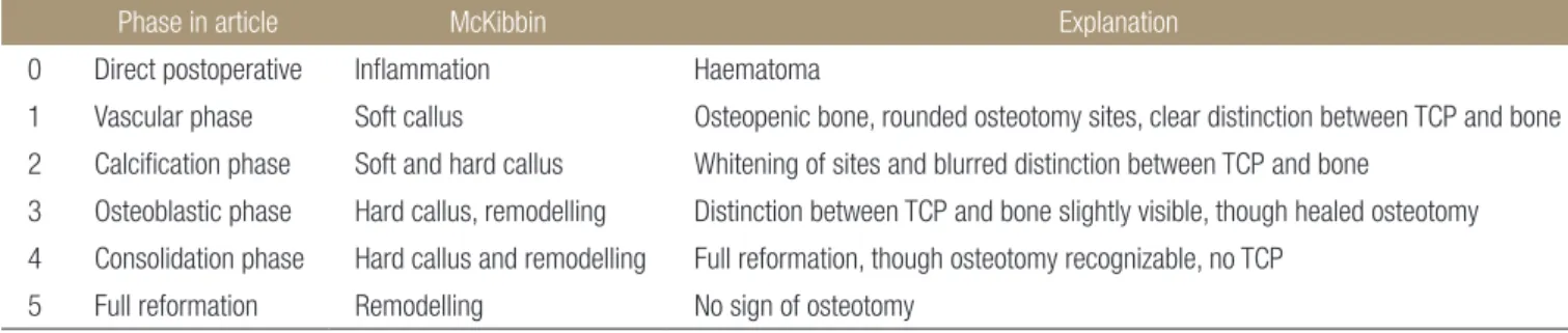
Table 2. Grading of van Hemert10)
Phase in article McKibbin Explanation
0 Direct postoperative Inflammation Haematoma
1 Vascular phase Soft callus Osteopenic bone, rounded osteotomy sites, clear distinction between TCP and bone 2 Calcification phase Soft and hard callus Whitening of sites and blurred distinction between TCP and bone
3 Osteoblastic phase Hard callus, remodelling Distinction between TCP and bone slightly visible, though healed osteotomy 4 Consolidation phase Hard callus and remodelling Full reformation, though osteotomy recognizable, no TCP
5 Full reformation Remodelling No sign of osteotomy TCP, tricalcium phosphate.
환자 관련 인자들에 따른 골유합의 정도를 확인하기 위해 추 시 기간 동안 van Hemert 등18)이 소개한 분류법을 이용하여 평가 하였다(Table 2). 모든 환자들에서 술 후 6주, 3개월, 6개월, 1년에 grading of van Hemert를 평가하기 위해 체중부하 방사선적 검사 를 시행하였다. 추가로 외측 피질골 골절이 있는 하위 집단에서 절골 부위의 골이식 종류에 따른 골유합 정도를 비교하기 위해 grading of van Hemert를 사용하였고, 임상적 평가를 위해 Oxford 점수 및 knee injury and osteoarthritis outcome score를 술 전, 술 후 관찰 시에 평가하였다.
환자들의 진료 기록지와 수술 기록지는 한 명의 관찰자에 의 해서 분석되었다. 진료 기록과 방사선적 분석을 통해서 골유합과 관련된 인자로 흡연, 비만, 교정각도, 환자의 키, 몸무게, 나이, 절 골 부위 이식물의 종류, 외측 피질골 골절 유무 등이 포함되었다.
환자의 수는 예비연구를 통하여 계산하였다. p<0.05에서 80%의 statical power를 위해서는 각각의 집단에 22명의 환자에 대한 실 험으로 충분하다고 결정되었다. 통계분석은 MedCalc ver. 15.2.2 (MedCalc Software, Ostend, Belgium)를 사용하여 independent samples t-test, one-way analysis of variance 방법으로 결과를 비교 하였으며, 통계적으로 유의한 값은 p-value가 0.05 미만일 경우로 정하였다.
결 과
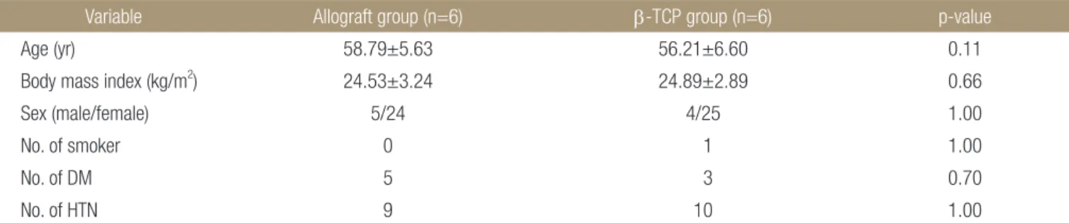
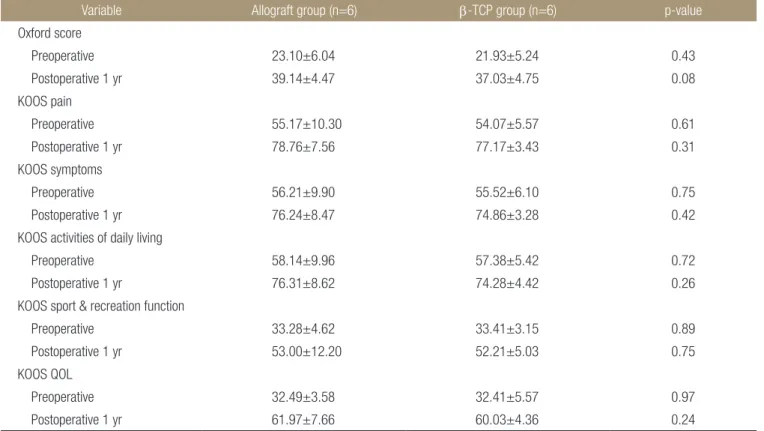
전체 환자에서 절골 부위 이식물의 종류는 동종골 이식군과 β-TCP 이식군의 비교에서 유의한 차이는 없었다(Table 3). 외측 피질골 손상이 있는 환자군 12예의 하위 집단에서(동종골 이식군 6예, β-TCP 이식군 6예) 두 군 간의 환자 관련 인자(Table 4) 및 방사선적(Table 5), 임상적 결과(Table 6)에서는 통계적으로 유의 한 차이는 없었지만, grading of van Hemert를 비교한 결과에서는 술 후 6개월, 1년에 동종골 이식군에서 유의하게 높은 결과를 보 였다(Table 7).
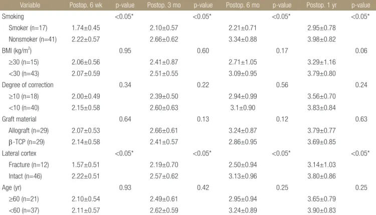
환자 관련 인자에서 흡연하는 경우와 외측 피질골이 손상된 경 우는 골유합에 부정적인 영향을 미칠 가능성이 더 높을 것으로 보였다. 비흡연 환자군과 외측 피질골 손상이 없는 환자군에서 모든 추시 기간 동안 대조군보다 골유합의 grading of van Hemert 가 통계적으로 유의하게 더 높은 결과를 보였으며(Table 3), 이는 흡연과 외측 피질골 손상이 지연유합 및 불유합의 가능성을 높이 는 인자일 것으로 생각된다. 그러나 술 전 내반슬, BMI, 교정각도, 환자 나이는 본 연구에서 골유합과 연관성이 없는 것으로 나타났 다.
Table 3. Demographic Data and Surgical Factors in Patients with Bone Union Grade
Variable Postop. 6 wk p-value Postop. 3 mo p-value Postop. 6 mo p-value Postop. 1 yr p-value
Smoking <0.05* <0.05* <0.05* <0.05*
Smoker (n=17) 1.74±0.45 2.10±0.57 2.21±0.71 2.95±0.78
Nonsmoker (n=41) 2.22±0.57 2.66±0.62 3.34±0.88 3.98±0.82
BMI (kg/m2) 0.95 0.60 0.17 0.06
≥30 (n=15) 2.06±0.56 2.41±0.87 2.71±1.05 3.29±1.16
<30 (n=43) 2.07±0.59 2.51±0.55 3.09±0.95 3.79±0.80
Degree of correction 0.34 0.22 0.56 0.24
≥10 (n=18) 2.00±0.49 2.39±0.50 2.94±0.99 3.56±0.70
<10 (n=40) 2.15±0.58 2.60±0.63 3.1±0.90 3.83±0.84
Graft material 0.64 0.13 0.12 0.63
Allograft (n=29) 2.07±0.53 2.66±0.61 3.24±0.87 3.79±0.77
β-TCP (n=29) 2.14±0.58 2.41±0.57 2.86±0.95 3.69±0.85
Lateral cortex <0.05* <0.05* <0.05* <0.05*
Fracture (n=12) 1.57±0.51 2.19±0.70 2.50±0.94 3.14±1.03
Intact (n=46) 2.22±0.51 2.57±0.62 3.13±0.96 3.80±0.86
Age (yr) 0.93 0.42 0.25 0.25
≥60 (n=21) 2.10±0.54 2.49±0.61 2.95±0.94 3.65±0.79
<60 (n=37) 2.11±0.57 2.62±0.59 3.24±0.89 3.90±0.83
Values are presented as mean±standard deviation. *This data is statistically significance. Postop., postoperative; BMI, body mass index; TCP, tricalcium phosphate.
고 찰
이 연구에서 개방형 내측 근위 경골 절골술에서 흡연하는 경우 와 외측 피질골이 손상된 경우 골유합이 지연될 가능이 더 높을 것으로 나타났다. 다른 연구에서도 개방형 내측 근위 경골 절골 술은 폐쇄형 외측 절골술과 달리 외측 경골 근위부에서 골을 제 거하지 않고 정상적인 해부학적 구조를 유지하는 장점이 있으나, 절골 부위에 지연유합 및 불유합의 위험이 증가하는 단점이 보고 되었다.15) 따라서 본 저자들은 개방형 내측 근위 경골 절골술에서 절골 부위의 골유합에 영향을 미치는 관련인자를 분석하고자 본 연구를 시행하였다. 술 전 흡연 유무와 외측 피질골 골절이 골유
합과 관련된 인자로 보였으며, 외측 피질골 골절이 있는 경우는 절골 부위 이식물의 종류가 동종골인 경우 더 나은 결과를 보였 다.
본 연구에서 흡연이 절골 부위 골유합과 관련된 인자 중 하나 였다. 흡연은 말초 혈류를 감소시키고,19) 말초 산소 장력에 악영 향을 미치게 하여,20) 상처 치유를 방해하고 골절 유합을 저해한다 고 알려져 있다.21,22) 또한 과거에 Meidinger 등14)도 역시 흡연이 불 유합의 위험 인자라고 보고하였다. 따라서 Lobenhoffer 등16)은 흡 연을 하는 환자에서 개방형 내측 근위 경골 절골술을 추천하지 않는다고 보고하였다. 본 연구의 결과에서도 환자가 흡연을 하는 경우 지연유합 및 불유합의 위험성이 높아질 수 있음을 보여주고 Table 4. Demographic Data of Patients in the Allograft Group and β-TCP Group with Lateral Cortex Fracture
Variable Allograft group (n=6) β-TCP group (n=6) p-value
Age (yr) 58.79±5.63 56.21±6.60 0.11
Body mass index (kg/m2) 24.53±3.24 24.89±2.89 0.66
Sex (male/female) 5/24 4/25 1.00
No. of smoker 0 1 1.00
No. of DM 5 3 0.70
No. of HTN 9 10 1.00
Values are presented as mean±standard deviation or number only. TCP, tricalcium phosphate; DM, diabetes mellitus; HTN, hypertension.
Table 5. Radiologic Assessment of the Allograft Group and β-TCP Group in Damaged Lateral Cortex
Variable Allograft group (n=6) β-TCP group (n=6) p-value
Mechanical FTA (°)
Preoperative 186.28±2.84 185.75±4.03 0.57
Postoperative 6 wk 176.88±1.78 177.58±2.59 0.24
Postoperative 3 mo 177.33±1.72 178.14±2.60 0.17
Postoperative 6 mo 177.62±1.70 178.40±2.59 0.18
Postoperative 1 yr 177.79±1.71 178.86±3.16 0.12
Mechanical MPTA (°)
Preoperative 85.15±2.12 85.99±1.74 0.10
Postoperative 6 wk 92.10±2.16 91.65±2.44 0.46
Postoperative 3 mo 90.97±1.83 90.69±2.08 0.59
Postoperative 6 mo 90.68±1.76 90.31±1.96 0.45
Postoperative 1 yr 90.51±1.66 90.11±1.90 0.39
Posterior slop of tibia (°)
Preoperative 11.42±3.12 12.47±3.49 0.23
Postoperative 6 wk 7.76±3.40 8.62±1.57 0.22
Postoperative 3 mo 8.49±3.17 9.41±1.70 0.18
Postoperative 6 mo 8.76±3.18 9.71±1.72 0.16
Postoperative 1 yr 8.95±3.26 9.88±1.76 0.18
Values are presented as mean±standard deviation. TCP, tricalcium phosphate; FTA, femorotibial angle; MPTA, medial proximal tibial angle.
있다.
외측 피질골 골절이 있는 경우 역시 골유합이 지연된 결과를 보였는데, Miller 등23)은 피질골 골절이 절골 부위에 미세 움직임 을 야기하여 이로 인한 불안정성이 지연유합 및 불유합의 발생률 에 기여한다고 하였다. 또한 Yacobucci와 Cocking24)은 개방형 내 측 근위 경골 절골술 후 외측 피질골 손상이 발생한 경우 불유합 이 나타난 증례를 통하여, 외측 피질골이 절골 부위의 안정성 및 골유합에 중요한 역할을 한다고 보고하였다. 외측 피질골 손상 시에 Esenkaya와 Elmali25)는 외측 피질골의 부가적인 고정 없이 내측 피질골 고정만으로도 교정각 소실이나 불안정성 등 합병증
이 발생하지 않는다고 보고하였다. 술 중에 전체 58예 중 12예에 서 외측 피질골 골절의 손상이 있었으며, 저자들도 추가적인 고 정을 시행하지는 않았다. 임상적 평가와 방사선적 평가 요소, 즉 대퇴 경골간 각, 후방 경골 경사에서는 외측 피질골 골절군과 대 조군 사이에 의미 있는 차이는 관찰되지 않았다. 그러나 모든 추 시 기간 중에 측정한 grading of van Hemert에서 외측 피질골 군이 통계적으로 유의하게 낮은 결과를 보였다. 따라서 외측 피질골 손상이 발생한 경우 지연유합 및 불유합의 위험성이 대조군보다 더 높아질 수 있음을 보여주고 있다.
절골 부위 이식물의 종류에 따른 골유합의 비교에서는 동종골 이식군과 β-TCP 이식군으로 분류하여 비교하였다. Santic 등26)은 개방형 내측 근위 경골 절골술 후 동종골 이식과 절골부 골유합 에 대한 연구를 통하여 osteotomy size가 9 mm 이하인 경우 12주 이내에 절골부 골유합이 관찰되었고, 10 mm 이상인 경우는 골유 합에 12주 이상 소요될 수 있음을 보고하였다. Cho 등27)은 개방형 내측 근위 경골 절골술 후 절골부 이식물을 비교하는 연구를 통 하여 동종골 이식이 자가골 이식과 비교하여 술 후 2년에 임상적, 방사선적으로 동등한 결과를 보였으며, 술 후 자가골 이식에 발 생할 수 있는 공여부 통증을 방지할 수 있어 술 후 통증은 더 감소 한다고 보고하였다. Gouin 등28)은 TCP 이식과 자가골 이식을 비 교하는 연구를 통하여, 자가골 이식군은 평균 2.6개월, TCP 이식 Table 6. Clinical Assessment of the Allograft Group and β-TCP Group in Damaged Lateral Cortex
Variable Allograft group (n=6) β-TCP group (n=6) p-value
Oxford score
Preoperative 23.10±6.04 21.93±5.24 0.43
Postoperative 1 yr 39.14±4.47 37.03±4.75 0.08
KOOS pain
Preoperative 55.17±10.30 54.07±5.57 0.61
Postoperative 1 yr 78.76±7.56 77.17±3.43 0.31
KOOS symptoms
Preoperative 56.21±9.90 55.52±6.10 0.75
Postoperative 1 yr 76.24±8.47 74.86±3.28 0.42
KOOS activities of daily living
Preoperative 58.14±9.96 57.38±5.42 0.72
Postoperative 1 yr 76.31±8.62 74.28±4.42 0.26
KOOS sport & recreation function
Preoperative 33.28±4.62 33.41±3.15 0.89
Postoperative 1 yr 53.00±12.20 52.21±5.03 0.75
KOOS QOL
Preoperative 32.49±3.58 32.41±5.57 0.97
Postoperative 1 yr 61.97±7.66 60.03±4.36 0.24
Values are presented as mean±standard deviation. TCP, tricalcium phosphate; KOOS, knee injury and osteoarthritis outcome score; QOL, quality of life.
Table 7. Bone Union Grade of the Allograft Group and β-TCP Group in Damaged Lateral Cortex
Variable Allograft group (n=6)
β-TCP group
(n=6) p-value
Postop. 6 wk 1.83±0.41 1.50±0.55 0.26 Postop. 3 mo 2.67±0.52 2.17±0.41 0.09 Postop. 6 mo 3.17±0.75 2.33±0.52 0.04*
Postop. 1 yr 3.83±0.41 3.17±0.41 0.01*
Values are presented as mean±standard deviation. *This data is stati sti- cally significance. TCP, tricalcium phosphate; Postop., postoperative.
군은 평균 5.8개월에 각각 골유합을 얻었다고 보고하였다. 저자들 은 방사선적 검사를 시행하여 grading of van Hemert로 두 군의 골 유합 등급을 측정하였으며, 그 결과 동종골 이식군이 β-TCP 이 식군보다 상대적으로 골유합 등급이 더 높은 경향성을 보였으나, 통계적으로 유의한 차이는 관찰되지 않았다. 그러나 외측 피질골 손상이 있는 하위 집단 12예(동종골 이식군 6예, β-TCP 이식군 6 예)에서 각각 이식물의 종류에 따른 골유합 등급을 비교한 결과 에서 동종골 이식군이 술 후 6개월, 1년에 β-TCP 이식군보다 통 계적으로 유의하게 높은 결과를 보였다. 따라서 외측 피질골 골 절이 발생한 경우는 절골 부위에 β-TCP 이식보다는 동종골 이식 이 술 후 6개월, 1년에 더 나은 골유합을 기대할 수 있을 것으로 생각된다.
본 연구에는 제한점이 있다. 연구를 시행한 환자군의 수가 적 었으며 또한 모든 환자군에서 TomoFix 금속판만 사용하여 다른 금속판을 사용한 개방형 절골술과 비교하여 일반화한다는 것에 무리가 있을 것으로 생각된다. 또한 외측 피질골 골절이 발생한 경우의 증례가 적었으며, 통계적으로 유의한 차이가 발생한 근거 에 대한 연구가 추가적으로 필요할 것으로 생각된다.
결 론
저자는 개방형 내측 경골 근위부 절골술에서 흡연과 외측 피질골 골절이 골유합을 지연시키는 위험 인자임을 관찰하였다. 절골 부 위 이식물에서 동종골과 β-TPC는 골유합과 유의한 관련성은 보 이지는 않았지만, 동종골 이식이 더 나은 골유합의 경향성을 보 였으며, 외측 피질골 손상이 있는 경우에 β-TCP 이식과 비교하 여 술 후에 더 나은 골유합을 보이는 것으로 생각된다. 이런 결과 를 토대로 저자는 개방형 내측 경골 근위부 절골술 시행하기 전 에 흡연을 하는 환자들에게 술 후 금연의 중요성에 대해 설명하 고 있으며, 술 중 외측 피질골 골절이 있는 경우에는 β-TCP보다 는 동종골 이식물로 절골 부위 이식을 시행하고 있다.
CONFLICTS OF INTEREST
The authors have nothing to disclose.
REFERENCES
1. Ministry for Health and Welfare. Elderly real state survey.
Seoul: Ministry for Health and Welfare; 2009.
2. Jackson JP, Waugh W. Tibial osteotomy for osteoarthritis of the knee. J Bone Joint Surg Br. 1961;43:746-51.
3. Coventry MB. Osteotomy about the knee for degenerative and rheumatoid arthritis. J Bone Joint Surg Am. 1973;55:23-
48.
4. Benzakour T, Hefti A, Lemseffer M, El Ahmadi JD, Bouyar- mane H, Benzakour A. High tibial osteotomy for medial osteoarthritis of the knee: 15 years follow-up. Int Orthop.
2010;34:209-15.
5. Keene JS, Dyreby JR Jr. High tibial osteotomy in the treat- ment of osteoarthritis of the knee. The role of preoperative arthroscopy. J Bone Joint Surg Am. 1983;65:36-42.
6. Staeheli JW, Cass JR, Morrey BF. Condylar total knee arthro- plasty after failed proximal tibial osteotomy. J Bone Joint Surg Am. 1987;69:28-31.
7. Brouwer RW, Bierma-Zeinstra SM, van Raaij TM, Verhaar JA. Osteotomy for medial compartment arthritis of the knee using a closing wedge or an opening wedge controlled by a Puddu plate. A one-year randomised, controlled study. J Bone Joint Surg Br. 2006;88:1454-9.
8. Spahn G. Complications in high tibial (medial opening wedge) osteotomy. Arch Orthop Trauma Surg. 2004;124:649- 53.
9. van den Bekerom MP, Patt TW, Kleinhout MY, van der Vis HM, Albers GH. Early complications after high tibial osteot- omy: a comparison of two techniques. J Knee Surg. 2008;21:
68-74.
10. Agneskirchner JD, Freiling D, Hurschler C, Lobenhoffer P.
Primary stability of four different implants for opening wedge high tibial osteotomy. Knee Surg Sports Traumatol Arthrosc.
2006;14:291-300.
11. Stoffel K, Stachowiak G, Kuster M. Open wedge high tibial osteotomy: biomechanical investigation of the modified Ar- threx Osteotomy Plate (Puddu Plate) and the TomoFix Plate.
Clin Biomech (Bristol, Avon). 2004;19:944-50.
12. van Raaij TM, Brouwer RW, de Vlieger R, Reijman M, Ver- haar JA. Opposite cortical fracture in high tibial osteotomy:
lateral closing compared to the medial opening-wedge tech- nique. Acta Orthop. 2008;79:508-14.
13. Brown CW, Orme TJ, Richardson HD. The rate of pseud- arthrosis (surgical nonunion) in patients who are smokers and patients who are nonsmokers: a comparison study. Spine (Phila Pa 1976). 1986;11:942-3.
14. Meidinger G, Imhoff AB, Paul J, Kirchhoff C, Sauerschnig M, Hinterwimmer S. May smokers and overweight patients be treated with a medial open-wedge HTO? Risk factors for non-union. Knee Surg Sports Traumatol Arthrosc. 2011;19:
333-9.
15. Sloan A, Hussain I, Maqsood M, Eremin O, El-Sheemy M.
The effects of smoking on fracture healing. Surgeon. 2010;8:
111-6.
16. Lobenhoffer P, Agneskirchner J, Zoch W. Open valgus align- ment osteotomy of the proximal tibia with fixation by medial plate fixator (in German). Orthopade. 2004;33:153-60.
17. Staubli AE, De Simoni C, Babst R, Lobenhoffer P. TomoFix:
a new LCP-concept for open wedge osteotomy of the medial proximal tibia: early results in 92 cases. Injury. 2003;34 Suppl 2:B55-62.
18. van Hemert WL, Willems K, Anderson PG, van Heer- waarden RJ, Wymenga AB. Tricalcium phosphate granules or rigid wedge preforms in open wedge high tibial osteotomy:
a radiological study with a new evaluation system. Knee.
2004;11:451-6.
19. Jensen JA, Goodson WH, Hopf HW, Hunt TK. Cigarette smoking decreases tissue oxygen. Arch Surg. 1991;126:1131-4.
20. Sarin CL, Austin JC, Nickel WO. Effects of smoking on digi- tal blood-flow velocity. JAMA. 1974;229:1327-8.
21. Jones JK, Triplett RG. The relationship of cigarette smoking to impaired intraoral wound healing: a review of evidence and implications for patient care. J Oral Maxillofacial Surg.
1992;50:237-9; discussion 239-40.
22. Harvey EJ, Agel J, Selznick HS, Champman JR, Henley MB.
Deleterious effect of smoking on healing of open tibia-shaft fractures. Am J Orthop (Belle Mead NJ). 2002;31:518-21.
23. Miller BS, Dorsey WO, Bryant CR, Austin JC. The effect of lateral cortex disruption and repair in the stability of the me- dial opening wedge high tibial osteotomy. Am J Sports Med.
2005;33:1552-7.
24. Yacobucci GN, Cocking MR. Union of medial opening- wedge high tibial osteotomy using a corticocancellous proxi- mal tibial wedge allograft. Am J Sports Med. 2008;36:713-9.
25. Esenkaya I, Elmali N. Proximal tibia medial open-wedge osteotomy using plates with wedges: early results in 58 cases.
Knee Surg Sports Traumatol Arthrosc. 2006;14:955-61.
26. Santic V, Tudor A, Sestan B, Legovic D, Sirola L, Rakovac I.
Bone allograft provides bone healing in the medial opening high tibial osteotomy. Int Orthop. 2010;34:225-9.
27. Cho SW, Kim DH, Lee GC, Lee SH, Park SH. Comparison between autogenous bone graft and allogenous cancellous bone graft in medial open wedge high tibial osteotomy with 2-year follow-up. Knee Surg Relat Res. 2013;25:117-25.
28. Gouin F, Yaouanc F, Waast D, Melchior B, Delecrin J, Passuti N. Open wedge high tibial osteotomies: calcium-phosphate ceramic spacer versus autologous bonegraft. Orthop Trauma- tol Surg Res. 2010;96:637-45.
개방형 근위 경골 절골술에서 골유합과 관련된 인자와 외측 피질골 골절 시 이식물에 따른 비교
서진혁 • 김도훈 • 서승석 • 김연구 • 김옥걸 • 박병윤
부민병원 정형외과
목적: 개방형 경골 근위부 절골술에서 환자 관련 인자들과 외측 피질골 골절이 발생한 경우에 골이식물 종류가 골유합에 미치는 영 향에 대하여 연구하였다.
대상 및 방법: Kallgren-Lawrence 2단계 이하의 환자 54명, 58예를 대상으로 2012년 5월부터 2014년 6월까지 후향적 연구를 시행 하였다. 평균 추시 기간은 22개월(14-38개월)이었다. 환자 관련 인자들이 골유합과 연관성이 있는지를 분석하고, 외측 피질골 골절 이 발생한 하위 집단에서 골이식물 종류(동종골, n=6; beta-tricalcium phosphate [β-TCP], n=6)에 따라 두 군으로 분류하여 술 후 6주, 3개월, 6개월, 1년에 방사선적, 임상적 평가 및 grading of van Hemert를 비교하였다.
결과: 하위 집단의 두 군 간에 임상적, 방사선적 결과는 유의한 차이가 없었고, grading of van Hemert는 술 후 6개월, 1년에 동종골 이식군에서 유의하게 높은 결과를 보였다. 비흡연 환자군과 외측 피질골 손상이 없는 환자군에서 유의하게 더 높은 골유합 등급을 보 였다.
결론: 개방형 경골 근위부 절골술에서 외측 피질골 손상 시에 동종골 이식이 β-TCP보다 술 후에 더 나은 골유합을 보이며, 흡연과 외측 피질골 손상이 골유합을 지연시키는 위험 인자임을 관찰하였다.
색인단어: 슬관절, 골관절염, 개방형 근위 경골 절골술, 동종골 이식, 베타-삼인산 칼슘
접수일 2016년 3월 7일 수정일 2016년 4월 26일 게재확정일 2016년 5월 7일 책임저자 김도훈
46555, 부산시 북구 만덕대로 59, 부민병원 정형외과
TEL 051-330-3082, FAX 051-330-3242, E-mail [email protected]
Copyright © 2016 by The Korean Orthopaedic Association
“This is an Open Access article distributed under the terms of the Creative Commons Attribution Non-Commercial License (http://creativecommons.org/licenses/by-nc/4.0/) which permits unrestricted non-commercial use, distribution, and reproduction in any medium, provided the original work is properly cited.”