Invasive Breast Cancer Presenting as a Mass
Replaced by Calcification
on Mammography: A Report of Two Cases
석회화로 대체된 종괴로 관찰된 침윤성 유방암:
2개의 증례 보고
Joo Hee Jeun, MD1 , Jin Hwa Lee, MD1* , Eun Cho, MD1, Su Jin Kim, MD2, Eun Hwa Park, MD3, Kyung Do Byun, MD3
Departments of 1Radiology, 3Surgery, Dong-A University Medical Center, Dong-A University College of Medicine, Busan, Korea
2Pathology Reference Lab, Seegene Medical Foundation, Busan, Korea
A distinct calcification pattern is one of the criteria for determining the malignancy of breast can- cer according to the Breast Imaging Reporting and Data System. A mass almost entirely replaced by calcification, however, is difficult to categorize and likely to be misdiagnosed. We present the report of two patients with invasive carcinoma of the breast that presented as a mass replaced by calcification on mammography. In the first case, the mass was confirmed as a mixed carcino- ma comprising mucinous and micropapillary carcinoma, and in the second case, the mass was a mucinous carcinoma. Diagnosis of cancer in the latter case was missed as the mass had been assessed as a category 2 typically benign calcification at the first screening mammography 2 years ago. This report merits publication because it shows that a mass replaced by calcification on mammography can be misdiagnosed as a benign finding.
Index terms Breast Cancer; Mammography; Pathologic Calcification
INTRODUCTION
Various morphologic features of calcifications are one of the important criteria deter- mining malignancy of breast tumor according to the American College of Radiology Breast Imaging Reporting and Data System (ACR-BIRADS) 5th edition (1). The interpre- tation of these calcifications, however, is partly dependent on the descriptors, and dif-
Received January 15, 2019 Revised March 6, 2019 Accepted March 12, 2019
*Corresponding author Jin Hwa Lee, MD Department of Radiology, Dong-A University Medical Center, Dong-A University
College of Medicine, 26 Daesingongwon-ro, Seo-gu, Busan 49201, Korea.
Tel 82-51-240-5368 Fax 82-51-253-4931 E-mail [email protected] This is an Open Access article distributed under the terms of the Creative Commons Attribu- tion Non-Commercial License (https://creativecommons.org/
licenses/by-nc/4.0) which permits unrestricted non-commercial use, distribution, and reproduc- tion in any medium, provided the original work is properly cited.
ORCID iDs Jin Hwa Lee https://
orcid.org/0000-0003-0843-9862 Joo Hee Jeun
https://
orcid.org/0000-0002-4012-8338
ferent diagnostic confirmation can be demonstrated. A mass with almost exclusive portion of calcification can be easily considered as a dense benign calcification. In the ACR-BIRADS, classic and large (> 2–3 mm in greatest diameter) coarse or “popcorn-like” calcification is de- fined as category 2 benign finding, and the recommended management is only regular fol- low-up. Subsequently, it can be more extensive and confluent and when so densely calcified, the underlying mass usually is not visible (1). However, there have been a few reported cases of primary and secondary breast cancer presenting as a mass replaced by calcification, which could be one of the causes of misdiagnosis on mammography. We present two cases of a mass replaced by calcification that resulted in malignant breast tumor. The imaging fea- tures of mammography, ultrasound (US), and magnetic resonance imaging (MRI) are pre- sented and correlated pathologically.
CASE REPORT CASE 1
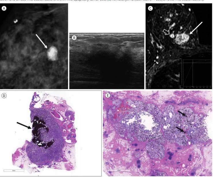
A 50-year-old woman presented to our breast center for evaluation of a palpable lump in the upper inner portion of right breast that was increasing in size for a year. Right mammo- gram showed extremely dense composition and a 24 mm-sized, calcified mass with an oval shape and circumscribed margins in the clinically palpable upper inner area. Coarse calcifi- cation was also noted in the upper outer portion (Fig. 1A). The mammographic BI-RADS as- sessment was category 2, but additional breast US was performed because of the growing palpable breast lump. US revealed a 26 mm-sized, irregular, and markedly hypoechoic mass with indistinct margins and strong posterior shadowing in the palpable area, which was a suspicious finding for malignancy (Fig. 1B). The BI-RADS assessment between the mammog- raphy and US findings was discordant, and thus US-guided 14-gauge core needle biopsy was performed. The pathologic result was consistent with invasive micropapillary carcinoma.
The subtraction image of dynamic contrast-enhanced breast MRI showed an oval shaped mass with circumscribed margins and heterogeneous enhancement and a satellite enhanc- ing nodule. The kinetic curve showed initial fast and delayed plateau enhancement pattern (Fig. 1C). The mass had a portion with low signal intensity on both T1 and T2-weighted im- age, suggesting calcification (not shown). She underwent total mastectomy, and the gross specimen measured 2.2 × 1.8 cm and showed a grayish granular appearance with a myxoid cut surface. Microscopically, it was diagnosed as mixed carcinoma that was composed of mucinous carcinoma (60%) with micropapillary carcinoma (40%) (Fig. 1D, E). Calcifications were noted in both cancers.
CASE 2
A 46-year-old woman visited an another clinic for screening, and the mammogram had shown heterogeneously dense composition and a 13 mm-sized mass almost entirely substi- tuted by calcification with circumscribed margins in the left central area (Fig. 2A). Consid- ered that no findings suspicious of malignancy were noted, she was recommended only for regular follow-up. After 2 years, she visited the clinic because of a palpable lump in the left breast. Left magnification mammogram showed that the pre-existing mass increased in size
A
B
C
D E
to 25 mm; new grouped calcifications in the subareolar area were also noted (Fig. 2B). Breast US showed that the mass was an oval-shaped hyperechoic mass with a few internal echogenic calcifications and circumscribed margins (Fig. 2C). Pathological findings of excisional biopsy indicated mucinous carcinoma, measuring 2.0 × 1.5 cm (Fig. 2D). She was referred to our breast center for operative treatment and underwent breast conservation surgery. There was no residual cancer, and axillary sentinel node biopsy was negative. This was an interval breast cancer that had been assessed as category 2 benign finding at the first screening mammography.
Fig. 1. A 50-year-old woman with a mixed-type mucinous carcinoma.
A. Right mediolateral oblique mammogram shows a calcified mass, measuring 24 mm, with an oval shape and circumscribed margins in the clinically palpable upper inner area (arrow).
B. Breast ultrasound image shows an irregular, indistinctly marginated, markedly hypoechoic mass with posterior shadowing.
C. Subtraction image of dynamic contrast-enhanced breast MRI shows a heterogeneously enhancing mass (arrow) and a satellite enhancing nodule (open arrow). The kinetic curve shows initial fast and delayed plateau enhancement pattern (inset).
D, E. In the surgical specimen, the mass shows collisional features with two histologically different types of carcinoma. The area on the right comprises a mucinous carcinoma with an abundant extracellular mucin pool, floating tumor cells, and multifocal microcalcifications (D, he- matoxylin & eosin stain, × 10, arrow: microcalcification). The area on the left comprises a micropapillary carcinoma with central dense calcifi- cation and diffuse microcalcifications within micropapillary tumor cells (E, hematoxylin & eosin stain, × 200, arrows: microcalcification).
A
C
B
D Fig. 2. A 46-year-old woman with a pure-type mucinous carcinoma.
A. Left craniocaudal screening mammogram obtained at another hospital shows a calcified mass, measuring 13 mm, with circumscribed margins in the left central area. The mass had been assessed as a category 2 typically benign finding.
B. Follow-up left magnification mammogram obtained after 2 years for a palpable lump in the left breast shows a mass with an increased size and a new group of calcifications in the left subareolar area (arrow).
C. Breast US image obtained at another hospital shows an oval-shaped, circumscribed, and hyperechoic mass (open arrows) with suspected echogenic calcifications (arrow).
D. In the excised specimen, a well-defined mass consisting of an abundant extracellular mucin pool with some floating carcinoma cells is ob- served. Diffuse microcalcification is noted within the mucin pool and tumor cells [hematoxylin & eosin stain, × 10 (inset: × 200); arrows: micro- calcification; asterisk: mucin pool]
DISCUSSION
Some cases of primary and secondary malignant breast tumor that appeared as a mass mimicking benign finding with extensive portion of calcification on mammography have been reported. To our knowledge, there have been cases of metaplastic breast carcinoma, mucinous carcinoma, primary and secondary osteosarcoma, and metastasis from ovarian cancer.
Metaplastic carcinoma of the breast is rare, accounting for only 5% of all breast carcino- mas. It shows metaplastic change in the form of mesenchymal elements producing osseous matrix and has squamous cell and spindle cell components (2). Meanwhile, osseous or chon- droid metaplasia can appear as a densely calcified mass. Lee et al. (3) reported a case of meta- plastic breast carcinoma with osseous differentiation that as appeared as a largely calcified mass with a partially spiculated margin. Although it could have been mistaken for as a be- nign mass because of the dense calcification, it could be assessed as suspicion for malignan- cy due to the spiculated margin. The dense calcification was confirmed as ossifying osteoid matrix. Another case reported by Evans et al. (4) showed a densely calcified mass with a spongy, bonelike appearance along the periphery of the calcification on mammogram. An additional image obtained using a higher kilovoltage setting showed trabeculations, which are consistent with an osteoid matrix.
Mucinous carcinoma is even rarer and accounts for only approximately 1.5–2.0% of all breast carcinoma (5). It has two subtypes according to histologic features: the pure type con- taining typical extracellular mucin-producing cells mostly and the mixed type with much lower portion of this cells (50–90%) (3, 5). The typical imaging finding is a well-circumscribed round or oval-shaped mass on mammography and rarely shows calcification. When present, the calcification shows pleomorphic and clustered form, and they also appear clumped and amorphous (6). In the current report, both cases of mixed- and pure-type mucinous carcino- ma presented as a more exclusively calcified mass than the usual calcification patterns re- ported previously. Although there was an atypical case of breast mucinous carcinoma with coarse calcification (6), it has not been reported to have almost entirely calcified portion of the mass. As a mass replaced by calcification or a mass accompanying benign coarse calcifi- cation is generally considered as benign lesion, the malignant breast tumor with these char- acteristics are prone to be a missed cancer. The first case of this report was also assessed as category 2 benign finding on mammography. However, she had a palpable breast lump, and subsequent breast US showed suspicious features for malignancy. The second case was an interval cancer, as shown above, because of the circumscribed marginated mass replaced by calcification on the first screening mammography.
Primary osteosarcoma of the breast is extremely rare and usually reveals a heavily calci- fied mass as a result of intermembranous ossification with irregular margin (7). Metastatic osteosarcoma to the breast was also described to have dense calcification in a case reported by Kim et al., (8) even though metastatic osteosarcoma usually involves lung and skeleton.
Calcifications in metastatic tumor of the breast are uncommon, except in ovarian metas- tasis. Metastatic lesion in the breast represents clusters of smooth and irregular dense calcifi- cation from the ovarian tumor. They are suggested to be related with psammoma bodies of
malignant tumor (9).
In conclusion, a mass replaced by calcification on mammography can be easily consid- ered as benign finding, however, we should keep in mind the possibility of malignancy such as mucinous breast cancer and assess the mass thoroughly with other suspicious findings for malignancy such as partially non-circumscribed margins or clinical palpability to prevent missed cancer.
Conflicts of Interest
The authors have no potential conflicts of interest to disclose.
REFERENCES
1. American College of Radiology. ACR BI-RADS atlas: breast imaging reporting and data system. 5th ed. Res- ton, VA: American College of Radiology 2013
2. Erguvan-Dogan B, Yazgan C, Atasoy C, Sak SD, Tukel S, Ceyhan K, et al. Radiologic-pathologic conference of the University of Ankara Medical School: metaplastic breast carcinoma with osteochondrosarcomatous differentiation. AJR Am J Roentgenol 2005;185:1593-1594
3. Lee JH, Kim EK, Choi SS, Nam KJ, Kim DC, Cho SH. Metaplastic breast carcinoma with extensive osseous differentiation: a case report. Breast 2008;17:314-316
4. Evans HA, Shaughnessy EA, Nikiforov YE. Infiltrating ductal carcinoma of the breast with osseous metapla- sia: imaging findings with pathologic correlation. AJR Am J Roentgenol 1999;172:1420-1422
5. Wilson DA, Kalisher L, Port JE, Titus JM, Kirzner HL. Breast imaging case of the day. Pure mucinous carci- noma with calcifying matrix. Radiographics 1997;17:800-804
6. Tani H, Murakami R, Yoshida T, Kumita S, Yanagihara K, Iida S, et al. Mucinous carcinoma of the breast ac- companied by coarse calcification. Open J Med Imaging 2012;2:125-127
7. Alghofaily KA, Almushayqih MH, Alanazi MF, Salamah AAB, Benediktsson H. Primary osteosarcoma of the breast arising in an intraductal papilloma. Case Rep Radiol 2017;2017:5787829
8. Kim JH, Woo HY, Kim EK, Kim MJ, Moon HJ, Yoon JH. Metastatic osteosarcoma to the breast presenting as a densely calcified mass on mammography. J Breast Cancer 2016;19:87-91
9. Ozsaran AA, Dikmen Y, Terek MC, Ulukus M, Ozdemir N, Orgüc S, et al. Bilateral metastatic carcinoma of the breast from primary ovarian cancer. Arch Gynecol Obstet 2000;264:166-167
석회화로 대체된 종괴로 관찰된 침윤성 유방암:
2개의 증례 보고
전주희1 · 이진화1* · 조 은1 · 김수진2 · 박은화3 · 변경도3
Breast Imaging Reporting and Data System에 의하면 유방 종양의 석회화 양상은 악성 종 양을 결정짓는 중요한 기준 중 하나이다. 하지만 종양의 대부분이 석회화로 대체된 경우 이 를 분류하는 데 한계가 있으며, 따라서 악성 종양을 놓치기 쉽다. 본 저자들은 유방촬영술에 서 석회화로 대체된 종양으로 발견된 침윤성 유방암의 증례 2개를 경험하였고 첫 번째 증례 는 혼합형 점액성과 미세유두성 암종이며, 두 번째 증례는 점액암으로 확인되었다. 특히 후 자는 2년 전에 시행되었던 검진 유방촬영술에서 양성 석회화로 오인되어 암 진단을 놓친 증 례이다. 유방촬영술에서 대부분이 석회화로 대체된 종양인 경우 악성의 가능성을 면밀히 검 토해야 함을 보여주는 증례들을 보고한다.
동아대학교의료원 1영상의학과, 3외과, 2씨젠의료재단 병리과