Copyrights © 2016 The Korean Society of Radiology
218
Case Report
pISSN 1738-2637 / eISSN 2288-2928 J Korean Soc Radiol 2016;75(3):218-221 http://dx.doi.org/10.3348/jksr.2016.75.3.218
INTRODUCTION
While uncommon, pulmonary artery aneurysms and pseudo- aneurysms are associated with high mortality. Overall they are considered rare entities with prevalence rates of around 1 case per 14000 to 100000 autopsies (1, 2). Most pulmonary artery an- eurysms and pseudoaneurysms are acquired and associated with cardiovascular disease, infection, iatrogenic causes, trauma, neo- plasm, and connective tissue disease. To our knowledge, while there are many cases of Rasmussen aneurysm in patients with cavitary tuberculosis, there have been no reports of pseudoaneu- rysm due to fungus ball in the progressive massive fibrosis (PMF) of a patient with pneumoconiosis. We described a case of pseu- doaneurysm detected by contrast-enhanced chest CT with three- dimensional (3D) reconstruction. The pseudoaneurysm arose in
the upper lobar branch of the right pulmonary artery and caused by a fungus ball within the PMF in a patient with pneumoconio- sis, who underwent transcatheter endovascular embolization of the aneurysm.
CASE REPORT
The patient was a 51-year-old man with a history of pneumo- coniosis, tuberculosis, and hypertension. He was a coal miner for 12 years and had a 30 pack-year smoking history. He initially presented to another hospital with an episode of pneumonia and was transferred to our hospital for recurrent episodes of massive hemoptysis of more than 1 liter per day. On hospital admission, the patient was afebrile and hemodynamically stable (134/85 mm Hg, 106 beats per minute, 20 breaths per minute, and tempera-
A Case of Pseudoaneurysm Due to Fungus Ball within the Progressive Massive Fibrosis in a Patient with Pneumoconiosis:
Computed Tomography-Pathologic Correlation
진폐증 환자에서 진행성거대섬유화증 내에 생긴 진균구에 의한 가성동맥류 일 예: 전산화단층촬영과 병리소견
Sang Geun Lee, MD, Dae Shick Ryu, MD*, Man Soo Park, MD, Soo Jung Choi, MD, Chae Hoon Kang, MD, Dong Rock Shin, MD, Jae Hong Ahn, MD
Department of Radiology, Gangneung Asan Hospital, College of Medicine, University of Ulsan, Gangneung, Korea
We reported a case of pseudoaneurysm in the upper lobar branch of the right pul- monary artery, which was caused by a fungus ball within the progressive massive fibrosis (PMF) in a patient with pneumoconiosis. Coil embolization of the pseudoan- eurysm initially stopped the bleeding. After right upper lobe lobectomy to prevent the recurrent hemoptysis, pathology confirmed pseudoaneurysm within the PMF due to aspergilloma.
Index terms Aneurysm Chest CT
Aspergillosis Pneumoconiosis
Received December 17, 2015 Revised January 19, 2016 Accepted April 2, 2016
*Corresponding author: Dae Shick Ryu, MD Department of Radiology, Gangneung Asan Hospital, College of Medicine, University of Ulsan, 38 Bangdong-gil, Sacheon-myeon, Gangneung 25440, Korea.
Tel. 82-33-610-3483 Fax. 82-33-610-3490 E-mail: [email protected]
This is an Open Access article distributed under the terms of the Creative Commons Attribution Non-Commercial License (http://creativecommons.org/licenses/by-nc/3.0) which permits unrestricted non-commercial use, distri- bution, and reproduction in any medium, provided the original work is properly cited.
219
Sang Geun Lee, et al
jksronline.org J Korean Soc Radiol 2016;75(3):218-221
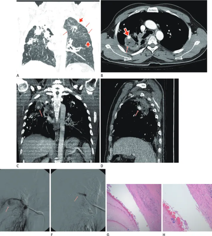
Fig. 1. Pseudoaneurysm due to fungus ball within the progressive massive fibrosis in a 51-year old patient, presenting with hemoptysis.
A. Coronal reconstruction image (lung window) shows multiple well-defined centrilobular nodules, mainly distributed in the corticomedullary junction of the lung (small arrows) suggestive of pneumoconiosis. Also in addition, multiple small costal pleural and fissural nodules (large ar- rows) indicate perilymphatic distribution of pneumoconiotic nodules.
B. Contrast-enhanced CT scan (mediastinal settings, 2.5 mm collimation) shows focus of enhancement suggesting pseudoaneurysm of segmen- tal pulmonary artery in right upper lobe (large arrow). Spongiform air bubble suggesting a fungus ball within progressive massive fibrosis (PMF) (small arrow).
C, D. Coronal and sagittal reconstruction images show pseudoaneurysm (arrow) of right upper lobe by fungus ball in PMF.
E. Right pulmonary artery selective angiography shows a pseudoaneurysm (arrow) of segmental pulmonary artery in right upper lobe.
F. Transcatheter embolization was performed with use of 4–5 mm coils (arrow) via the right femoral venous approach.
G, H. Hematoxylin & eosin stain (× 40) and B (× 100) show fungus ball in the cavity of PMF.
A
E
B
F C
G D
H
220
A Case of Pseudoaneurysm Due to Fungus Ball within the PMF in a Patient with Pneumoconiosis
jksronline.org
J Korean Soc Radiol 2016;75(3):218-221 ture of 36.8°C). The initial laboratory results showed anemia,
mild leukocytosis (hemoglobin: 7.6 g/dL, hematocrit: 22.7%, and white blood cell count: 10300/μL) and negative three sputum acid fast bacillus smears and culture. Contrast-enhanced chest CT with 3D reconstruction (LightSpeed 16; GE Medical Systems, Milwaukee, WI, USA) showed multiple perilymphangitic nod- ules suggestive of pneumoconiosis (Fig. 1A), spongiform air bubbles indicating a fungus ball within a PMF, and a tuberculous scar with volume loss and traction bronchiectasis. There was no three-in-bud or centrilobular nodule, cavitary nodule, lymph- adenopathy, or effusion that was suggestive of active tuberculosis.
Within the fungus ball, a pseudoaneurysm was noted in the up- per lobar branch of the right pulmonary artery (Fig. 1B-D). The lack of a draining vein ruled out an arteriovenous malformation.
Emergent pulmonary artery angiogram and transcatheter embo- lization were performed with 4–5 mm coils (Tornado; Cook, Bloomington, IN, USA) via a right femoral venous approach (Fig.
1E, F). Particle embolization (Contour; Boston Scientific, Natick, MA, USA) of the right intercostobronchial trunk and right 1st- 2nd intercostal artery arising from the costocervical trunk were also performed due to arterial hypertrophy. Following these pro- cedures, the hemoptysis stopped, but recurred at not more than 20 cc per day after one week. Because of recurrent hemoptysis and deteriorating pulmonary function test results, a right upper lobe lobectomy was performed for the pulmonary aspergilloma with pulmonary artery pseudoaneurysm. Although the presence of cavitation in a conglomerate mass is an important indication of associated tuberculosis, the pathologic specimen of this case showed fungal hyphae within the PMF cavity without chronic granulomatous inflammation suggesting tuberculosis (Fig. 1G, H). Polarized light images showed scattered interstitial silica par- ticles. The postoperative period was uneventful and the patient was discharged in the fourth postoperative week.
DISCUSSION
Pulmonary artery aneurysms and pseudoaneurysms can be congenital or acquired. Common congenital causes include ves- sel wall deficiency, valvular and postvalvular stenosis, and in- creased flow due to left-to-right shunts. Common acquired causes include trauma, pulmonary artery hypertension, vasculitis (Be- hçet’s syndrome and Hughes-Stovin syndrome), infection (my-
cotic aneurysms and pseudoaneurysms), neoplasm, iatrogenic causes (malpositioned Swan-Ganz catheters), and connective tis- sue abnormalities such as Marfan syndrome, Ehlers-Danlos syn- drome, and cystic medial necrosis (3, 4). There have been many reports of pulmonary artery pseudoaneurysm due to tuberculo- sis or fungus infection. Also, fungus infection is well known in patients with pneumoconiosis. To the best of our knowledge, there have been no reports of pseudoaneurysm due to fungus ball in the PMF of a patient with pneumoconiosis. Therefore, we reported the first case of pseudoaneurysm due to fungus ball in the PMF of a patient with pneumoconiosis.
The cavity formation within the PMF suggests ischemic necro- sis due to deficient blood supply (5). PMF greater than 4 cm may show cavitation due to ischemic necrosis. Tuberculosis is another possible cause of cavitation in PMF. The presence of cavitation in a conglomerate mass (PMF) is an important indication of associ- ated tuberculosis (6). Kato et al. (7) proposed that chronic persis- tent or progressive upper lobe infiltrates and cavities in patients with pneumoconiosis should raise the possibility of chronic nec- rotizing pulmonary aspergillosis. Preexisting diseases associated with aspergilloma include tuberculosis, bronchiectasis, pneumo- coniosis, and sarcoidosis (8). Aspergillomas occur predominantly in the upper lobes, reflecting a predilection for cavity formation at this site, because the relative imbalance in perfusion and venti- lation in the lung apices provide an oxygen-rich environment for micro-organisms (9).
Our case showed spongiform air bubbles in the PMF cavity suggestive of fungus ball. Ischemic necrosis of PMF due to defi- cient blood supply might cause cavitation of PMF and subse- quent fungal hyphae growth in the PMF cavity. Mycotic pulmo- nary artery pseudoaneurysms are caused by several mechanisms such as direct extension of inflammation to involve the vessel wall, endovascular seeding of the vessel wall from bronchial ar- teries in septicemia and intimal invasion of the vessel wall from septic embolism. In our case, it is likely that the fungal hyphae eroded and weakened the branch of the pulmonary artery, lead- ing to the vessel pseudoaneurysm.
CT is useful for diagnosing diseases that cause massive hemop- tysis, localizing bleeding sites, and selecting vessels to be emboli- zed (10). Contrast-enhanced chest CT with 3D reconstruction scan helped us to detect pseudoaneurysm of the pulmonary ar- tery in PMF, because the enhancement density of the pseudoan-
221
Sang Geun Lee, et al
jksronline.org J Korean Soc Radiol 2016;75(3):218-221 eurysm of pulmonary artery was similar to that of the aorta. After detecting the cause of massive hemoptysis such as pseudoaneu- rysm, we suggest that embolization of pseudoaneurysm due to fungus ball within the PMF is necessary to stop abrupt massive pulmonary bleeding. After that, surgical removal of PMF with fungus ball is the best way to prevent recurrent hemoptysis.
In conclusion, we reported a case of pseudoaneurysm due to fungus ball in the PMF of a patient with pneumoconiosis. As- sessment with contrast-enhanced CT with coronal reconstruc- tion images allowed accurate evaluation of pulmonary artery pseudoaneurysm, facilitating prompt diagnosis and treatment.
REFERENCES
1. Seguchi M, Wada H, Sakakura K, Kubo N, Ikeda N, Suga- wara Y, et al. Idiopathic pulmonary artery aneurysm. Circu- lation 2011;124:e369-e370
2. Ting P, Jugdutt BI, Le Tan J. Large pulmonary artery aneu- rysm associated with Marfan syndrome. Int J Angiol 2010;
19:e48-e50
3. Nguyen ET, Silva CI, Seely JM, Chong S, Lee KS, Müller NL.
Pulmonary artery aneurysms and pseudoaneurysms in adults:
findings at CT and radiography. AJR Am J Roentgenol 2007;
188:W126-W134
4. Moon JI, Lee JW, Jeong YJ, Song SH. A mycotic pulmonary
artery aneurysm associated with Candida endocarditis:
case report. J Korean Soc Radiol 2014;70:205-208 5. Kim KI, Kim CW, Lee MK, Lee KS, Park CK, Choi SJ, et al. Im-
aging of occupational lung disease. Radiographics 2001;21:
1371-1391
6. Martins P, Marchiori E, Zanetti G, Muccillo A, Ventura N, Brandão V, et al. Cavitated conglomerate mass in silicosis indicating associated tuberculosis. Case Rep Med 2010 Aug 5 [Epub]. http://dx.doi.org/10.1155/2010/293730
7. Kato T, Usami I, Morita H, Goto M, Hosoda M, Nakamura A, et al. Chronic necrotizing pulmonary aspergillosis in pneu- moconiosis: clinical and radiologic findings in 10 patients.
Chest 2002;121:118-127
8. Kang EY, Kim DH, Woo OH, Choi JA, Oh YW, Kim CH. Pul- monary aspergillosis in immunocompetent hosts without underlying lesions of the lung: radiologic and pathologic findings. AJR Am J Roentgenol 2002;178:1395-1399 9. Roberts CM, Citron KM, Strickland B. Intrathoracic asper-
gilloma: role of CT in diagnosis and treatment. Radiology 1987;165:123-128
10. Yoon W, Kim JK, Kim YH, Chung TW, Kang HK. Bronchial and nonbronchial systemic artery embolization for life- threatening hemoptysis: a comprehensive review. Radio- graphics 2002;22:1395-1409
진폐증 환자에서 진행성거대섬유화증 내에 생긴 진균구에 의한 가성동맥류 일 예: 전산화단층촬영과 병리소견
이상근 · 류대식* · 박만수 · 최수정 · 강채훈 · 신동락 · 안재홍
저자들은 진폐증 환자에서 진행성거대섬유화증 내에 생긴 진균구에 의한 우상엽 폐동맥의 가성동맥류 증례를 보고하는 바이다. 코일 색전술로 가성동맥류에 의한 객혈은 일시적으로 지혈되었다. 진행성거대섬유화증 내에 국균종에 의해 생긴 동맥류는 반복적으로 재발한 객혈치료를 위해 시행된 우상엽 절제술을 통해 병리적으로 확진되었다.
울산대학교 의과대학 강릉아산병원 영상의학과