Effects of Transglutaminase 2 Inhibition on Ventilator-Induced Lung Injury
This study was performed to examine the role of transglutaminase 2 (TG2) in ventilator- induced lung injury (VILI). C57BL/6 mice were divided into six experimental groups: 1) control group; 2) lipopolysaccharide (LPS) group; 3) lung protective ventilation (LPV) group; 4) VILI group; 5) VILI with cystamine, a TG2 inhibitor, pretreatment (Cyst+VILI) group; and 6) LPV with cystamine pretreatment (Cyst+LPV) group. Acute lung injury (ALI) score, TG2 activity and gene expression, inflammatory cytokines, and nuclear factor-κB (NF-κB) activity were measured. TG2 activity and gene expression were significantly increased in the VILI group (P < 0.05). Cystamine pretreatment significantly decreased TG2 activity and gene expression in the Cyst+VILI group (P < 0.05). Inflammatory cytokines were higher in the VILI group than in the LPS and LPV groups (P < 0.05), and significantly lower in the Cyst+VILI group than the VILI group (P < 0.05). NF-κB activity was increased in the VILI group compared with the LPS and LPV groups (P < 0.05), and significantly decreased in the Cyst+VILI group compared to the VILI group (P = 0.029). The ALI score of the Cyst+VILI group was lower than the VILI group, but the difference was not statistically significant (P = 0.105). These results suggest potential roles of TG2 in the pathogenesis of VILI.
Keywords: Acute Lung Injury; Respiratory Distress Syndrome, Adult; Respiration, Artificial; Ventilator-Induced Lung Injury; Inflammation; Transglutaminase 2 In Bum Suh,1 Dae Wui Yoon,2
Won-Oak Oh,3 Eun Joo Lee,4 Kyung Hoon Min,5 Gyu Young Hur,5 Seung Heon Lee,2 Sung Yong Lee,5 Sang Yeub Lee,4 Chol Shin,2 Jae Jeong Shim,5 Kwang Ho In,4 Kyung Ho Kang,5 and Je Hyeong Kim2
1Department of Laboratory Medicine, College of Medicine, Kangwon National University, Chuncheon;
2Division of Pulmonary, Sleep and Critical Care Medicine, Department of Internal Medicine, Korea University Ansan Hospital, Ansan; 3College of Nursing, Korea University, Seoul; 4Division of Respiratory and Critical Care Medicine, Department of Internal Medicine, Korea University Anam Hospital, Seoul; 5Division of Pulmonary, Allergy and Critical Care Medicine, Department of Internal Medicine, Korea University Guro Hospital, Seoul, Korea
Received: 28 October 2013 Accepted: 11 February 2014 Address for Correspondence:
Je Hyeong Kim, MD
Division of Pulmonary, Sleep and Critical Care Medicine, Department of Internal Medicine, Korea University Ansan Hospital, 123 Jeokgeum-ro, Danwon-gu, Ansan 425-707, Korea Tel: +82.31-412-5950, Fax: +82.31-413-5950
E-mail: [email protected]
This research was supported by the Basic Science Research Program through the National Research Foundation of Korea (NRF) funded by the Ministry of Education, Science and Technology (2009-0075596).
http://dx.doi.org/10.3346/jkms.2014.29.4.556 • J Korean Med Sci 2014; 29: 556-563
INTRODUCTION
Ventilator-induced lung injury (VILI) refers to deleterious lung injury produced or worsened by mechanical ventilation (MV) (1). Acute respiratory distress syndrome (ARDS) patients re- ceiving MV are at significant risk for developing VILI. Lung pro- tective ventilation (LPV) with low tidal volume has been shown to reduce VILI and mortality in ARDS patients (2). However, the effectiveness of LPV may be limited because of the heterogene- ity of lung involvement, resulting in an inability to completely prevent regional alveolar distension (3). To prevent VILI in ef- fective, adjunctive therapeutic strategies based on a precise un- derstanding of the pathophysiology of VILI need to be evaluated.
Transglutaminases (TGs) are enzymes that catalyze the post- translational modification of proteins through the formation of isopeptide bonds (4). They play a pivotal role in several bioche-
mical processes such as blood clotting, skin formation, and apop- tosis through the modification of various substrate proteins (5- 7). Of the TGs, transglutaminase 2 (TG2) is considered a key factor in the protection from injury and in the promotion of re- pair (8). Aberrant induction of TG2 activity, however, contrib- utes to various disease pathologies, including neurodegenera- tive diseases, atherosclerosis, inflammatory diseases, autoim- mune diseases, and fibrosis (9). Increased TG2 activity is com- monly detected in diseased tissues with inflammation and in cells undergoing inflammatory stress (10), and has been report- ed to induce or exacerbate inflammation via nuclear factor-κB (NF-κB) activation (10, 11). While several studies have explored the role of TG2 in lung disease, including in non-small cell lung cancer (12), pulmonary fibrosis (13), allergic asthma (14), and acute lung injury (15), this is the first study to assess the poten- tial role of TG2 in VILI.
Respiratory Diseases
The purpose of this study was to examine: 1) the TG2 activity and gene expression in the inflammation which could be over- exaggerated by the injurious MV strategy, 2) the effect of TG2 inhibition on TG2 activity, gene expression, and inflammatory parameters, and 3) the additional effect of TG2 inhibition in the LPV strategy in a mouse VILI model.
MATERIALS AND METHODS Animals and mechanical ventilation
Five-week-old specific-pathogen-free male C57BL/6 mice (Ori- entBio, Sungnam, Korea), each weighing 20-25 g, were random- ly divided into the following six experimental groups: 1) a con- trol group (n = 24), in which mice were tracheostomized and instilled with 50 μL of saline; 2) a lipopolysaccharide group (LPS group, n = 24), in which mice were instilled with 0.5 mg/kg of LPS (Escherichia coli O127:B8, Sigma, St. Louis, MO, USA) in 50 μL of saline through the tracheostomy; 3) a lung protective ven- tilation group (LPV group, n = 24), in which mice were ventilat- ed with low tidal volumes (VT) and positive end-expiratory pres- sure (PEEP) after instillation of 0.5 mg/kg of LPS in 50 μL of sa- line; 4) a ventilator-induced lung injury group (VILI group, n = 24), in which mice were ventilated with a high tidal volume with- out PEEP after instillation of 0.5 mg/kg of LPS in 50 μL of saline;
5) a VILI with cystamine pretreatment group (Cyst+VILI group, n = 24), in which mice were pretreated with the TG2 inhibitor cystamine and ventilated with the same settings as in the VILI group after LPS instillation; and 6) a LPV with cystamine pre- treatment group (Cyst+LPV group, n = 24), in which mice were pretreated with the TG2 inhibitor cystamine and ventilated with the same settings as in the LPV group after LPS instillation. Each group was subdivided into four experimental subgroups: 1) a tissue subgroup (n = 6) for histopathologic examination; 2) a bronchoalveolar lavage (BAL) subgroup (n = 6) for measure- ment of tumor necrosis factor-α (TNF-α), interleukin (IL)-1β, and IL-6 in BAL fluid (BALF); 3) a tissue homogenate subgroup (n = 6) for measurement of NF-κB activity in lung tissue homo- genates; and 4) a TG2 subgroup (n = 6) for TG2 quantitative re- al-time polymerase chain reaction (RT-PCR) analysis and mea- surement of TG2 activity.
Tracheostomy and intubation were performed under anes- thesia with an intraperitoneal injection of 65 mg/kg of pento- barbital sodium. MV was performed using a rodent ventilator (Harvard Apparatus, Holliston, MA, USA). The mice in the LPV group were ventilated with 7 mL/kg tidal volume, a PEEP of 3 cmH2O, and a respiratory rate of 90 breaths/min for 4 hr. An ad- equate setting for the VILI model with increase in TG2 activity has been determined by preliminary studies using various MV settings (16-19). The mice in the VILI group were ventilated with 35 mL/kg tidal volume, a PEEP of 0 cmH2O, and a respiratory rate of 90 breaths/min for 4 hr. To maintain deep anesthesia,
half of the initial dose of pentobarbital sodium was adminis- tered once every 1 hr of MV.
Evaluation of ventilator-induced lung injury
After MV, mice from the tissue, tissue homogenate, and TG2 subgroups were rapidly exsanguinated by dissection of the ab- dominal aorta. The heart and lungs were excised en bloc through a midsternal incision. The lungs of the tissue subgroup were immediately instilled with 4% paraformaldehyde through the trachea at a hydrostatic pressure of 15 cmH2O and fixed in 4%
paraformaldehyde for 48 hr. Paraffin blocks were prepared by dehydrating samples with ethanol and embedding in paraffin.
The posterior portions of the right lower lobe were sectioned at a thickness of 5 μm, placed on glass slides, and stained with he- matoxylin-eosin (H-E). A pathologist blinded to the protocol and the experimental groupings examined the degree of lung injury and graded the specimens by acute lung injury (ALI) score based on: 1) alveolar capillary congestion; 2) hemorrhage; 3) infiltration or aggregation of neutrophils in the airspace or the vessel wall; and 4) the thickness of the alveolar wall and hyaline membrane formation. Each item was graded according to the following five-point scale: 0 = minimal damage; 1 = mild dam- age; 2 = moderate damage; 3 = severe damage; and 4 = maxi- mal damage. The degree of VILI was assessed by the sum of the scores of items 0 to 16 in five randomly selected high-power fields (HPF, × 400). The average of the total field score was com- pared among groups.
BALF analysis and NF-κB activity in lung tissue homogenates
For the BAL subgroup, the thorax was opened following exsan- guination, and three BAL procedures were performed, each us- ing 1 mL of phosphate-buffered saline (PBS). The retrieval fluid was centrifuged (2,000 g at 4°C) for 10 min and the supernatants were divided into aliquots and stored at -70°C until measure- ment of the concentrations of inflammatory cytokines. The con- centrations of TNF-α, IL-1β, and IL-6 in BALF were measured by enzyme-linked immunosorbent assay (ELISA) (R&D Systems, Minneapolis, MN, USA). The mean minimum detectable dose of ELISA kits of TNF-α, IL-1β, and IL-6 were 1.88 pg/mL, 2.31 pg/mL, and 1.6 pg/mL, respectively. Nuclear protein from the tissue homogenate subgroup was prepared using a Nuclear Ex- tract Kit (Active Motif, Carlsbad, CA, USA). Activation of the NF- κB p65 subunit was measured in 5 μg of nuclear extracts using an NF-κB p65 ELISA-based transcription factor assay kit (Trans- AMTM NF-κB p65 Transcriptional Factor Assay Kit; Active Motif) (20).
TG2 activity assay
For the TG2 activity assay, lung tissue was homogenized in PBS (pH 7.4) containing a protease inhibitor cocktail (Calbiochem, Darmstadt, Germany), then centrifuged at 14,000 rpm for 15 min
at 4°C. The supernatant was stored at -80°C until analysis. TG2 activity was measured using a chip activity assay (21). Briefly, protein arrays were fabricated by immobilizing fibrinogen on the 3-aminopropyl-trimethoxy-silane surface of well-type ar- rays. Transamidating activity was determined by probing bioti- nylated fibrinogen with Cy3-conjugated streptavidin. Amine arrays were prepared by cleaning glass slides (75 × 25 mm) with H2O2/NH4OH/H2O (1:1:5 v/v/v) at 70°C for 10 min and incubat- ing the slides in 1.5% (v/v) 3-aminopropyltrimethoxysilane solu- tion in 95% ethanol for 2 hr. Teflon tape (75 × 25 mm) with 200 holes (25 × 8) 1.5 mm in diameter was attached to the amine- modified slides. Fibrinogen arrays were fabricated by incubat- ing these well-type amine arrays with 1 μL/well of 50 μg/mL fi- brinogen solution in 9.3 mM phosphate buffer (pH 7.4) for 1 hr at 37°C. Fibrinogen arrays were blocked with 3% bovine serum albumin containing 0.1% Tween 20 in phosphate buffered sa- line (PBS; 8.1 mM Na2HPO4, 1.2 mM KH2PO4, pH 7.4, 2.7 mM KCl, and 138 mM NaCl) for 30 min at 37°C and washed with 0.1%
Tween 20 in PBS and Milli-Q water.
To measure TG2 specific activity, samples and standard mix- tures were prepared in 30 μL of buffer containing 40 mM Bis- Tris-HCl pH 6.0, 2 mM CaCl2, 10 mM DTT, 5 mM BAPA, 0.01%
Triton X-100, and 140 mM NaCl. For the activity assay, 0.9 μL from each set of reaction mixtures was applied to the fibrinogen arrays described above and incubated at 37°C for 15 min. Trans- amidating activity-catalyzed incorporation of BAPA into fibrin- ogen was probed by incubating the arrays with 10 μg/mL Cy3- conjugated streptavidin at 37°C for 30 min. Following washing, the arrays were scanned with a fluorescence scanner using a 543-nm laser (ScanArray Express GM; Perkin-Elmer, Waltham, MA, USA). The fluorescence intensities of array spots were mea- sured using a ScanArray Express program (Perkin-Elmer). TG2 specific activity was calculated according to a standard curve obtained from the fluorescence intensities of a TG2-specific stan- dard. TG2 activity for each standard control and experimental sample were measured three times and a mean TG2 activity value was calculated. TG2 activity was expressed as miliunits (mU) per mg of protein (nanomoles per minute per mg of pro- tein) (21).
TG2 quantitative RT-PCR
For RT-PCR, isolated lung tissue was immediately preserved in RNA stabilization reagent (RNAlater, Qiagen, Hilden, Germany) and stored at -20°C until RNA extraction. Total RNA was extract- ed from the lung tissue using the Qiagen RNeasy Mini kit (Qia- gen) according to the manufacturer’s protocol. Total RNA was quantified from the optical density at 260 nm as measured us- ing a NanoDropTM 1,000 spectrophotometer (Thermo Fisher Scientific, Wilmington, DE, USA). cDNA was synthesized from 1 μg of total RNA in 20 μL using a Transcriptor First Strand cDNA synthesis kit (Roche Diagnostics, Indianapolis, IN, USA) as rec-
ommended by the manufacturer. RT-PCR was performed with a LightCycler 2.0 instrument (Roche Applied Science, Mannheim, Germany) using LightCycler FastStart DNA MasterPLUS SYBR Green 1 (Roche Diagnostics). The thermocycler parameters were as follows: 95°C for 10 min, followed by 95°C for 15 sec, 60°C (TG2) and 63°C (GAPDH) for 1 min, and 72°C for 40 sec.
Real-time PCR signal was monitored by measuring the fluores- cence signal during each cycle. The relative expression of each mRNA level was normalized against the corresponding GAP- DH level and expressed as a relative change from the control group. Primers for TG2 and GAPDH were synthesized by Bion- ner Inc. (Daejeon, Korea). The following primer sequences were used in this study: for TG2, forward 5´-TTCGTGTTTGCCGAG- GTCAACG-3´, reverse 5´-CTTTCTCTGCCAGTTTGTTCAGGTG- 3´; and for GAPDH, forward 5´-TGTGATGGGTGTGAACCAC- GAGAA-3´, reverse 5´-GAGCCCTTCCACAATGCCAAAGTT-3´.
Administration of TG2 inhibitor
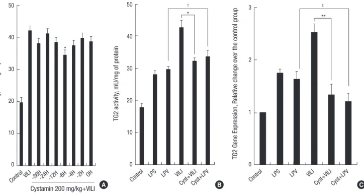
Mice in the Cyst+VILI and Cyst+LPV group received intraperi- toneal treatment with 200 mg/kg of cystamine dihydrochloride (Sigma, St. Louis, MO, USA) dissolved in PBS. This dose was found to be effective in LPS-induced acute lung inflammation (15). To determine the optimal pretreatment time, cystamine was administered intraperitoneally to six mice at 36, 24, 12, 6, 4, 2, and 0 hr before LPS instillation and 4 hr of MV. The lowest TG2 activity and TG2 RT-PCR were observed at the 6-hr cysta- mine pretreatment time (Fig. 1A). Therefore, the Cyst+VILI and Cyst+LPV group mice were pretreated with cystamine at 6 hr prior to LPS instillation and MV and the mice in other groups were pretreated with 200 μL of PBS at 6 hr prior to tracheosto- my, LPS instillation, or LPS instillation and MV.
Statistical analysis
All data are expressed as mean ± standard error of the mean (SEM). Statistical analysis was performed using IBM SPSS Sta- tistics for Windows® (Release 20.0; SPSS Inc., Chicago, IL, USA).
Inter-group differences were determined by non-parametric Mann-Whitney U and Kruskal-Wallis tests. Statistical signifi- cance was defined as P < 0.05.
RESULTS
TG2 activity and TG2 quantitative RT-PCR
TG2 activity (Fig. 1B) was significantly different between groups (P = 0.002 by Kruskal-Wallis test). The LPS (28 ± 1.13 mU/mg of protein) and LPV (29.7 ± 0.84 mU/mg of protein) groups showed higher TG2 activity than the control group (17.9 ± 1.14 mU/mg of protein) (P < 0.05). TG2 activity was significantly increased in the VILI group (41.6 ± 2.16 mU/mg of protein) compared to the LPS and LPV groups (P < 0.05). Cystamine pretreatment decreas- ed TG2 activity significantly in the Cyst+VILI group (32.2 ± 0.92
mU/mg of protein) compared with the VILI group (P = 0.029).
However, TG2 activity in the Cyst+VILI group was not signifi- cantly different from the LPS and LPV groups (P > 0.05). In the Cyst+LPV group (33.6 ± 1.85 mU/mg of protein), cystamine pre- treatment had no additional effect on TG2 activity when com- pared to the LPV group (P = 0.114).
In the analysis of quantitative RT-PCR of TG2 (Fig. 1C), TG2 gene expression in the VILI group (2.52 ± 0.16 relative change over the control group) was significantly higher than the LPS (1.75 ± 0.07) and LPV (1.63 ± 0.15) groups (P < 0.05). In com- paring the VILI and Cyst+VILI groups, TG2 gene expression in the Cyst+VILI group (1.34 ± 0.18) was significantly lower than in the VILI group (P = 0.016). There was no difference in TG2 gene expression in the Cyst+VILI group compared to the LPS and LPV groups (P > 0.05). TG2 gene expression in the Cyst+LPV group (1.21 ± 0.15) was not significantly lower than in the LPV group (P = 0.19).
Inflammatory cytokines and NF-κB activity
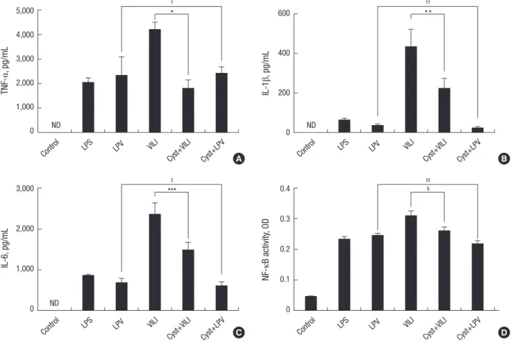
TNF-α, IL-1β, and IL-6 were not detected in the BALF of the con- trol group. The concentration of TNF-α was higher in the VILI group (4,207.17 ± 300.73 pg/mL) than in the LPS (2,053.12 ± 189.26 pg/mL) and LPV (2,348.79 ± 744.5 pg/mL) groups (P <
0.05) (Fig. 2A). The concentration of TNF-α was significantly lower in the Cyst+VILI group (1,814.43 ± 325.14 pg/mL) than in
the VILI group (P = 0.016), but was not different from the LPS, LPV, and Cyst+LPV (2,427.79 ± 244.42 pg/mL) groups (P > 0.05).
The concentration of IL-1β (Fig. 2B) was higher in the VILI group (432.29 ± 87.99 pg/mL) than in the other groups (P < 0.05). The concentration of IL-1β was significantly lower in the Cyst+VILI group (225.78 ± 47.02 pg/mL) than in the VILI group (P = 0.026), and higher in than the LPV (35.48 ± 7.28 pg/mL) and Cyst+LPV (22.95 ± 6.73 pg/mL) groups (P < 0.05). The Cyst+LPV group had a lower concentration of IL-1β than the LPV group, but this dif- ference was not significant (P = 0.19). The concentration of IL-6 (Fig. 2C) was also significantly higher in the VILI group (2,365.77
± 275.97 pg/mL) than in the other groups (P < 0.05). Cystamine pretreatment lowered the concentration of IL-6 in the Cyst+VILI group (1,489.11 ± 186.86 pg/mL) more than in the VILI group (P = 0.015). The Cyst+LPV group (611.03 ± 91.01 pg/mL) showed insignificant lower IL-6 concentration than the LPV group (677.71
± 105.56 pg/mL) (P = 0.429).
NF-κB activity was increased in the VILI group (0.3101 ± 0.0152 optical density, OD) compared with the LPS (0.2349 ± 0.0065 OD) and LPV (0.2465 ± 0.0055 OD) groups (P < 0.05) (Fig. 2D).
Cystamine pretreatment significantly decreased NF-κB activity in the Cyst+VILI group (0.2615 ± 0.0107 OD) compared to the VILI group (P = 0.029). NF-κB activity was lower in the Cyst+LPV group (0.2178 ± 0.011 OD) than the LPV group, but this differ- ence was not statistically significant (P = 0.114).
TG2 activity, mU/mg of protein
Control
Cystamin 200 mg/kg+VILI VILI -36H-24H-12H -6H -4H -2H OH 50
40
30
20
10
0
*
TG2 activity, mU/mg of protein
Control LPS LPV VILI
Cyst+VILICyst+LPV 50
40
30
20
10
0
*
†
TG2 Gene Expression, Relative change over the control group
Control LPS LPV VILI
Cyst+VILI Cyst+LPV 3
2
1
0
**
‡
A B C
Fig. 1. Determination pretreatment time, transglutaminase 2 (TG2) activity and gene expression. To determine the optimal pretreatment time, cystamine was administered in- traperitoneally to six mice at 36, 24, 12, 6, 4, 2, and 0 hr before LPS instillation and 4 hr of MV. The lowest TG2 activity and TG2 RT-PCR are observed at the 6-hr cystamine pretreatment time (P = 0.001 by Kruskal-Wallis test, *P < 0.05, compared with the VILI and other time points) (A). TG2 activity (B) and gene expression (C) are significantly higher in the VILI group than in other groups (P < 0.05). In the Cyst+VILI group, TG2 activity and gene expression are lower than in the VILI group (*P = 0.029 and **P = 0.016, respectively). Cystamine pretreatment has no additional effect on TG2 activity and gene expression in the Cyst+LPV group comparing with the LPV group (†P = 0.114 and ‡P = 0.19, respectively).
Histopathology and acute lung injury (ALI) score
Histopathologic examination revealed increases in the ALI pa- rameters in the LPS group (Fig. 3B), including increased alveo- lar capillary congestion, aggregation of inflammatory cells, and thickening of the alveolar wall compared to the control group (Fig. 3A). A higher level of ALI was seen in the VILI group (Fig.
3C) compared to the LPS and LPV groups (Fig. 3E). In the Cyst+
VILI group (Fig. 3D), the ALI parameters were attenuated with cystamine pretreatment. In a quantitative comparison of ALI scores (Fig. 3G), the VILI group (12.88 ± 0.29) showed signifi- cantly higher scores than the control (2.13 ± 0.29), LPS (8.88 ± 0.58), LPV (8.13 ± 1.29), or Cyst+LPV (8.5 ± 1.09) groups (P <
0.05). Although the ALI score of the Cyst+VILI group (10.88 ± 0.97) was lower than the VILI group, this was not statistically sig nificant (P = 0.105). In comparing the Cyst+VILI group to the LPS, LPV, and Cyst+LPV groups, the Cyst+VILI group had a high- er ALI score, but this difference was not statistically significant (P > 0.05).
DISCUSSION
In the present study, TG2 activity and gene expression were sig- nificantly increased in a VILI mouse model. The increase in TG2 activity and gene expression was accompanied by increases in the concentration of inflammatory cytokines, NF-κB activity, and ALI score. The pretreatment with cystamine significantly decreased TG2 activity and gene expression, the concentration of inflammatory cytokines, and NF-κB activity, but did not sig- nificantly decrease ALI scores.
In terms of managing MV in ALI and ARDS patients, LPV is not always possible due to the heterogeneity of lung injury in some patients (3). A more thorough understanding of the mech- anisms that mediate lung injury might permit the development of potential strategies for preventing VILI. TG2 is the most ubiq- uitously expressed and most studied member of the TG family.
This enzyme catalyzes thiol- and calcium-dependent transami- dation reactions. TG2 has many functions related to the protec- tion and prevention of injury, as well as tissue remodeling and repair (8). Aberrantly activated TG2 has been implicated in neu-
Control ND
LPS LPV VILI
Cyst+VILI Cyst+LPV 5,000
4,000 3,000 2,000 1,000 0
†
*
TNF-α, pg/mL
Control ND
LPS LPV VILI
Cyst+VILI Cyst+LPV 600
400
200
0
††
**
IL-1β, pg/mL
A B
Control ND
LPS LPV VILI
Cyst+VILI Cyst+LPV 3,000
2,000
1,000
0
‡
***
IL-6, pg/mL
Control LPS LPV VILI
Cyst+VILI Cyst+LPV 0.4
0.3 0.2 0.1 0
‡‡
§
NF-κB activity, OD
C D
Fig. 2. Inflammatory cytokines and nuclear factor-κB (NF-κB) activity. The concentration of tumor necrosis factor-α (TNF-α) (A), interleukin (IL)-1β (B), and IL-6 (C) and NF-κB activity (D) are significantly increased in the VILI group compared to other groups (P < 0.05). The Cyst+VILI group demonstrates significantly decreased inflammatory cytokine levels and NF-κB activity than the VILI group (*P = 0.016, **P = 0.026, ***P = 0.015, and §P = 0.029, respectively). In the comparisons between the LPV and Cyst+LPV groups, the inflammatory cytokine levels and NF-κB activity are not different (†P = 1.000, ††P = 0.19, ‡P = 0.429, and ‡‡P = 0.114, respectively). ND, not detectable; OD, optical density.
Fig. 3. Histopathologic findings and acute lung injury (ALI) scores. The LPS group (B) shows typical ALI findings, including intra-alveolar exudates, inflammatory cell infiltration, intra-alveolar hemorrhage, and interstitial edema that were not found in the control group (A). These findings are exacerbated in the VILI group (C). In the Cyst+VILI group (D), the degree of ALI is mildly decreased compared to the VILI group (C). The findings of LPV (E) and Cyst+LPV (F) are similar to the LPS group (B). The ALI score (G) of the VILI group is significantly higher than those of the control, LPS, LPV, and Cyst+LPV groups (*P < 0.05). The Cyst+VILI group has non-significantly lower ALI scores than the VILI group (†P = 0.105), and is not different compared to the LPS, LPV, and Cyst+LPV groups (‡P > 0.05).
Control LPS
VILI Cyst+VILI
LPV Cyst+LPV
×100 ×400
A B
C D
E F
ALI score
Control LPS LPV VILI
Cyst+VILI Cyst+LPV 14
12
10
8
6
4
2
0
*
†, ‡
G
rodegenerative diseases, atherosclerosis, inflammatory diseas- es, autoimmune diseases, and fibrosis (9). TG2 is induced by various stressors including LPS, reactive oxygen species, UV, calcium ionophores, retinoic acid, and viral infection (10). Sev- eral studies have found that TG2 is involved in the initial phase of inflammation (22, 23). Cytokines and growth factors secreted during the early phases of cell injury regulate TG2 expression.
The expression of TG2 is regulated by transforming growth fac- tor-β1 (TGF-β1), TNF-α, IL-1β, and IL-6 (24-27). Increased TG2 activity induces and exacerbates inflammatory processes via NF-κB activation (10, 11). In studies of ALI models in mice, TG activity was increased, along with TNF-α and IL-6, by the intra- peritoneal injection of LPS (28). Intratracheal instillation of LPS also increased TG2 activity along with TGF-β1, TNF-α, IL-1β, IL-6, myeloperoxidase activity, and NF-κB activity (15). Howev- er, to date, there have not been any studies examining the role of TG2 in the pathogenesis of VILI, in which injurious MV strat- egies with high tidal volumes and low PEEP aggravate preexist-
ing acute lung inflammation and induce additional lung injury.
Similar to previous studies, this study found that intratracheal LPS instillation resulted in increased TG2 activity and gene ex- pression and increased TNF-α, IL-1β, IL-6, and ALI scores when compared to the control group (15, 28). TG2 activity and gene expression, concentrations of inflammatory cytokines, and ALI scores were significantly elevated in the VILI group compared to the LPS group in response to MV with a high tidal volume and zero PEEP. These results suggest that injurious mechanical ven- tilation strategies induce aberrant TG2 activation and eventual- ly lead to acute lung inflammation.
TG2-mediated NF-κB activation has been reported to have an important role in inflammatory disease processes. Increases in TG2 activity induce or exacerbate inflammation via NF-κB activation without I-κBα kinase signaling (11). An increase in TG2 activity reduces free I-κBα in the cytosol via I-κBα polymer- ization, resulting in the translocation of free NF-κB into the nu- cleus. In turn, TG2 can be directly induced by NF-κB activation
as the TG2 promoter contains a NF-κB binding motif (29). In this study, the LPS-induced inflammatory reaction and the in- crease in TG2 activity and gene expression were also accompa- nied by an increase in NF-κB activity. Injurious MV strategies also exacerbated inflammation and increased TG2 activity and gene expression and concentrations of NF-κB. However, this study was limited by the fact that I-κBα polymerization was not examined.
The therapeutic possibilities of TG inhibitors have been eval- uated in various disease models. TG inhibitors have the poten- tial to reverse inflammatory damage in brain injury, conjuncti- vitis, and uveitis (10). In the present study, pretreatment with cystamine, which is a unique inhibitor of TG2 with multiple in- hibition mechanisms (30), significantly attenuated TG2 activity and gene expression in mice which had experienced injurious MV. Furthermore, cystamine decreased concentrations of in- flammatory cytokine and the activity of NF-κB. Cystamine pre- treatment resulted in a trend towards decreased ALI scores; how- ever, the changes were not statistically significant, suggesting that TG2 inhibition was not sufficient and has limitations in the histopathologic reduction of VILI. The LPV strategies showed a protective effect by decreasing TG2 activity and gene expres- sion, concentrations of inflammatory cytokines, NF-κB activity, and ALI score. The addition of cystamine pretreatment to LPV did not significantly change the parameters compared with the LPV group. TG2 gene expression, IL-1β and IL-6 concentrations, and NF-κB activity showed a downward trend in the Cyst+LPV group, more than in the LPV group. There may be limited bene- fits from the addition of TG2 inhibition to LPV. Cystamine pre- treatment only with LPS instillation (Cyst+LPS) group was not experimented in the present study. According to the study of the time response of TG2 activity after 0.5 mg/kg of LPS intra- tracheal instillation (15), TG2 activity was reported to be high- est 36 hr post LPS instillation. The increase of TG2 activity 3 and 6 hr post LPS instillation was modest compared to the control group. It was judged to be inappropriate to examine TG2 activi- ty and the effects of TG2 inhibition at the time of 4 hr after LPS instillation without additional VILI. In the preliminary study, the TG2 activity was experimented in the model using an inju- rious MV strategy alone without LPS instillation. However, in- crease in the TG2 activity was not significant in this model. This study was therefore performed in the model with both LPS in- stillation and injurious MV strategy, which might be more clini- cally relevant with ARDS.
There are several limitations in this study. The most impor- tant limitation is omission of data about pulmonary dynamics and physiologies. Under the mechanical ventilator setting with high tidal volume, intrinsic PEEP could develop and influence pulmonary dynamics of the animal. However, pulmonary dy- namics could not be measured in this experiment, and other data such as arterial partial pressure of oxygen and lung water
content which reflect the degree of lung injury were not mea- sured. In the inflammatory disease, the pro/anti-inflammatory balance is important. Therefore, measurement of anti-inflam- matory cytokines could be necessary in observing the balance.
However, none of anti-inflammatory cytokines was examined in this study.
The aberrant activation of TG2 plays an important role in the inflammatory processes that underlie the development of VILI.
The inhibition of TG2 had therapeutic effects on the inflamma- tory parameters of VILI in mice, suggesting potential clinical usefulness in the prevention and treatment of VILI in ARDS pa- tients.
DISCLOSURE
All authors have disclosed no financial or personal relationship with organizations that could potentially be perceived as influ- encing the described research. All authors have read the jour- nal’s policy on disclosure of potential conflicts of interest.
ORCID
In Bum Suh http://orcid.org/0000-0001-7012-0305 Dae Wui Yoon http://orcid.org/0000-0001-8875-2255 Won-Oak Oh http://orcid.org/0000-0003-0156-3422 Eun Joo Lee http://orcid.org/0000-0001-7884-9045 Kyung Hoon Min http://orcid.org/0000-0003-0610-2182 Gyu Young Hur http://orcid.org/0000-0001-5039-0199 Seung Heon Lee http://orcid.org/0000-0002-7180-3877 Sung Yong Lee http://orcid.org/0000-0002-8693-5792 Sang Yeub Lee http://orcid.org/0000-0001-7565-1076 Chol Shin http://orcid.org/0000-0002-2928-8576 Jae Jeong Shim http://orcid.org/0000-0002-3095-1021 Kwang Ho In http://orcid.org/0000-0003-3010-6423 Kyung Ho Kang http://orcid.org/0000-0002-9778-1725 Je Hyeong Kim http://orcid.org/0000-0002-8995-7460 REFERENCES
1. Matthay MA, Bhattacharya S, Gaver D, Ware LB, Lim LH, Syrkina O, Eyal F, Hubmayr R. Ventilator-induced lung injury: in vivo and in vitro mech- anisms. Am J Physiol Lung Cell Mol Physiol 2002; 283: L678-82.
2. Ventilation with lower tidal volumes as compared with traditional tidal volumes for acute lung injury and the acute respiratory distress syndro- me: the Acute Respiratory Distress Syndrome Network. N Engl J Med 2000; 342: 1301-8.
3. Gattinoni L, Caironi P, Pelosi P, Goodman LR. What has computed to- mography taught us about the acute respiratory distress syndrome? Am J Respir Crit Care Med 2001; 164: 1701-11.
4. Beninati S, Piacentini M. The transglutaminase family: an overview:
minireview article. Amino Acids 2004; 26: 367-72.
5. Candi E, Schmidt R, Melino G. The cornified envelope: a model of cell
death in the skin. Nat Rev Mol Cell Biol 2005; 6: 328-40.
6. Fésüs L, Szondy Z. Transglutaminase 2 in the balance of cell death and survival. FEBS Lett 2005; 579: 3297-302.
7. Hitomi K. Transglutaminases in skin epidermis. Eur J Dermatol 2005;
15: 313-9.
8. Folk JE, Finlayson JS. The epsilon-(gamma-glutamyl)lysine crosslink and the catalytic role of transglutaminases. Adv Protein Chem 1977; 31: 1-133.
9. Kim SY, Jeitner TM, Steinert PM. Transglutaminases in disease. Neuro- chem Int 2002; 40: 85-103.
10. Kim SY. Transglutaminase 2 in inflammation. Front Biosci 2006; 11: 3026- 35.
11. Lee J, Kim YS, Choi DH, Bang MS, Han TR, Joh TH, Kim SY. Transgluta- minase 2 induces nuclear factor-kappaB activation via a novel pathway in BV-2 microglia. J Biol Chem 2004; 279: 53725-35.
12. Choi CM, Jang SJ, Park SY, Choi YB, Jeong JH, Kim DS, Kim HK, Park KS, Nam BH, Kim HR, et al. Transglutaminase 2 as an independent prog- nostic marker for survival of patients with non-adenocarcinoma sub- type of non-small cell lung cancer. Mol Cancer 2011; 10: 119.
13. Oh K, Park HB, Byoun OJ, Shin DM, Jeong EM, Kim YW, Kim YS, Meli- no G, Kim IG, Lee DS. Epithelial transglutaminase 2 is needed for T cell interleukin-17 production and subsequent pulmonary inflammation and fibrosis in bleomycin-treated mice. J Exp Med 2011; 208: 1707-19.
14. Kim DY, Park BS, Hong GU, Lee BJ, Park JW, Kim SY, Ro JY. Anti-inflam- matory effects of the R2 peptide, an inhibitor of transglutaminase 2, in a mouse model of allergic asthma, induced by ovalbumin. Br J Pharmacol 2011; 162: 210-25.
15. Kim JH. The role of transglutaminase-2 in fibroproliferation after lipo- polysaccharide-induced acute lung injury. Tuberc Respir Dis 2010; 69:
337-47.
16. Francis RC, Vaporidi K, Bloch KD, Ichinose F, Zapol WM. Protective and detrimental effects of sodium sulfide and hydrogen sulfide in murine ven- tilator-induced lung injury. Anesthesiology 2011; 115: 1012-21.
17. Huang CS, Kawamura T, Lee S, Tochigi N, Shigemura N, Buchholz BM, Kloke JD, Billiar TR, Toyoda Y, Nakao A. Hydrogen inhalation amelio- rates ventilator-induced lung injury. Crit Care 2010; 14: R234.
18. Takahashi K, Saha D, Shattino I, Pavlov VI, Stahl GL, Finnegan P, Melo MF. Complement 3 is involved with ventilator-induced lung injury. Int Immunopharmacol 2011; 11: 2138-43.
19. Wilson MR, O’Dea KP, Zhang D, Shearman AD, van Rooijen N, Takata
M. Role of lung-marginated monocytes in an in vivo mouse model of ven- tilator-induced lung injury. Am J Respir Crit Care Med 2009; 179: 914-22.
20. Kim JH, Suk MH, Yoon DW, Kim HY, Jung KH, Kang EH, Lee SY, Lee SY, Suh IB, Shin C, et al. Inflammatory and transcriptional roles of poly (ADP- ribose) polymerase in ventilator-induced lung injury. Crit Care 2008; 12:
R108.
21. Kwon MH, Jung SH, Kim YM, Ha KS. Simultaneous activity assay of two transglutaminase isozymes, blood coagulation factor XIII and transglu- taminase 2, by use of fibrinogen arrays. Anal Chem 2011; 83: 8718-24.
22. Iismaa SE, Mearns BM, Lorand L, Graham RM. Transglutaminases and disease: lessons from genetically engineered mouse models and inherited disorders. Physiol Rev 2009; 89: 991-1023.
23. Verderio EA, Johnson T, Griffin M. Tissue transglutaminase in normal and abnormal wound healing: review article. Amino Acids 2004; 26: 387- 404.
24. George MD, Vollberg TM, Floyd EE, Stein JP, Jetten AM. Regulation of transglutaminase type II by transforming growth factor-beta 1 in normal and transformed human epidermal keratinocytes. J Biol Chem 1990; 265:
11098-104.
25. Ikura K, Shinagawa R, Suto N, Sasaki R. Increase caused by interleukin-6 in promoter activity of guinea pig liver transglutaminase gene. Biosci Bio- technol Biochem 1994; 58: 1540-1.
26. Kuncio GS, Tsyganskaya M, Zhu J, Liu SL, Nagy L, Thomazy V, Davies PJ, Zern MA. TNF-alpha modulates expression of the tissue transgluta- minase gene in liver cells. Am J Physiol 1998; 274: G240-5.
27. Suto N, Ikura K, Sasaki R. Expression induced by interleukin-6 of tissue- type transglutaminase in human hepatoblastoma HepG2 cells. J Biol Chem 1993; 268: 7469-73.
28. Suh GY, Ham HS, Lee SH, Choi JC, Koh WJ, Kim SY, Lee J, Han J, Kim HP, Choi AM, et al. A peptide with anti-transglutaminase activity de- creases lipopolysaccharide-induced lung inflammation in mice. Exp Lung Res 2006; 32: 43-53.
29. Mirza A, Liu SL, Frizell E, Zhu J, Maddukuri S, Martinez J, Davies P, Sch- warting R, Norton P, Zern MA. A role for tissue transglutaminase in he- patic injury and fibrogenesis, and its regulation by NF-kappaB. Am J Phy- siol 1997; 272: G281-8.
30. Siegel M, Khosla C. Transglutaminase 2 inhibitors and their therapeutic role in disease states. Pharmacol Ther 2007; 115: 232-45.