Original Article
Corresponding author:
Sun Goo Kim
Address: 7-206, Sinheung-dong, Jung-gu, Incheon, Korea [400-711]
Tel : +82-32-890-3514, Fax : +82-32-890-2918, E-mail : jokerhg@inha.
ac.kr
Th is is an Open Access article distributed under the terms of the Creative Commons Attribution Non-Commercial License (http://creativecommons.org/licenses/by-nc/3.0/) which permits unrestricted non-commercial use, distribution, and reproduction in any medium, provided the original work is properly cited.
Copyright © 2010. Anatomy and Cell Biology
doi: 10.5115/acb.2010.43.2.165 pISSN 2093-3665 eISSN 2093-3673
Shape and innervation of popliteus muscle
Kun Hwang
1, Kyung Moo Lee
2, Seung Ho Han
3, Sun Goo Kim
1,41Department of Plastic Surgery, and Center for Advanced Medical Education by BK21 Project, Inha University School of Medicine, Incheon, 2Department of Physical Medicine and Rehabilitation, Chungbuk University Medical School, Chungju, 3Department of Anatomy and Institute for Applied Anatomy, Th e Catholic Universty of Korea, Seoul, 4Department of Plastic Surgery, Gacheon Gil Hospital, Incheon, Korea
Abstract: Th e aim of this study was to delineate the shape of the popliteus muscle and determine the correct motor point site for treating spasticity. A total of 22 legs from 13 fresh Korean cadavers were evaluated. Th e x-axis was set as a transverse line across the lateral and medial epicondyle of the femur and the y-axis as a vertical line at the midpoint of the medial malleolus of the tibia and lateral malleolus of the fi bula. Th e popliteus muscle is an obtuse triangle in shape. Superior, medial, and inferior angles were 27.2±4.3o, 114.8±19.8o, and 38.0±18.8o respectively. The lengths of the superior, medial, and lateral sides of the triangle were 7.6±1.0 cm, 6.2±1.0 cm, and 11.9±1.5 cm respectively. Nerve branches ran superfi cially on the periosteum of the tibia and entered the popliteus on its superfi cial surface. Th e diverging point of the nerve branch entered the popliteus from the tibial nerve located at the midline of the popliteal fossa and 17% of the leg length above the intercondylar line. Most nerve entry points (83.3%) were within a 2.0×3.0 cm rectangle with the center located at -1.0 cm (-7%) on the x-axis and -3.3 cm (-9%) on the y-axis.
Key words: Popliteus muscle, muscle spasticity, denervation, anatomy
Received January 9, 2010; Revised February 11, 2010; Accepted February 12, 2010
A chemical agent, 5 to 12% phenol, is typically used for neurolysis. It is effective for several months following application (Glenn 1990). Another agent with neurolytic potency effective against spasticity and pain is 50% ethanol (Kong & Chua, 2002).
The aim of this study was to delineate the shape of the popliteus muscle and determine the correct motor point for chemodenervation or neurolysis application to treat spasticity and in-toeing deformity of the leg.
Materials and Methods
Materials
A total of 22 legs from 13 fresh Korean cadavers (5 males, 8 females; 50~80 years of age) were used for this study.
Dissection
Th rough a vertical midline incision on the popliteal fossa, the tibial nerve was exposed and traced. The triceps surae
Introduction
The popliteus rotates the tibia medially under the femur and the femur laterally on the tibia in repose (Standring 2005). Th e popliteus muscle is innervated by tibial nerves (L4, 5 and S1).
Muscular spasticity is common in post-stroke patients.
The spastic lower extremity manifests with in-toeing of the lower leg due to shortening of the popliteus muscle acting as the medial rotator of the tibia. A motor point block or intramuscular neurolysis is used to relieve focal spasticity by interrupting the stretch refl ex arc at its terminal motor output.
Knowledge of the precise locale of the muscle motor point is necessary to avoid blocking sensory or larger nerves.
Anat Cell Biol 43:165~168, 2010 Kun Hwang, et al
166
www.acbjournal.com doi: 10.5115/acb.2010.43.2.165
muscles were removed and the popliteus muscle exposed.
Measurement
Th e angles of the triangular popliteus and the motor points were mapped on rectangular coordinates. Th e x-axis was set as a transverse line across the lateral and medial epicondyle of the femur and the y-axis as a vertical line on the midpoint between the medial malleolus of the tibia and lateral malleolus
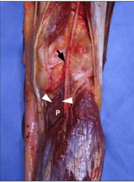
of the fi bula (Fig. 1). Each angle point of the popliteal triangle was marked and lengths of the three sides of the triangle measured. The diverging point of the nerve branch to the popliteus from the tibial nerve was observed along with the entering site of the nerve branch to the muscle (Fig. 2).
Each diverging and entering site of a nerve branch was marked on a rectangular coordinate and estimated. The location of the nerve branch entering the muscle was also identifi ed.
Results
Triangular popliteus
The popliteus muscle is an obtuse triangle in shape. The three angles were 114.8±19.8°, 27.2±4.3°, and 38.8±18.8° and the three sides of the triangle were 7.6±1.0 cm, 11.9±1.5 cm, and 6.2±1.0 cm (Fig. 3).
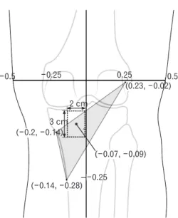
The proximal angle of the triangular popliteus was located laterally at 25% of the leg width on the intercondylar line: +0.23 on the x-axis and -0.02 on the y-axis. The intermediate angle was situated medially at 1/5 of the leg width and at the proximal 14% of the leg length below the intercondylar line: -0.2 on the x-axis and -0.14 on the y-axis. Th e distal angle was medially located at 15% of the leg width and approximately 30% of the leg length below the intercondylar line: -0.14 on the x-axis and -0.29 on the y-axis (Fig. 3).
Fig. 1. Rectangular coordinates: x-axis crosses over the lateral and medial epicondyle of the femur; y-axis accords with a vertical mid-line between the medial malleous of the tibia and lateral malleolus of the fi bula.
Fig. 2. Diverging point (arrow) of the nerve branch to the popliteus from the tibial nerve: nerve entry points (arrow heads) in the popliteus muscle (P).
Fig. 3. Triangular popliteus: superior, medial, and inferior angles were 27.2±4.3o, 114.8±19.8o, and 38.0±18.8o respectively; superior, medial, and lateral sides of the triangle were 7.6±1.0 cm, 6.2±1.0 cm, and 11.9
±1.5 cm respectively.
Popliteus muscle
www.acbjournal.com doi: 10.5115/acb.2010.43.2.165
Anat Cell Biol 43:165~168, 2010
167
Branching site from the tibial nerve
Th e diverging site of the nerve branch entered the popliteus from the tibial nerve located on the midline of the popliteal fossa at 17% of the leg length above the intercondylar line:
+6.1 cm on the y-axis and 0 on the x-axis.
Entering site of nerve branches into the popliteus The number of diverging nerve branches from the tibial
nerve was 2.2 on average (range 1~3).
Th e site (motor point) where the nerve branches entered the popliteus together was located at -0.7±0.9 cm (-5±6%) on the x-axis and -3.1±1.1 cm (-9±3%) on the y-axis. Most motor points (83.3%) were located within a 2.0×3.0 cm rectangle with the center located at -1.0 cm (-7%) on the x-axis and -3.3 cm (-9%) on the y-axis (Fig. 4).
Deep nerve branches to the popliteus
Th e nerve branches ran superfi cially on the periosteum of the tibia and entered the popliteus on its superficial surface about 1 cm distal to its superior border (Fig. 5).
Discussion
The Gray's anatomy textbook depicts the tibial nerve branch to the popliteus as descending obliquely across the popliteal vessel and curling around the distal border of the muscle on its anterior surface (Standring 2005).
From our observation, the nerves innervating the popliteus muscle were fine and torturous, running superficially on the periosteum of the posterior surface of the tibia, entering its superfi cial surface about 1 cm distal to its superior border (Fig. 5).
The popliteus is an internal rotator of the leg on the femur or an external rotator of the femur upon the leg and also a flexor of the leg at the knee. When standing with the knee partly flexed, it contracts to help prevent forward displacement of the femur on the tibia (Hollinshed 1986).
In the normal gait cycle, from the toe-off of the swing phase, the lower limb moves in the direction of internal rotation until a peak is reached at mid-stance at which point the rotation begins to reverse towards external rotation until push off, especially during the very last part of the stance (Lehmann & De Lateur, 1990). Popliteus activity begins shortly before the heel-strike and continues throughout three- quarters of the stance phase (Mann & Hagy, 1977). It appears that the popliteus acts to prevent the foot from rotating in the external direction in the terminal swing and three-quarter stance phases.
Common gait variations in children include in-toeing, out-toeing, and angular problems such as genu varum or valgum (Sass & Hassan, 2003). In the normal infant, the most common cause of in-toeing gait is metatarsus adductus. In this condition, the entire forefoot is adducted at the tarsometatarsal level but the hindfoot is in its normal Fig. 4. Most nerve entry points (83.3%) were within a 2.0 by 3.0 cm
rectangle with the center located at -1.0 cm (-7%) on the x-axis and -3.3 cm (-9%) on the y-axis.
Fig. 5. Nerve branches (arrow heads) running superficially on the periosteum of the tibia entering the popliteus on its superfi cial surface about 1 cm distal to its superior border. T: tibia, P: popliteus, TN:
tibial nerve.
Anat Cell Biol 43:165~168, 2010 Kun Hwang, et al
168
www.acbjournal.com doi: 10.5115/acb.2010.43.2.165
position. When present in the second year of life, in-toeing is commonly due to internal tibial torsion. Aft er three years of age, this problem is usually due to excessive femoral anteversion (Li & Leong, 1999). When a child toes-in severely, a combination of causes should be suspected. Sometimes a rotational problem is a manifestation of an underlying disorder such as cerebral palsy or hip dysplasia (Li & Leong, 1999).
In children with cerebral palsy, in-toeing is a frequent gait problem. The most common causes of in-toeing in subjects with bilateral involvement are internal hip rotation, internal tibial torsion, and internal pelvic rotation; while the most common causes of in-toeing in hemiplegic children are internal tibial torsion, pes varus, internal hip rotation, and metatarsus adductus.
Internal tibial torsion was assessed statically by measuring the internal thigh-foot angle over 0 degrees (Rethlefsen et al., 2006). Increased thigh-foot angle can be caused by various reasons. In patients with cerebral palsy, one cause of increased thigh-foot angle may be hypertonus of the internal rotator of the leg. Since the popliteus is an important internal rotator of the tibia, motor point block of the popliteus with alcohol or phenol can be used to relieve in-toeing gait in those patients.
To relieve spasticity, it is suggested to insert a needle deep against the tibial bone, retreat a few millimeters, and inject alcohol. Identification of the exact location of the motor point in the popliteus is very important in the nerve blocking process; hence the information obtained in this study is of value.
References
Glenn MB. (1990). Nerve blocks. In Glenn MB, Whyte J, eds.
The practical management of spasticity in children and adults. Philadelphia, Lea & Febiger, 227-258
Hollinshed WH. (1986). Functional anatomy of the limbs and back, 4th ed. Philadelphia, W.B. Saunders Co., 318-322 Kong KH, Chua KS. (2002). Intramuscular neurolysis with
alcohol to treat post-stroke finger flexor spasticity. Clin Rehabilit 16: 378-381
Lehmann JF, De Lateur BJ. (1990). Gait analysis: diagnosis and management. In Kottke FJ, Lehmann JF, eds. Krusen's handbook of physical medicine and rehabilitation, 4th ed.
Philadelphia, W.B. Saunders Co., 108-111
Li YH, Leong JC. (1999). Intoeing gait in children. Hong Kong Med J 5: 360-366
Mann RA, Hagy JL. (1977). The popliteus muscle. J Bone Joint Surg Am 59: 924-927
Rethlefsen SA, Healy BS, Wren TA, Skaggs DL, Kay RM.
(2006). Causes of intoeing gait in children with cerebral palsy. J Bone Joint Surg Am 88: 2175-2180
Sass P, Hassan G. (2003). Lower extremity abnormalities in children. Am Fam Physician 68: 461-468
Standring S. (2005). Gray's anatomy, 39th ed. Edinburgh, Elsevier, 1485