Original Article
골절 유발
Rat
에 대한血府逐瘀湯
의 전임상 연구허건⋅오민석
대전대학교 한의과대학 한방재활의학교실
The Preclinical Study of Hyeolbuchugeo-tang (Xuefuzhuyu-tang) on Bone Healing in Rats with Rib Fracture
Gun Huh, K.M.D., Min-Seok Oh, K.M.D.
Department of Korean Medicine Rehabilitation, College of Korean Medicine, Daejeon University
RECEIVED June 17, 2020 REVISED June 22, 2020 ACCEPTED June 23, 2020 CORRESPONDING TO Min-Seok Oh, Department of Korean Medicine Rehabilitation, College of Korean Medicine, Daejeon University, 75 Daedeok-daero, 176beon-gil, Seo-gu, Daejeon 35235, Korea TEL (042) 470-9424 FAX (042) 470-9005 E-mail [email protected] Copyright © 2020 The Society of Korean Medicine Rehabilitation
Objectives The purpose of this study is to evaluate the healing effect of Hyeolbuchugeo- tang (HC) in rats with rib fracture.
Methods Rats were randomly divided into 5 groups (naive, control, positive control, HC-L and HC-H). All groups except naive group were subjected to bone fracture of rib. Naive group received no treatment at all. Control group was fed with phosphate buffered saline. Positive control group was orally medicated with tramadol. Experimental group was orally medicated with HC extract (50 mg/kg for low concentration [HC-L], 100 mg/kg for high concentration [HC-H]). X-ray and micro-computed tomography (micro-CT) were conducted to assess the effect of HC. We analysed the level of 2) transforming growth factor-β1 (TGF-β1), Ki67, alkaline phosphatase (ALP), receptor activator of nuclear factor kappa-β, runt-related transcription factor 2 (Runx2) and tartrate resistant acid phosphatase (TRAP) on 7 and 14 days after fracture. ALP, ala- nine aminotransferase, aspartate aminotransferase, blood urea nitrogen, creatinine was measured for safety assessment.
Results X-ray and micro-CT, showed HC enhance bone repair process. Callus for- mation was increased in experimental group at 7 days after fracture, but decreased at 14 days after fracture. 7 days after fracture, the level of TGF-β1 in experimental group was decreased. The level of Ki67, Runx2 in HC-H, TRAP in HC-L was increased. 14 days after fracture, the level of Ki67 in HC-L and HC-H was decreased. The level of ALP, Runx2, BUN in HC-L, TRAP in HC-L and HC-H was increased.
Conclusions Taken together the results, HC promoted healing of bone fracture. In conclusion, HC has a potential to promote healing of bone fracture. (J Korean Med Rehabil 2020;30(3):23-44)
Key words Herbal medicine, Rib fractures, Bone fractures, Gene expression
서론»»»
골절이란 과도한 힘이 뼈나 연골에 가해져 그 연속성 이 일부 혹은 전체에서 상실된 상태를 말하며 피부, 피 하조직, 근막, 혈관 및 신경 등의 손상을 동반한다1).
골절의 치료는 수술적 치료와 비수술적 치료로 나눌 수 있으며 환자의 전신상태, 다른 손상의 동반 유무, 골 절의 폐쇄성 혹은 개방성 여부, 골절의 위치와 전위 정 도 등의 사항을 고려하여 치료의 방향을 결정한다2).
골절의 치유 과정은 골절 직후 1~5일 사이의 염증기,
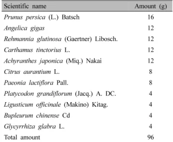
Scientific name Amount (g)
Prunus persica (L.) Batsch 16
Angelica gigas 12
Rehmannia glutinosa (Gaertner) Libosch. 12
Carthamus tinctorius L. 12
Achyranthes japonica (Miq.) Nakai 12
Citrus aurantium L. 8
Paeonia lactiflora Pall. 8
Platycodon grandiflorum (Jacq.) A. DC. 4 Ligusticum officinale (Makino) Kitag. 4
Bupleurum chinense Cd 4
Glycyrrhiza glabra L. 4
Total amount 96
Table Ⅰ. The Herbal Composition of Hyeolbuchugeo-tang 4~40일 사이의 복원기, 이후 수 주~수 개월간의 재형성
기 과정 등 크게 세 단계로 나눌 수 있는데3) 이 기간에 적절한 치료가 이루어지지 않을 경우 관절 변형, 외상 성 관절염, 불유합, 지연유합 등의 합병증이 유발될 수 있기 때문에 적절한 치료 계획을 세우는 것은 매우 중 요하다4).
한의학적 골절 치료는 골절을 초기, 중기, 후기로 나 누어 접근을 하는데 골절 초기에는 筋脈이 손상되어 瘀 血阻滯, 腫脹疼痛한 상태로 보고 活血化瘀, 消腫止痛하 는 약물을 사용하며, 중기에는 腫脹 및 瘀血이 줄어드는 단계로 接骨續斷하는 약물을, 후기에는 골이 유합되었 으나 氣血이 부족해 근육의 정상적인 기능이 회복되지 못한 상태로 補氣養血, 補益肝腎하는 약물을 사용한다5).
혈부축어탕은 淸代 王淸任이 저술한 ≪醫林改錯≫6) 에 처음 기록되었고 瘀血證을 치료하는 대표적인 처방 으로 瘀血과 관련된 여러 증상에 광범위하게 활용되고 있으며7) 자궁근종8), 골다공증9), 다낭성난소증후군10), 전뇌허혈11), 신경보호 및 재생효과12), 동맥경화13)에 효 과가 있음이 실험적으로 연구되었지만 아직 골절 유합 과 관련된 보고는 없었다.
이에 저자는 혈부축어탕이 瘀血과 관련된 골절에 미 치는 영향을 확인하고자 rat의 늑골에 골절을 유발시킨 후 골절 유합과 관련된 형태조직학적 검사 및 안전성 검사를 실시하여 유의한 결과를 얻었으므로 보고하는 바이다.
재료 및 방법»»»
1. 재료
1) 약재
실험에 사용한 혈부축어탕(Hyeolbuchugeo-tang, HC) 의 구성 약재는 桃仁, 當歸, 生地黃, 紅花, 牛膝, 枳殼, 赤 芍藥, 桔梗, 川芎, 柴胡, 甘草이며 약재별 용량은 ≪醫林 改錯≫6)을 기준으로 하였다. 약재는 옴니허브(Daegu, Korea)에서 구입하였으며 그 내용은 Table Ⅰ과 같다.
2) 실험동물
실험동물은 대한바이오링크(Eumseong, Korea)에서 7
주령 수컷 Sprague Dawley rat (300~350 g)를 분양받아 5주일간 사육한 뒤 실험에 사용하였다. 실험동물은 실 험 당일까지 고형사료와 물을 자유롭게 섭취할 수 있도 록 하였다. 사육환경은 자유식이하면서 온도는 23±1℃
로 맞추었고 12시간 light-dark cycle의 환경에 7일간 적 응시킨 후 실험에 사용하였다. 본 연구의 동물실험에 관 한 모든 사항은 대전대학교 동물실험윤리위원회의 승인 을 받은 후 진행하였다(승인번호: DJUARB2019-006).
2. 방법
1) 약재 추출
2첩 분량의 한약재 192 g에 1 L의 증류수를 넣고 약 탕기(Daewoong, Naju, Korea)를 이용하여 약 2시간 동 안 열수추출하였다. 끓인 약재는 Whatman No. 2 filter (Maidstone, UK)로 여과해 용액에 추출된 성분만을 분리 하였다. 여액은 rotary evaporator (Buchi, Switzerland)로 70℃에서 감압증발한 뒤 동결건조하여 38.8 g의 건조분 말을 얻어 추출효율은 20.2%였다. 분말은 -80℃에 분주 하여 보관하였으며 사용하기 전 phosphate buffered saline (PBS)로 희석하여 0.45 μm필터로 거른 후 사용하였다.
2) 골절 유발 방법
실험 동물에게 60 mg/kg의 ketamine (Yuhan Corporation, Seoul, Korea)과 500 μL/kg의 rompun (Bayer, Leverkusen, Germany)을 섞어 근육 주사하여 마취하였다. 골절 유발
을 위한 방법으로 가슴을 삭모한 다음 피부와 근육을 절개한 뒤 수술용 가위를 이용하여 왼쪽 9번 늑골을 절 단하여 골절을 유발하였다. 골절 유발 후 근육, 피부 순 서로 봉합사를 이용하여 5 mm 간격으로 단순 결찰 봉 합하였다.
3) 실험군 분류 및 약물 투여
실험동물은 무작위로 각각의 그룹으로 나누었다. 정 상군(Nor)과 대조군(Con), 양성대조군(Tra)은 6마리씩, 약물 투여군은 저농도와 고농도로 나누어 각각 10마리 씩 배속시키고 골절 후 1주와 2주에 치사하였다. 정상 군은 늑골 골절 손상을 주지 않았다. 그 외 모든 그룹의 실험동물은 늑골에 골절 손상을 주었고 실험 약물은 1 주 혹은 2주 동안 1일 1회 일정 시간에 경구 투여하였 다. 양성 대조군은 임상에서 골절 혹은 수술 후 진통제 로 사용하는 tramadol (20 mg/kg; S igma Aldrich Co., Ltd., St. Louis, MO, USA)을 사용하였다. 실험군(HC-L, HC-H)은 HC (50 mg/kg, 100 mg/kg)을 사용하였다.
4) Plasma 분리
마취된 실험동물의 심장에서 직접 채혈한 뒤 곧장 ethylenediaminetetraacetic acid가 들어 있는 BD vacutainer tube (BD, Franklin Lakes, NJ, USA)에 넣고 가볍게 흔들 어 혈액 응고를 억제하였다. 이후 3,000 rpm에서 10분간 원심분리한 뒤 상등액을 취하여 plasma를 확보하였다.
5) 골절 부위의 영상학적 관찰 (1) X-ray
골절된 뼈와 재생된 뼈를 확인하기 위하여 X-ray (ROTANODE-E7239X; Toshiba, Tokyo, Japan)를 사용 하여 늑골을 촬영하였다. X-ray 소스는 관전압 36 ㎸p, 관전류 5 mA로 촬영하였다. X-ray 촬영 후 이미지는 Fire-CR (3DIHC, 3DISC, Herndon, VA, USA)을 사용하 여 영상을 분석하였다.
(2) Micro-computed tomography (Micro-CT)
재생된 뼈를 확인하기 위하여 micro-CT (Quantum FX micro-CT; Perkin Elmer Inc., Waltham, MA, USA)를 사 용하여 늑골을 스캔하였다. X-ray 소스는 관전압 90 ㎸ p, 관전류 160 ㎂로 3분간 스캔하였다. 8.8 μm의 공간 분해능에서 0.7o의 회전 단계로 360o에 걸쳐 스캔을 수
행하였다. Micro-CT 스캔 후 이미지는 Dataview (Bruker, Ettlingen, Germany)를 사용하여 정리하였다.
6) 골절 조직 분석 (1) 조직슬라이드 제작
치사된 실험동물에서 골절을 유발한 늑골 부위를 적 출한 뒤 10% formalin에 1주일동안 조직을 고정하고 조 직처리 과정을 거쳐 파라핀에 포매한 후 파라핀 블록을 4 μm 두께로 박절하여 절편을 만들었다.
(2) Hematoxylin & Eosin (H&E) staining
탈 파라핀 후 함수, 수세 과정을 거쳐 Hematoxylin 과정을 10분간 처리하고, 수세 후 Eosin에 1분 40초간 처리하였다. 그 다음 함수, 청명과정을 거쳐 cover glass 를 덮고 봉입하였다. 조직슬라이드는 40배의 배율로 bright field microscope (Nikon, Tokyo, Japan)로 관찰하 였다.
(3) Safranin O staining
탈 파라핀 후 Weigert’s iron hematoxylin 과정을 5분 간 처리하였다. 수세 후 0.02% fast green 5분, 1% acetic aced 10초, 0.1% Safranin O 5분의 처리과정을 순서대로 거쳤다. 그 다음 함수, 청명과정을 거쳐 cover glass를 덮고 봉입하였다. 조직슬라이드는 40배의 배율로 bright field microscope (Nikon)로 관찰하였다.
7) Immunohistochemistry (IHC) 분석 (1) IHC 슬라이드 제작
치사된 실험동물에서 골절을 유발한 늑골 부위를 적 출한 뒤 10% formalin에 1주일동안 조직을 고정하였다.
고정된 조직을 조직처리 과정(processing)을 거쳐 파라핀 에 포매(embedding)한 후 파라핀 블록을 4 μm 두께로 박 절(cutting)하여 절편(section)을 만들고 탈 파라핀, 함수 과정을 거쳐 증류수로 세척하였다. 내인성 peroxidase의 활성을 없애기 위해 Peroxide Blocking (DAKO, Glostrup, Denmark)을 실온에서 10분간 처리하고 PBS로 2회 세 척하였다.
(2) Transforming growth factor-β1 (TGF-β1), Ki67 항체 TGF-β1 (Dilution 1:200; Abcam, Cambridge, UK), Ki67 (Dilution 1:100; Abcam)를 4℃에서 over night동안 반응시킨 후 Wash buffer (DAKO)로 세척하 고 Envision+ Rabbit (DAKO)으로 30분간 반응시켰다.
Score 0 1 2 3 4 5
Staining intensity No staining WeakMild weakModerate Mild strong Strong
Table Ⅱ. Grade of Immunohistochemistry Score
반응 후 반응용액은 Wash buffer (DAKO)로 세척하고, 3,3-diaminibenzidine tetrahydrochloride (DAB)으로 약 3 분간 발색하였다. 발색 후 증류수로 중화하고, Mayer hematoxylin으로 대조 염색 후 수돗물에 세척하여 남은 염색 시약을 제거한 뒤 함수, 청명과정을 거쳐 cover glass 를 덮고 봉입하였다. 조직슬라이드는 200배, 400배의 배율로 bright field microscope (Nikon)로 관찰하였다.
(3) Alkaline phosphatase (ALP), Runt-related tran- scription factor 2 (Runx2)
항체 ALP (1:200; MyBioSource, San Diego, CA, USA), Runx2 (Dilution 1:100; Abcam)를 4℃에서 over night동안 반응시킨 후, Wash buffer (Thermo Fisher Scientific, Rockford, IL, USA)로 세척하고 Biotinylated antibody-Rabbit (Vector Laboratories, Burlingame, CA, USA)으로 30분간 반응시켰다. 반응 후 반응용액은 Wash buffer (Thermo Fisher Scientific)로 세척하고, Avidin Biotin HRP complex (Vector Laboratories)로 반응 시켰다. Wash buffer로 세척 후 DAB로 약 3분간 발색하 였다. 발색 후 증류수로 중화하고, Harris hematoxylin으 로 대조 염색 후 수돗물에 세척하여 남은 염색 시약을 제거한 뒤 함수, 청명과정을 거쳐 cover glass를 덮고 봉 입하였다. 조직슬라이드는 200배의 배율로 bright field microscope (Nikon)로 관찰하였다.
(4) Receptor activator of nuclear factor kappa-β (RANK) 항체 RANK (Dilution 1:100; Abcam)를 4℃에서 over night동안 반응시킨 후 Wash buffer (Thermo Fisher Scientific) 로 세척하고 Biotinylated antibody-Mouse (Vector Laboratories) 로 30분간 반응시켰다. 반응 후 반응용액은 Wash buffer (Thermo Fisher Scientific)로 세척하고, Avidin Biotin HRP complex (Vector Laboratories)로 반응시켰다. Wash buf- fer로 세척 후 DAB로 약 3분간 발색하였다. 발색 후 증 류수로 중화하고, Harris hematoxylin으로 대조 염색 후 수돗물에 세척하여 남은 염색 시약을 제거한 뒤 함수, 청 명과정을 거쳐 cover glass를 덮고 봉입하였다. 조직슬라 이드는 200배의 배율로 bright field microscope (Nikon)
로 관찰하였다.
(5) Tartrate resistant acid phosphatase (TRAP) 항체 TRAP (Dilution 1:100; Abcam)를 4℃에서 over night동안 반응시킨 후 Wash buffer (Thermo Fisher Scientific) 로 세척하고 Biotinylated antibody-Rabbit (Vector Laboratories) 으로 30분간 반응시켰다. 반응 후 반응용액은 Wash buf- fer (Thermo Fisher Scientific)로 세척하고, Avidin Biotin HRP complex (Vector Laboratories)로 반응시켰다. Wash buffer로 세척 후 DAB로 약 3분간 발색하였다. 발색 후 증류수로 중화하고, Harris hematoxylin으로 대조 염색 후 수돗물에 세척하여 남은 염색 시약을 제거한 뒤 함수, 청 명과정을 거쳐 cover glass를 덮고 봉입하였다. 조직슬라 이드는 200배의 배율로 bright field microscope (Nikon)로 관찰하였다.
8) IHC staining score
조직슬라이드의 정보를 블라인드 처리 후 발현량 정 도를 0부터 5까지 Scoring하였다. 발현량 score 점수는 Table Ⅱ와 같다.
9) 혈액생화학적 분석
혈액생화학적 분석하기 위하여 COBAS 8000 C702 analyzer (Roche Diagnostic System, Basel, Switzerland)를 사용하였다. 시약은 Roche Diagnostics (Basel, Switzerland) 를 사용하였다. ALP, aminotransferase (AST), alanine aminotransferase (ALT)는 Colorimetry 검사법을 이용하 였고, blood urea nitrogen (BUN)은 Kinetic test, crea- tinine는 Enzyme법을 이용하였다. 실험동물에서 분리한 plasma를 사용하여 각각의 혈액생화학적 분석을 수행 하였다.
10) Osteocalcin 분석
Rat Gla-Osteocalcin ELISA kit (Takara Bio Inc., Kusatsu, Japan)를 이용하여 antibody coated microtiter plate에 plasma을 100 μL씩 분주하고 1시간 동안 반응시킨다.
Fig. 1. X-ray image of rib bone at 7 days after fracture. Sprague Dawley rats were subjected to rib fracture and orally administered with HC (50 mg/kg/day and 100 mg/kg/day) for 7 days. The fractured rib bone was isolated and analyzed by X-ray. HC:
Hyeolbuchugeo-tang, Nor: normal, no fracture, Con: control, rib fracture with vehicle, Tra: rib fracture with tramadol (20 mg/kg/day), HC-L: rib fracture with HC (50 mg/kg/day), HC-H: rib fracture with HC (100 mg/kg/day).
Fig. 2. X-ray image of rib bone at 14 days after fracture. Sprague Dawley rats were subjected to rib fracture and orally administered with HC (50 mg/kg/day and 100 mg/kg/day) for 14 days. The fractured rib bone was isolated and analyzed by X-ray. HC:
Hyeolbuchugeo-tang, Nor: normal, no fracture, Con: control, rib fracture with vehicle, Tra: rib fracture with tramadol (20 mg/kg/day), HC-L: rib fracture with HC (50 mg/kg/day), HC-H: rib fracture with HC (100 mg/kg/day).
Washing buffer를 이용하여 세척하고, antibody-POD conjugate를 각 well에 100 μL씩 넣어 1시간 동안 반응 시킨 뒤 다시 washing buffer를 이용하여 세척하고, tetra- methylbenzidine을 각 well에 100 μL씩 넣는다. 15분 뒤에 stop solution 100 μL를 넣은 후 흡광도 측정을 ELISA reader 450 nm에서 하였다.
3. 통계분석
모든 실험 결과는 mean±standard error of the mean으 로 기록하였으며, 통계처리는 GraphPad Prism 5 (version 5.01; GraphPad Software, Inc., San Diego, CA, USA) 프 로그램을 이용하였고, Mann-Whitney test로 유의성을 검증하였다. p값이 0.05 미만일 때 통계적으로 유의성 이 있다고 판정하였다.
결과»»»
1. 골절 부위의 영상학적 분석
1) X-ray
X-ray 관찰 결과 골절 유발 7일 후에는 대조군을 포 함한 모든 그룹에서 늑골의 골절 부위가 회복되지 않았 고, HC-L과 HC-H에서는 callus가 형성된 것을 확인할 수 있었다(Fig. 1).
골절 유발 후 14일이 지난 실험동물에서도 1주와 마 찬가지로 대조군을 포함한 모든 그룹에서 늑골의 골절 부위를 확인할 수 있었고, HC-H에서는 골 유합이 진행 되고 있는 것을 관찰할 수 있었다(Fig. 2).
Fig. 4. Micro-CT image of rib bone at 14 days after fracture. Sprague Dawley rats were subjected to rib fracture and orally administered with HC (50 mg/kg/day and 100 mg/kg/day) for 14 days. The fractured rib bone was isolated and analyzed by micro-CT. Micro-CT: micro-computed tomography, HC: Hyeolbuchugeo-tang, Nor: normal, no fracture, Con: control, rib fracture with vehicle, Tra: rib fracture with tramadol (20 mg/kg/day), HC-L: rib fracture with HC (50 mg/kg/day), HC-H: rib fracture with HC (100 mg/kg/day).
Fig. 3. Micro-CT image of rib bone at 7 days after fracture. Sprague Dawley rats were subjected to rib fracture and orally administered with HC (50 mg/kg/day and 100 mg/kg/day) for 7 days. The fractured rib bone was isolated and analyzed by micro-CT. Micro-CT: micro-computed tomography, HC: Hyeolbuchugeo-tang, Nor: normal, no fracture, Con: control, rib fracture with vehicle, Tra: rib fracture with tramadol (20 mg/kg/day), HC-L: rib fracture with HC (50 mg/kg/day), HC-H: rib fracture with HC (100 mg/kg/day).
2) Micro-CT
Micro-CT 관찰 결과 골절 유발 후 7일 후에는 대조군 을 포함한 모든 그룹에서 늑골의 골절 부위가 회복되지 않았고, HC-H에서는 골 유합이 진행되고 있었다(Fig. 3).
골절 유발 후 14일이 지난 실험동물에서도 1주와 마 찬가지로 대조군을 포함한 모든 그룹에서 늑골의 골절 부위를 확인할 수 있었고, HC-L에서 골 유합이 진행되 고 있는 것을 관찰할 수 있었다(Fig. 4).
Fig. 5. The histological analysis of rib fracture with hematoxylin and Eosin staining after 7 days. Sprague Dawley rats were subjected to rib fracture and orally administered with HC (50 mg/kg/day and 100 mg/kg/day) for 7 days. The damaged rats ribs were isolated and used for tissue slide specimen. The tissue was stained with H&E and visulized with bright field microscope.
Magnification, ×40. HC: Hyeolbuchugeo-tang, Nor: normal, no fracture, Con: control, rib fracture with vehicle, Tra: rib fracture with tramadol (20 mg/kg/day), HC-L: rib fracture with HC (50 mg/kg/day), HC-H: rib fracture with HC (100 mg/kg/day).
Fig. 6. The histological analysis of rib fracture with Hematoxylin and Eosin staining after 14 days. Sprague Dawley rats were subjected to rib fracture and orally administered with HC (50 mg/kg/day and 100 mg/kg/day) for 14 days. The damaged rats ribs were isolated and used for tissue slide specimen. The tissue was stained with H&E and visulized with bright field microscope.
Magnification, ×40. HC: Hyeolbuchugeo-tang, Nor: normal, no fracture, Con: control, rib fracture with vehicle, Tra: rib fracture with tramadol (20 mg/kg/day), HC-L: rib fracture with HC (50 mg/kg/day), HC-H: rib fracture with HC (100 mg/kg/day).
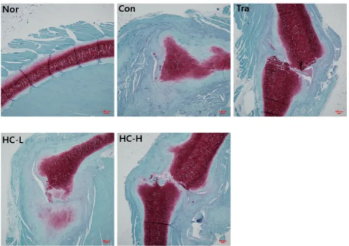
Fig. 7. The histological analysis of rib fracture with safranin O staining after 7 days. Sprague Dawley rats were subjected to rib fracture and orally administered with HC (50 mg/kg/day and 100 mg/kg/day) for 7 days. The damaged rats ribs were isolated and used for tissue slide specimen. The tissue was stained with safranin O and visulized with bright field microscope.
Magnification, ×40. HC: Hyeolbuchugeo-tang, Nor: normal, no fracture, Con: control, rib fracture with vehicle, Tra: rib fracture with tramadol (20 mg/kg/day), HC-L: rib fracture with HC (50 mg/kg/day), HC-H: rib fracture with HC (100 mg/kg/day).
2. 골절 조직 슬라이드 분석
1) H&E staining
H&E로 염색하고 현미경으로 관찰한 결과 골절 유발 후 7일 후에는 대조군 및 양성대조군 그리고 HC-L 및 HC-H에서 모두 골절이 관찰되었고, HC-L와 HC-H의 골절 부위에서의 callus 형성이 대조군과 양성대조군에 비해 증가하였다(Fig. 5).
골절 유발 후 14일 후에는 대조군 및 양성대조군 그 리고 HC-L 및 HC-H에서 모두 골절이 관찰되었고, 대 조군에 비해 양성대조군 및 HC-L과 HC-H의 골절 부위 에서의 callus 형성이 감소되는 것이 관찰되었다(Fig. 6).
2) Safranin O staining
Safranin O로 염색하고 현미경으로 관찰한 결과 골절 유발 후 7일 후에는 대조군 및 양성대조군 그리고 HC-L 및 HC-H에서 모두 골절이 있었고, HC-L의 골절 부위 에서의 callus 형성이 대조군에 비해 증가하였지만 양성 대조군과 HC-H의 골절 부위에서의 callus가 대조군과 비슷하게 형성되는 것이 관찰되었다(Fig. 7).
골절 유발 후 14일 후에는 대조군 및 양성대조군 그
리고 HC-L 및 HC-H에서 모두 골절이 관찰되었고, 대 조군에 비해 양성대조군 및 HC-L과 HC-H의 골절 부위
Fig. 8. The histological analysis of rib fracture with safranin O staining after 14 days. Sprague Dawley rats were subjected to rib fracture and orally administered with HC (50 mg/kg/day and 100 mg/kg/day) for 14 days. The damaged rats ribs were isolated and used for tissue slide specimen. The tissue was stained with safranin O and visulized with bright field microscope.
Magnification, ×40. HC: Hyeolbuchugeo-tang, Nor: normal, no fracture, Con: control, rib fracture with vehicle, Tra: rib fracture with tramadol (20 mg/kg/day), HC-L: rib fracture with HC (50 mg/kg/day), HC-H: rib fracture with HC (100 mg/kg/day).
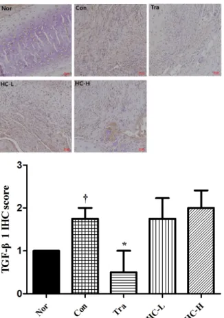
Fig. 9. IHC staining with TGF-β1 in rib fracture after 7 days.
Sprague Dawley rats were subjected to rib fracture and orally administered with HC (50 mg/kg/day and 100 mg/kg/day) for 7 days. The damaged rats ribs were isolated and used for tissue slide specimen. The sliced bone tissue were incubated with TGF-β1 antibody and subjected to IHC. Magnification, ×200.
IHC: immunohistochemistry, TGF-β1: transforming growth factor-β1, HC: Hyeolbuchugeo-tang, Nor: normal, no fracture, Con: control, rib fracture with vehicle, Tra: rib fracture with tramadol (20 mg/kg/day), HC-L: rib fracture with HC (50 mg/kg/day), HC-H: rib fracture with HC (100 mg/kg/day).
†††Significantly different from normal (p<0.001), *Significantly different from control (p<0.05), **Significantly different from control (p<0.01), ***Significantly different from control (p<0.001).
에서의 callus 형성이 감소되는 것이 관찰되었다(Fig. 8).
3. IHC 분석
1) TGF-β1
TGF-β1에 대한 항체를 결합한 뒤 면역조직염색을 실 시하고 현미경으로 관찰한 결과 골절 유발 후 7일 후에 는 대조군(4.3±0.50)의 병변 부위에서 확연하게 증가한 TGF-β1 발현세포들을 확인할 수 있었다. 그러나 HC-L (2.8±0.50)와 HC-H (3.5±0.58)에서 실험동물의 병변 부 위에서 대조군에 비해 TGF-β1 발현세포들이 감소하였 다(Fig. 9).
골절 유발 후 14일 후에는 양성대조군(0.5±1.00)을 제외한 모든 군에서 실험동물의 병변 부위에 확연하게 증가한 TGF-β1 발현세포들을 확인할 수 있었다. 그러 나 대조군(1.8±0.50)에 비해 HC-L (1.8±0.96)과 HC-H (2.0±0.82)의 골절 부위에서의 TGF-β1 발현세포들의 변 화는 거의 없는 것으로 관찰되었다(Fig. 10).
2) Ki67
Ki67에 대한 항체를 결합한 뒤 면역조직염색을 실시
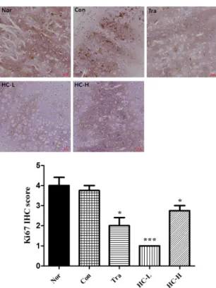
하고 현미경으로 관찰한 결과 골절 유발 후 7일 후에는 대조군(1.0±0.82)과 양성대조군(1.8±0.96)의 골절이 일어 난 병변 부위에서 Ki67의 발현이 감소하는 것을 확인할 수 있었다. 그러나 HC-L (1.8±0.50)과 HC-H (3.8±0.50) 에서 실험동물의 골절병변부위에서 대조군에 비해 Ki67 발현 세포들이 증가하는 것이 관찰되었고, 그 중 HC-H 에서 확연하게 증가하였다(Fig. 11).
골절 유발 14일 후에는 대조군(3.8±0.50)을 포함하여 골절이 일어난 실험동물의 병변 부위에서 확연하게 감소
Fig. 10. IHC staining with TGF-β1 in rib fracture after 14 days. Sprague Dawley rats were subjected to rib fracture and orally administered with HC (50 mg/kg/day and 100 mg/kg/day) for 14 days. The damaged rats ribs were isolated and used for tissue slide specimen. The sliced bone tissue were incubated with TGF-β1 antibody and subjected to IHC. Magnification, ×200.
IHC: immunohistochemistry, TGF-β1: transforming growth factor-β 1, HC: Hyeolbuchugeo-tang, Nor: normal, no fracture, Con:
control, rib fracture with vehicle, Tra: rib fracture with tramadol (20 mg/kg/day), HC-L: rib fracture with HC (50 mg/kg/day), HC-H: rib fracture with HC (100 mg/kg/day). †Significantly different from normal (p<0.05), *Significantly different from control (p<0.05).
Fig. 11. IHC staining with Ki67 in rib fracture after 7 days.
Sprague Dawley rats were subjected to rib fracture and orally administered with HC (50 mg/kg/day and 100 mg/kg/day) for 7 days. The damaged rats ribs were isolated and used for tissue slide specimen. The sliced bone tissue were incubated with Ki67 antibody and subjected to IHC. Magnification, ×200. IHC:
immunohistochemistry, HC: Hyeolbuchugeo-tang, Nor: normal, no fracture, Con: control, rib fracture with vehicle, Tra: rib fracture with tramadol (20 mg/kg/day), HC-L: rib fracture with HC (50 mg/kg/day), HC-H: rib fracture with HC (100 mg/kg/day).
†††Significantly different from normal (p<0.001), ***Significantly different from control (p<0.001).
한 Ki67 발현을 확인할 수 있었다. 또한 HC-L (1.0±0.00) 과 HC-H (2.8±0.50)의 실험동물 골절병변부위에서 대 조군에 비해 Ki67 발현세포들이 감소하였다(Fig. 12).
3) ALP
ALP에 대한 항체를 결합한 뒤 면역조직염색을 실시 하고 현미경으로 관찰한 결과 골절 유발 7일 후에는 대 조군(1.8±0.50), 양성대조군(1.8±0.50), HC-L (1.8±0.50), HC-H (2.8±0.50)에서 실험동물의 골절이 일어난 병변 부위에서 ALP의 발현이 증가하는 것을 확인할 수 있었
다. HC-H에서 실험동물의 골절병변부위에서 대조군에 비해 ALP 발현세포들이 증가되는 것이 관찰되었지만 HC-L에서는 변화가 거의 없었다(Fig. 13).
골절 유발 14일 후에는 대조군(0.8±0.50)과 양성대조 군(1.5±0.58)에서 골절이 일어난 실험동물의 병변 부위 에서 큰 변화가 거의 없었다. 그러나 HC-L (2.3±0.96)과 HC-H (2.0±0.82)에서 실험동물의 골절병변 부위에서 대조군에 비해 ALP 발현이 증가되는 것이 관찰되었다 (Fig. 14).
Fig. 12. IHC staining with Ki67 in rib fracture after 14 days.
Sprague Dawley rats were subjected to rib fracture and orally administered with HC (50 mg/kg/day and 100 mg/kg/day) for 14 days. The damaged rats ribs were isolated and used for tissue slide specimen. The sliced bone tissue were incubated with Ki67 antibody and subjected to IHC. Magnification, ×200.
IHC: immunohistochemistry, HC: Hyeolbuchugeo-tang, Nor:
normal, no fracture, Con: control, rib fracture with vehicle, Tra: rib fracture with tramadol (20 mg/kg/day), HC-L: rib fracture with HC (50 mg/kg/day), HC-H: rib fracture with HC (100 mg/kg/day). *Significantly different from control (p<0.05),
***Significantly different from control (p<0.001).
Fig. 13. IHC staining with ALP in rib fracture after 7 days.
Sprague Dawley rats were subjected to rib fracture and orally administered with HC (50 mg/kg/day and 100 mg/kg/day) for 7 days. The damaged rats ribs were isolated and used for tissue slide specimen. The sliced bone tissue were incubated with ALP antibody and subjected to IHC. Magnification, ×200.
IHC: immunohistochemistry, ALP: alkaline phosphatase, HC:
Hyeolbuchugeo-tang, Nor: normal, no fracture, Con: control, rib fracture with vehicle, Tra: rib fracture with tramadol (20 mg/kg/day), HC-L: rib fracture with HC (50 mg/kg/day), HC-H:
rib fracture with HC (100 mg/kg/day).
Fig. 14. IHC staining with ALP in rib fracture after 14 days. Sprague Dawley rats were subjected to rib fracture and orally administered with HC (50 mg/kg/day and 100 mg/kg/day) for 14 days. The damaged rats ribs were isolated and used for tissue slide specimen. The sliced bone tissue were incubated with ALP antibody and subjected to IHC. Magnification, ×200. IHC:
immunohistochemistry, ALP: alkaline phosphatase, HC: Hyeolbuchugeo-tang, Nor: normal, no fracture, Con: control, rib fracture with vehicle, Tra: rib fracture with tramadol (20 mg/kg/day), HC-L: rib fracture with HC (50 mg/kg/day), HC-H: rib fracture with HC (100 mg/kg/day). †Significantly different from normal (p<0.05), *Significantly different from control (p<0.05).
Fig. 15. IHC staining with RANK in rib fracture after 7 days.
Sprague Dawley rats were subjected to rib fracture and orally administered with HC (50 mg/kg/day and 100 mg/kg/day) for 7 days. The damaged rats ribs were isolated and used for tissue slide specimen. The sliced bone tissue were incubated with RANK antibody and subjected to IHC. Magnification, ×200.
IHC: immunohistochemistry, RANK: receptor activator of nuclear factor kappa-β, HC: Hyeolbuchugeo-tang, Nor: normal, no fracture, Con: control, rib fracture with vehicle, Tra: rib fracture with tramadol (20 mg/kg/day), HC-L: rib fracture with HC (50 mg/kg/day), HC-H: rib fracture with HC (100 mg/kg/day).
Fig. 16. IHC staining with RANK in rib fracture after 14 days.
Sprague Dawley rats were subjected to rib fracture and orally administered with HC (50 mg/kg/day and 100 mg/kg/day) for 14 days. The damaged rats ribs were isolated and used for tissue slide specimen. The sliced bone tissue were incubated with RANK antibody and subjected to IHC. Magnification, ×200.
IHC: immunohistochemistry, RANK: receptor activator of nuclear factor kappa-β, HC: Hyeolbuchugeo-tang, Nor: normal, no fracture, Con: control, rib fracture with vehicle, Tra: rib fracture with tramadol (20 mg/kg/day), HC-L: rib fracture with HC (50 mg/kg/day), HC-H: rib fracture with HC (100 mg/kg/day).
†Significantly different from normal (p<0.05).
4) RANK
RANK에 대한 항체를 결합한 뒤 면역조직염색을 실 시하고 현미경으로 관찰하였다. 그 결과 골절 유발 7일 후 에는 대조군(2.5±1.29)과 양성대조군(2.3±1.26) 및 HC-L (2.3±1.71), HC-H (3.3±0.96)을 포함하여 골절이 일어난 병변 부위에서 RANK의 발현이 증가하는 것을 확인할 수 있었다. HC-L에서 실험동물의 골절병변부위에서 대 조군에 비해 RANK의 발현이 큰 변화가 없는 것으로 관찰되었지만 HC-H에서는 RANK 발현이 증가하는 것 이 관찰되었다(Fig. 15).
골절 유발 14일 후에는 대조군(1.8±1.26)을 포함한 양 성대조군(1.3±0.96)과 HC-L (1.3±0.96), HC-H (2.0±1.41) 에서 골절이 일어난 실험동물의 병변 부위에 증가한 RANK 발현을 확인할 수 있었다. 또한 HC-L에서는 대 조군에 비해 RANK의 발현이 감소하였고 HC-H에서 실험동물의 골절병변부위에서 대조군에 비해 RANK의 발현이 증가되는 것이 관찰되었다(Fig. 16).
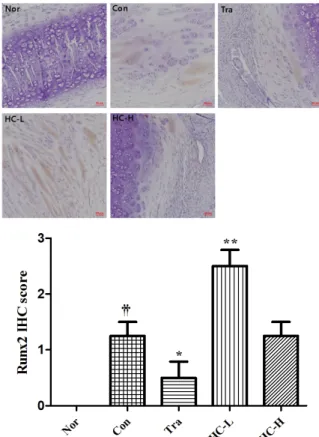
5) Runx2
Runx2에 대한 항체를 결합한 뒤 면역조직염색을 실 시하고 현미경으로 관찰한 결과 골절 유발 후 7일 후에
Fig. 17. IHC staining with Runx2 in rib fracture after 7 days.
Sprague Dawley rats were subjected to rib fracture and orally administered with HC (50 mg/kg/day and 100 mg/kg/day) for 7 days. The damaged rats ribs were isolated and used for tissue slide specimen. The sliced bone tissue were incubated with Runx2 antibody and subjected to IHC. Magnification, ×200.
IHC: immunohistochemistry, Runx2: runt-related transcription factor 2, HC: Hyeolbuchugeo-tang, Nor: normal, no fracture, Con: control, rib fracture with vehicle, Tra: rib fracture with tramadol (20 mg/kg/day), HC-L: rib fracture with HC (50 mg/kg/day), HC-H: rib fracture with HC (100 mg/kg/day).
†Significantly different from normal (p<0.05), **Significantly different from control (p<0.01).
Fig. 18. IHC staining with Runx2 in rib fracture after 14 days.
Sprague Dawley rats were subjected to rib fracture and orally administered with HC (50 mg/kg/day and 100 mg/kg/day) for 14 days. The damaged rats ribs were isolated and used for tissue slide specimen. The sliced bone tissue were incubated with Runx2 antibody and subjected to IHC. Magnification, ×200.
IHC: immunohistochemistry, Runx2: runt-related transcription factor 2, HC: Hyeolbuchugeo-tang, Nor: normal, no fracture, Con:
control, rib fracture with vehicle, Tra: rib fracture with tramadol (20 mg/kg/day), HC-L: rib fracture with HC (50 mg/kg/day), HC-H: rib fracture with HC (100 mg/kg/day). ††Significantly different from normal (p<0.01), *Significantly different from control (p<0.05), **Significantly different from control (p<0.01).
는 대조군(1.0±0.82)과 양성대조군(0.3±0.50)의 골절이 일어난 병변 부위에서 Runx2의 발현이 거의 변화가 없 었다. HC-L (1.0±1.15) 또한 대조군에 비해 발현 변화가 거의 없었으며, HC-H (2.3±0.96)에서는 실험동물의 골 절병변부위에서 대조군에 비해 Runx2 발현이 증가되는 것이 관찰되었다(Fig. 17).
골절 유발 후 14일 후에는 대조군(1.3±0.50)과 양성 대조군(0.5±0.58) 및 HC-L (2.5±0.58), HC-H (1.3±0.50) 을 포함하여 골절이 일어난 실험동물의 병변 부위에서 증가한 Runx2의 발현을 확인할 수 있었다. HC-L에서는
Runx2 발현이 증가되는 것이 관찰되었고, HC-H에서는 대조군에 비해 Runx2의 발현이 큰 변화는 없는 것으로 관찰되었다(Fig. 18).
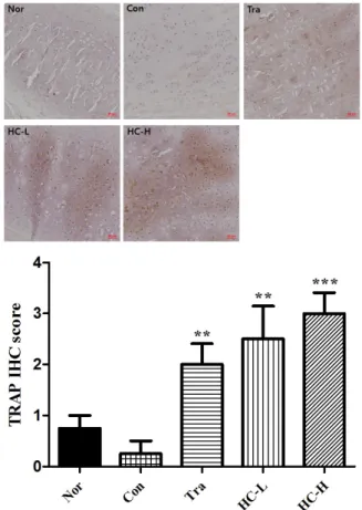
6) TRAP
TRAP에 대한 항체를 결합한 뒤 면역조직염색을 실시 하고 현미경으로 관찰한 결과 골절 유발 7일 후에는 대조 군(2.3±0.50)과 양성대조군(3.5±0.58) 및 HC-L (2.5±0.58), HC-H (1.3±0.50)을 포함한 실험동물의 골절이 일어난 병변 부위에서 TRAP의 발현이 증가하는 것을 확인할 수 있었다. 그러나 HC-H에서 실험동물의 골절병변부위
Fig. 19. IHC staining with TRAP in rib fracture after 7 days.
Sprague Dawley rats were subjected to rib fracture and orally administered with HC (50 mg/kg/day and 100 mg/kg/day) for 7 days. The damaged rats ribs were isolated and used for tissue slide specimen. The sliced bone tissue were incubated with TRAP antibody and subjected to IHC. Magnification, ×200. IHC:
immunohistochemistry, TRAP: tartrate resistant acid phosphatase, HC: Hyeolbuchugeo-tang, Nor: normal, no fracture, Con:
control, rib fracture with vehicle, Tra: rib fracture with tramadol (20 mg/kg/day), HC-L: rib fracture with HC (50 mg/kg/day), HC-H: rib fracture with HC (100 mg/kg/day). ††Significantly different from normal (p<0.01), *Significantly different from control (p<0.05).
Fig. 20. IHC staining with TRAP in rib fracture after 14 days.
Sprague Dawley rats were subjected to rib fracture and orally administered with HC (50 mg/kg/day and 100 mg/kg/day) for 14 days. The damaged rats ribs were isolated and used for tissue slide specimen. The sliced bone tissue were incubated with TRAP antibody and subjected to IHC. Magnification,
×200. IHC: immunohistochemistry, TRAP: tartrate resistant acid phosphatase, HC: Hyeolbuchugeo-tang, Nor: normal, no fracture, Con: control, rib fracture with vehicle, Tra: rib fracture with tramadol (20 mg/kg/day), HC-L: rib fracture with HC (50 mg/kg/day), HC-H: rib fracture with HC (100 mg/kg/day).
**Significantly different from control (p<0.01), ***Significantly different from control (p<0.001).
에서 대조군에 비해 TRAP 발현세포들이 감소되는 것 이 관찰되었다(Fig. 19).
골절 유발 후 14일 후에는 대조군(0.3±0.50)과 양성 대조군(2.0±0.82) 및 HC-L (2.5±1.29), HC-H (3.0±0.82) 을 포함하여 골절이 일어난 실험동물의 병변 부위에서 확연하게 감소한 TRAP 발현을 확인할 수 있었다. 또한, HC-L과 HC-H에서 실험동물의 골절병변부위에서 대조 군에 비해 TRAP 발현세포들이 증가되는 것이 관찰되 었다(Fig. 20).
4. 안전성 검사
1) 간독성 평가 (1) ALP
골절 유발 7일 후 ALP 수치는 정상군에서 320.3±31.73 이었을 때 대조군은 267.2±24.0으로 정상군에 비해 감 소하였다. 양성대조군은 229.5±20.66으로 정상군과 대 조군에 비해 감소하였으며, HC-L은 271.6±37.26으로 정상군에 비해서는 감소하였지만 대조군에 비해서는 변화가 거의 없었다. HC-H는 198.2±20.00으로 정상군
Fig. 21. Level of ALP in serum of rib fracture model of rats after 7 days. Sprague Dawley rats were subjected to rib fracture and orally administered with HC (50 mg/kg/day and 100 mg/kg/day) for 7 days. The level of ALP was measured by biochemical analyzer. The result were presented by the mean±standard error of the mean. ALP: alkaline phosphatase, HC: Hyeolbuchugeo-tang, Nor: normal, no fracture, Con: control, rib fracture with vehicle, Tra: rib fracture with tramadol (20 mg/kg/day), HC-L: rib fracture with HC (50 mg/kg/day), HC-H: rib fracture with HC (100 mg/kg/day). aSignificantly different from normal (p<0.05).
Fig. 22. Level of ALP in serum of rib fracture model of rats after 14 days. Sprague Dawley rats were subjected to rib fracture and orally administered with HC (50 mg/kg/day and 100 mg/kg/day) for 14 days. The level of ALP was measured by biochemical analyzer. The result were presented by the mean±standard error of the mean. ALP: alkaline phosphatase, HC: Hyeolbuchugeo-tang, Nor: normal, no fracture, Con:
control, rib fracture with vehicle, Tra: rib fracture with tramadol (20 mg/kg/day), HC-L: rib fracture with HC (50 mg/kg/day), HC-H: rib fracture with HC (100 mg/kg/day). aSignificantly different from normal (p<0.05), bSignificantly different from control (p<0.05).
Fig. 23. Level of ALT in serum of rib fracture model of rats after 7 days. Sprague Dawley rats were subjected to rib fracture and orally administered with HC (50 mg/kg/day and 100 mg/kg/day) for 7 days. The level of ALT was measured by biochemical analyzer. The result were presented by the mean±standard error of the mean. ALT: alanine aminotransferase, HC: Hyeolbuchugeo-tang, Nor: normal, no fracture, Con: control, rib fracture with vehicle, Tra: rib fracture with tramadol (20 mg/kg/day), HC-L: rib fracture with HC (50 mg/kg/day), HC-H: rib fracture with HC (100 mg/kg/day).
에 비해 유의적으로 감소하였고, 대조군에 비해 역시 감소하였다(Fig. 21).
골절 유발 14일 후 ALP 수치는 정상군에서 320.3±31.73 이었을 때 대조군은 281.2±13.33으로 정상군에 비해 감 소하였다. 양성대조군은 217.8±7.13으로 정상군과 대조 군에 비해 유의적으로 감소하였으며, HC-L과 HC-H는 각각 253.6±7.48, 263.0±31.69로 정상군과 대조군에 비 해 감소하였다(Fig. 22).
(2) ALT
골절 유발 7일 후 ALT 수치는 정상군에서 51.13±2.31 이었을 때 대조군은 48.30±3.39로 정상군에 비해 감소 하였다. 양성대조군은 56.47±2.0으로 정상군과 대조군 에 비해 증가하였지만 유의하지 않았다. HC-L과 HC-H 는 50.36±2.03, 52.42±4.11로 정상군과 대조군에 비해 거의 변화가 없었다(Fig. 23).
골절 유발 14일 후 ALT 수치는 정상군에서 51.13±2.31 이었을 때 대조군은 46.26±5.01로 정상군에 비해 감소 하였다. 양성대조군은 60.20±5.83으로 정상군과 대조군 에 비해 증가하였으며, HC-L은 43.24±5.58로 정상군과 대조군에 비해 감소하였고, HC-H는 50.86±5.82로 정상 군과 비슷하지만 대조군에 비해 증가하였다(Fig. 24).
(3) AST
골절 유발 7일 후 AST 수치는 정상군에서 73.83±7.21 이었을 때 대조군은 71.72±5.74로 정상군에 비해 감소
Fig. 24. Level of ALT in serum of rib fracture model of rats after 14 days. Sprague Dawley rats were subjected to rib fracture and orally administered with HC (50 mg/kg/day and 100 mg/kg/day) for 14 days. The level of ALT was measured by biochemical analyzer. The result were presented by the mean±standard error of the mean. ALT: alanine aminotransferase, HC: Hyeolbuchugeo-tang, Nor: normal, no fracture, Con:
control, rib fracture with vehicle, Tra: rib fracture with tramadol (20 mg/kg/day), HC-L: rib fracture with HC (50 mg/kg/day), HC-H: rib fracture with HC (100 mg/kg/day).
Fig. 25. Level of AST in serum of rib fracture model of rats after 7 days. Sprague Dawley rats were subjected to rib fracture and orally administered with HC (50 mg/kg/day and 100 mg/kg/day) for 7 days. The level of AST was measured by biochemical analyzer.
The result were presented by the mean±standard error of the mean. AST: aspartate aminotransferase, HC: Hyeolbuchugeo-tang, Nor: normal, no fracture, Con: control, rib fracture with vehicle, Tra: rib fracture with tramadol (20 mg/kg/day), HC-L: rib fracture with HC (50 mg/kg/day), HC-H: rib fracture with HC (100 mg/kg/day). aSignificantly different from normal (p<0.05),
bSignificantly different from control (p<0.05).
Fig. 26. Level of AST in serum of rib fracture model of rats after 14 days. Sprague Dawley rats were subjected to rib fracture and orally administered with HC (50 mg/kg/day and 100 mg/kg/day) for 14 days. The level of AST was measured by biochemical analyzer. The result were presented by the mean±standard error of the mean. AST: aspartate aminotransferase, HC: Hyeolbuchugeo-tang, Nor: normal, no fracture, Con:
control, rib fracture with vehicle, Tra: rib fracture with tramadol (20 mg/kg/day), HC-L: rib fracture with HC (50 mg/kg/day), HC-H: rib fracture with HC (100 mg/kg/day).
하였다. 양성대조군은 117.3±12.70으로 정상군과 대조 군에 비해 유의적으로 증가하였다. HC-L과 HC-H에서 는 각각 105.3±14.87과 89.08±5.82로 정상군과 대조군 에 비해 증가하였으나 유의하지는 않았다(Fig. 25).
골절 유발 14일 후 AST 수치는 정상군에서 73.83±7.21 이었을 때 대조군은 81.72±13.11로 정상군에 비해 증가 하였다. 양성대조군은 98.00±11.63으로 정상군과 대조군 에 비해 증가하였으나 유의하지 않았고, HC-L과 HC-H 또한 각각 102.2±11.72, 94.18±15.17로 정상군과 대조군 에 비해 증가하였으나 유의적이지는 않았다(Fig. 26).
2) 신독성 평가 (1) Creatinine
골절 유발 7일 후 creatinine 수치는 정상군에서 0.32±0.02 이었을 때 대조군은 0.26±0.01로 정상군에 비해 유의적으 로 감소하였다. 양성대조군은 0.20±0.02로 정상군과 대조 군에 비해 유의적으로 감소하였으며, HC-L은 0.28±0.03 으로 정상군에 비해 감소하였지만 대조군에 비해서는 증 가하였고, HC-H는 0.23±0.02로 정상군에 비해 유의적으 로 감소하였고, 대조군에 비해서도 감소하였다(Fig. 27).
골절 유발 14일 후 creatinine 수치는 정상군에서 0.32±0.02 이었을 때 대조군은 0.24±0.03으로 정상군에 비해 감소
하였으며, 양성대조군은 0.25±0.02로 정상군에 비해 유 의적으로 감소하였다. HC-L은 0.29±0.02으로 정상군에 비해서는 감소하였지만 대조군에 비해서는 증가하였고
Fig. 27. Level of creatinine in serum of rib fracture model of rats after 7 days. Sprague Dawley rats were subjected to rib fracture and orally administered with HC (50 mg/kg/day and 100 mg/kg/day) for 7 days. The level of creatinine was measured by biochemical analyzer. The result were presented by the mean±standard error of the mean. HC: Hyeolbuchugeo- tang, Nor: normal, no fracture, Con: control, rib fracture with vehicle, Tra: rib fracture with tramadol (20 mg/kg/day), HC-L:
rib fracture with HC (50 mg/kg/day), HC-H: rib fracture with HC (100 mg/kg/day). aSignificantly different from normal (p<0.05), bSignificantly different from control (p<0.05).
Fig. 28. Level of creatinine in serum of rib fracture model of rats after 14 days. Sprague Dawley rats were subjected to rib fracture and orally administered with HC (50 mg/kg/day and 100 mg/kg/day) for 14 days. The level of creatinine was measured by biochemical analyzer. The result were presented by the mean±standard error of the mean. HC: Hyeolbuchugeo- tang, Nor: normal, no fracture, Con: control, rib fracture with vehicle, Tra: rib fracture with tramadol (20 mg/kg/day), HC-L:
rib fracture with HC (50 mg/kg/day), HC-H: rib fracture with HC (100 mg/kg/day). aSignificantly different from normal (p<0.05).
Fig. 29. Level of BUN in serum of rib fracture model of rats after 7 days. Sprague Dawley rats were subjected to rib fracture and orally administered with HC (50 mg/kg/day and 100 mg/kg/day) for 7 days. The level of BUN was measured by biochemical analyzer. The result were presented by the mean±standard error of the mean. BUN: blood urea nitrogen, HC: Hyeolbuchugeo-tang, Nor: normal, no fracture, Con:
control, rib fracture with vehicle, Tra: rib fracture with tramadol (20 mg/kg/day), HC-L: rib fracture with HC (50 mg/kg/day), HC-H: rib fracture with HC (100 mg/kg/day). aSignificantly different from normal (p<0.05), bSignificantly different from control (p<0.05).
HC-H는 0.25±0.02로 대조군과 거의 비슷하였다(Fig. 28).
(2) BUN
골절 유발 7일 후 BUN 수치는 정상군에서 18.20±1.06 이었을 때 대조군은 16.68±0.76으로 정상군에 비해 감소 하였다. 양성대조군은 13.27±0.90으로 정상군과 대조군 에 비해 유의적으로 감소하였으며, HC-L은 17.64±0.42 로 대조군과 비슷하였고 HC-H는 14.80±1.27로 정상군 과 대조군에 비해 감소하였다(Fig. 29).
또한, 골절 유발 14일 후 BUN 수치는 정상군에서 18.20±1.06이었을 때 대조군은 17.98±0.26으로 정상군 에 비해 감소하였다. 양성대조군은 16.03±0.57로 정상군 과 대조군에 비해 감소하였으며, HC-L은 21.36±1.01로 정상군과 대조군에 비해 증가하였고, HC-H는 16.46±0.72 로 정상군과 대조군에 비해 매우 약하게 감소하였다 (Fig. 30).
5. Osteocalcin
Osteocalcin 수치는 정상군에서 5.71±0.74이었을 때 대 조군은 4.11±0.40으로 정상군에 비해 감소하였고, 양성 대조군 또한 4.146±0.51로 대조군과 비슷한 수치를 나
타내었다. HC-L은 3.74±0.29로 정상군과 대조군에 비해 감소하였다. HC-H는 4.59±0.32로 정상군에 비해서는 감소하였지만 대조군에 비해서는 증가하였다(Fig. 31).
Fig. 30. Level of BUN in serum of rib fracture model of rats after 14 days. Sprague Dawley rats were subjected to rib fracture and orally administered with HC (50 mg/kg/day and 100 mg/kg/day) for 14 days. The level of BUN was measured by biochemical analyzer. The result were presented by the mean±standard error of the mean. BUN: blood urea nitrogen, HC: Hyeolbuchugeo-tang, Nor: normal, no fracture, Con:
control, rib fracture with vehicle, Tra: rib fracture with tramadol (20 mg/kg/day), HC-L: rib fracture with HC (50 mg/kg/day), HC-H: rib fracture with HC (100 mg/kg/day). bSignificantly different from control (p<0.05).
Fig. 31. Level of osteocalcin in serum of rib fracture model of rats after 14 days. Sprague Dawley rats were subjected to rib fracture and orally administered with HC (50 mg/kg/day and 100 mg/kg/day) for 14 days. The level of osteocalcin was measured by commercially available ELISA kit. The result were presented by the mean±standard error of the mean. HC:
Hyeolbuchugeo-tang, Nor: normal, no fracture, Con: control, rib fracture with vehicle, Tra: rib fracture with tramadol (20 mg/kg/day), HC-L: rib fracture with HC (50 mg/kg/day), HC-H: rib fracture with HC (100 mg/kg/day). aSignificantly different from normal (p<0.05).
고찰»»»
골절이란 과부하 등 여러 원인으로 인해 뼈의 연속성 이 소실된 상태를 말한다. 뼈 주위의 연부조직 손상과 함께 통증을 유발하며 신경 및 혈관의 손상이 동반되기 도 하고 외관상 변형을 초래하기도 한다. 대부분은 방 사선 검사로 확진이 가능하며 정확한 진단을 내리기 어 려운 경우 전산화단층촬영(CT)를 사용하기도 한다1).
골절의 유합 과정은 염증기, 복원기, 재형성기 등 3단 계로 나눌 수 있으며 복원기는 연성 가골기와 경성 가 골기로 나눌 수 있다3). 염증기때 파열된 혈관에서 혈종 과 염증성 삼출물이 발생하여 골절 부위의 움직임을 급 격하게 감소시키고 혈소판이 모여 혈전 및 응고 작용이 진행된다. 이후 섬유모세포와 골모세포에 의해 육아조 직이 형성되며 이는 골절 부위의 혈종을 대체한다14). 연성가골기는 대체로 골절 후 3주 정도의 기간에 일어 나며 혈종으로 인한 통증과 부종이 감소하면서 골편이 움직이지 않는다15). 연골세포 발현유전자와 골모세포 발현유전자는 발현을 통해 각각 연골과 뼈로 분리되며 골모세포는 손상이 덜 심한 골절의 끝단에서 뼈의 표면
을 따라 직접 뼈를 형성하게 되고, 연골은 손상이 가장 심한 골절의 중심부인 저산소 영역에서 형성된다16). 경 성가골기에는 가골들이 연골 내 골화와 막내 골화를 통 해 견고한 석회성 조직으로 전환된다17).
쥐나 토끼는 동물모델로 다용되며 이들은 외상 후 7~9일에 연성가골의 형성이, 14일에 경성가골의 형성 이 최고치에 이른다18). 가골기가 지난 후에는 재형성기 가 진행된다. 경성가골이 층판골로 대치되고, 과잉 생 성된 골은 파골세포에 의해 흡수되며 파골세포와 조골 세포의 자가 교정을 통해 골이 재형성되고 본래의 형태 를 갖춘다2). 재형성기는 사람과 동물 모델 모두 3-4주 사이에 시작되나, 완전한 골격 구조로 돌아가기까지는 몇 년의 시간이 소요된다. 이 4가지 단계의 골 유합 과 정은 연속적으로 중첩되며 진행된다1,19).
한의학에서 골절은 ≪外臺秘要≫에서 처음으로 언급 되었으며 ≪諸病源候論≫과 ≪千金要方≫에서 정복과 고정의 방법을 제시했고, ≪聖濟總錄≫에서는 약물을 활 용하여 초기에는 活血祛瘀, 중기에는 接骨續筋, 후기에는 補氣養血, 健壯筋骨을 치료 방법으로 제시하였다17). 이