INTRODUCTION
Anteromedial surface of the leg does not have muscular bed and is covered with only skin and subcutaneous fat layer on the tibia. Furthermore, the skin and subcutaneous fat layer of anteromedial surface of the leg is thinner than other areas.
As a result, the soft tissue of this area is frequently damaged and once it is injured, it easily deteriorates to an intractable soft tissue defect with exposure of the cortex of the tibia. As a treatment method of reconstruction of the soft tissue defect on anteromedial surface of the leg, a local skin flap, a pedicled fasciocutaneous flap or a free flap could be considered. But,
it is difficult to obtain sufficient soft tissues with a local skin flap. To perform a free flap, microsurgical instruments and an operating microscope should be equipped and microvascular anastomosis should be performed. On the other hand, a local muscle flap provides soft tissues of enough size and good quality and can be performed easily without any special equipment. The gastrocnemius and soleus flap have been widely known. The gastrocnemius flap is usually used to reconstruct soft tissue defects of proximal 1/3 of the leg and soleus flap is used to reconstruct soft tissue defects of middle and distal 1/3. Recently, the medial hemisoleus flap has been reported to be superior to the classic proximally based entire
Reconstruction of the Soft Tissue Defect on Anteromedial Surface of the Leg Using Medial Hemisoleus Flap
Il-Jung Park1, Yoo-Joon Sur*, Sung-Lim You
Department of Orthopedic Surgery, Uijeongbu St. Mary’s Hospital, Uijeongbu,
1Bucheon St. Mary’s Hospital, Bucheon, The Catholic University of Korea College of Medicine, Korea
CC This is an open-access article distributed under the terms of the Creative Commons Attribution Non-Commercial License (http://creativecommons.org/licenses/by-nc/3.0) which permits unrestricted noncommercial use, distribution, and reproduction in any medium, provided the original work is properly cited.
Copyright © 2014 by the Korean Society for Microsurgery. All Rights Reserved.
Received September 20, 2014 Revised September 26, 2014 Accepted September 29, 2014
*Correspondence to: Yoo-Joon Sur Department of Orthopedic Surgery, Uijeongbu St. Mary’s Hospital, The Catholic University of Korea College of Medicine, 271 Cheonbo-ro, Uijeongbu 480-717, Korea
Tel: +82-31-820-3066 Fax: +82-31-847-3671 E-mail: [email protected]
Financial support: None.
Conflict of interest: None.
Purpose: Anteromedial surface of the leg is susceptible to trauma, which frequently induces soft tissue defect. When the size of a soft tissue defect is small to moderate, a local muscle flap is an easy and reliable alternative to a free flap. The authors performed medial hemisoleus flaps for reconstruction of soft tissue defects on the anteromedial surface of legs. The aim of this study was to evaluate clinical outcomes and effectiveness of the medial hemisoleus flap.
Materials and Methods: Twelve patients underwent the medial hemisoleus flap for reconstruction of a soft tissue defect on the anteromedial surface of the leg from February 2009 to December 2013. There were eight males and four females with a mean age of 47.8 years (15 to 69 years). The mean size of defects was 4.7×4.2 cm (2×2 to 9×6 cm). Flap survival and postoperative complications were evaluated.
Results: Mean follow-up period was 39.6 months (7 to 64 months) and all flaps survived.
There were two cases of negligible necrosis of distal margin of the flap, which were healed after debridement. All patients were capable of full weight bearing ambulation at the last follow-up.
Conclusion: The medial hemisoleus flap is a simple, reliable procedure for treatment of a small to moderate sized soft tissue defect on the anteromedial surface of the leg.
Key Words: Leg, Soft tissue injury, Medial hemisoleus, Flap
ARMS
Archives of Reconstructive Microsurgery http://dx.doi.org/10.15596/ARMS.2014.23.2.76soleus flap in English literature.1-10 However, there has been no domestic study about the medial hemisoleus flap. The authors performed medial hemisoleus flaps to reconstruct soft tissue defects on anteromedial surface of legs. The aim of this study was to evaluate clinical outcomes and effectiveness of the medial hemisoleus flap.
MATERIALS AND METHODS
Patients who underwent the medial hemisoleus flap for reconstruction of the soft tissue defect on anteromedial surface of the leg from February 2009 to December 2013 were included in this study. A total of 12 patients were available for this retrospective analysis. There were eight males and four females with a mean age of 47.8 years (15 to 69 years) (Table 1). The causes of the soft tissue defect were open fracture in 7 cases, chronic osteomyelitis in 4 cases, and soft tissue necrosis which occurred after open reduction and internal fixation of tibia fracture in 1 case. The mean size of the soft tissue defects was 4.7×4.2 cm (2×2 to 9×6 cm). The location of the soft tissue defect was middle 1/3 of the leg in 6 cases and distal 1/3 in 6 cases. The medial hemisoleus flap was harvested as a proximally or distally based depending on the size and location of the soft tissue defect. Six cases were proximally based and 6 cases were distally based. Patients were followed up at least 6 months postoperatively. As an assessment of the result of the procedure, flap survival and postoperative complications were analyzed.
Operation method
The patient was placed in a supine position and a pneumatic tourniquet was applied. A longitudinal skin incision was made about 2 cm medial to the medial edge of the tibia, but the location and length of the skin incision was modified to be connected to the soft tissue defect. The greater saphenous vein and saphenous nerve were preserved and subcutaneous fat layer and deep fascia were divided to expose the soleus. The soleus was separated from the gastrocnemius and the flexor digitorum longus. The locations of perforators of the posterior tibial artery to the soleus were identified. According to the locations of the soft tissue defect and perforators, proximally or distally based flap was designed and appropriate pivot point was determined.
The pivot point was the location of the most proximal or distal reliable perforator, which was chosen to be the pedicle of the flap. Then, the soleus was divided in half along with the midline raphe and cut at the planned proximal or distal end. The location of proximal or distal end of the flap was a few centimeters proximal or distal to the second perforator, which was the perforator immediate cephalad or caudad to the perforator chosen to be the pedicle of the flap. During the flap harvesting, perforators to the soleus were preserved as many as possible while allowing adequate rotation of flap to cover the soft tissue defect. The pneumatic tourniquet was deflated and circulation of flap was checked. Then, the flap was rotated and advanced to the soft tissue defect. If the length of flap was short, several shallow transverse incisions were made on the surface of flap to increase its length. Postoperatively, a long leg splint was applied
Table 1. Summary of patients
Case No. Age (yr)/sex Cause Site Size (cm) Type of flap Complication
1 15/male Tibia open fracture Middle 1/3 2×2 Proximally based -
2 60/female Chronic osteomyelitis Middle 1/3 5×2 Proximally based -
3 68/female Chronic osteomyelitis Middle 1/3 3×2 Proximally based -
4 31/male Chronic osteomyelitis Middle 1/3 3×4 Proximally based Necrosis of distal margin
5 46/male Tibia open fracture Distal 1/3 3×5 Distally based Necrosis of distal margin
6 67/male Tibia open fracture Middle 1/3 3×4 Proximally based -
7 69/female Wound necrosis Distal 1/3 7×4 Distally based -
8 33/male Tibia open fracture Distal 1/3 2×3 Distally based -
9 16/male Tibia open fracture Distal 1/3 7×4 Distally based -
10 62/male Chronic osteomyelitis Distal 1/3 4×4 Distally based -
11 44/male Tibia open fracture Distal 1/3 9×6 Distally based
12 62/female Tibia open fracture Middle 1/3 9×5 Proximally based -
and the leg was elevated to prevent edema. Two weeks after the operation, the flap was covered with a split-thickness skin graft.
RESULTS
The mean follow-up period was 39.6 months (7 to 64 months) and all flaps survived. Although negligible necrosis of distal margin occurred in 2 cases (1 proximally based, 1 distally based), they were healed after a debridement. All 12 patients were able to ambulate with full weight bearing and recovered their pre-injury physical activity levels at the last follow-up.
No patient complained of ambulation difficulty due to muscle weakness of the leg.
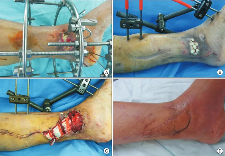
Case 1 (case No. 10)
A 62-year-old male patient sustained a severely comminuted
fracture of distal tibia by a falling accident. After initial treatment of external fixation, debridement, and primary wound closure, chronic osteomyelitis of the distal tibia and a 4×4 cm sized soft tissue defect developed at distal 1/3 of the leg (Fig. 1). As a next step, the distal tibia was excised and an antibiotics-impregnated bone cement spacer was inserted. After six week’s intravenous antibiotics therapy and wound care, distally based medial hemisoleus flap and split-thickness skin graft were performed.
Flap survived without complication. After the soft tissue reconstruction, distraction osteogenesis and ankle arthrodesis were performed. Thirty months after the initial trauma, ankle joint was fused and the soft tissue condition was favorable.
Case 2 (case No. 12)
A 62-year-old female patient underwent external fixation, debridement, and primary wound closure for the treatment of
Fig. 1. A 62-year-old male patient with open comminuted intraarticular fracture of the distal tibia. (A) A 4×4 cm open wound developed at the distal pin sites of the external fixator. The wound was connected to the ankle joint space. (B) Six weeks after insertion of the antibiotics impregnated polymethyl methacrylate cement beads. (C) Soft tissue defect was covered by the distally based medial hemisoleus flap. (D) Photograph taken 30 months after the surgery.
D
A B
C
an open comminuted fracture of the tibia shaft by a pedestrian traffic accident. Subsequently, a soft tissue defect developed on anteromedial surface of the middle 1/3 of the leg due to soft tissue necrosis (Fig. 2). For the treatment of the 9×5 cm sized soft tissue defect at the fracture site, proximally based medial hemisoleus flap and split-thickness skin graft were performed.
After the operations, flap survived without complications.
At postoperative 19 months, the soft tissue condition was favorable.
DISCUSSION
Reverse sural neurocutaneous flap or saphenous neurocu- taneous flap are frequently used to reconstruct soft tissue defects on anteromedial surface of legs, but these flaps must sacrifice sural or saphenous nerves and the width of pedicle should be kept to be wider than 3 cm in order to prevent venous congestion of flaps. Because the vascular pedicle is
relatively thick and the soft tissues of the leg is thin and tight without enough space underneath the subcutaneous tissue layer, when the vascular pedicle is passed through subcutaneous tunnel, venous congestion of the flap is likely to occur. If skin and subcutaneous tissues are incised to make pedicle flap pathway, primary closure is frequently impossible and the vascular pedicle need to be covered by skin graft. In this case, it’s not only inconvenient to manage wound postoperatively, but also unsatisfactory in aesthetic aspect. Furthermore, in high energy trauma such as an open facture, zone of injury is so broad that taking a reliable, healthy vascular pedicle is often impossible. Meanwhile, a free flap, one of another treatment options, has many limitations. It needs a microscope, microsurgery instruments and appropriate patient condition.
Not infrequently, soft tissue defects are too small to perform a free flap. Under the various aforementioned conditions, a local muscle flap could be a good alternative.
Among various muscle flaps, the gastrocnemius and soleus
Fig. 2. A 62-year-old female patient with open fracture of the tibial shaft and a soft tissue defect on middle 1/3 of the leg. (A) A 9×5 cm wound necrosis developed after external fixation and primary wound closure. (B) Flap dissection. Black arrows indicated perforating branches of the posterior tibial artery. (C) The soft tissue defect was covered by the proximally based medial hemisoleus flap. (D) Photograph taken 19 months after the surgery.
C D
A B
flap are frequently used for the reconstruction of soft tissue defects on anteromedial surface of legs. The gastrocnemius flap is usually used for proximal 1/3 of the leg and the soleus flap is used for the middle and distal 1/3 of the leg. The anatomical structure of soleus has been investigated in detail in many cadaveric studies11-14 and various soleus flaps have been developed based on its anatomical characteristics. The soleus is a bipennate muscle which receives blood supply from the popliteal, posterior tibial and peroneal artery. It is classified into type 2 of the classification of Mathes and Nahai,15 and the main source of blood supply is the posterior tibial artery.
The popliteal artery is in charge of blood supply in proximal area of the muscle, posterior tibial artery is in charge of medial 1/2, and peroneal artery is in charge of lateral 1/2. Three arteries give multiple perforators to the soleus. Raveendran and Kumaragama14 conducted a cadaveric study with 50 cadavers and reported that the mean numbers of perforators are 2.2 for the popliteal artery, 5.4 for the posterior tibial artery, and 4.1 for the peroneal artery. According to their study, perforators from the posterior tibial artery are evenly distributed in the entire soleus. The most proximal perforator of the posterior tibial artery is located at about 5.4 cm distal to the head of fibula and the most distal perforator is located at about 6.5 cm proximal to the ankle joint. However, the perforators of peroneal artery are mostly located at proximal area and they are not present in distal 1/2 of the leg. Because the soleus receives sufficient blood supply from many perforators of 3 arteries, it can be harvested as a proximally or distally based flap. And, because the soleus has independent medial and lateral neurovascular structures, it can also be harvested as a medial or lateral half flap.11 But, because lateral approach may damage the peroneal nerve and blood supply by the peroneal artery is unreliable, the lateral hemisoleus flap is hardly used.10,16
Because the proximal soleus is wide in width and distal port- ion of the soleus is merged into the Achilles tendon, proximally based soleus flap has relatively short length and narrow range of rotation. Therefore, the classic proximally based entire soleus flap has a clear limitation for the reconstruction of the soft tissue defect on anteromedial surface or distal 1/3 of the leg.17 The distally based soleus flap or hemisoleus flap can overcome these weaknesses. Because they are more slender and longer than proximally based entire soleus flap, they have a wide range of rotation and can reach more distant area. Additionally, the
hemisoleus flap has a functional advantage that it can preserve plantar flexion function of the soleus by remaining half.11 In 1978, Townsend17 reported the distally based soleus flap for the fist time, and in 1985, Tobin11 first reported the hemisoleus flap. However, they had not been popularized because of the concern for possibilities of the necrosis of distal margin of the flap.16 Recently, with the improvement of operation technique and introduction of the angiosome concept to the flap harvest technique, the usefulness of the medial hemisoleus flap has been reappraised.1-10 Pu1-7 reported good clinical results of proximally and distally based medial hemisoleus flaps for the middle and distal 1/3 of legs, stating that the necrosis of distal margin of the flap could be prevented if perforators which branched from the posterior tibial artery to the soleus were preserved as many as possible. Pu5 suggested that if size of a soft tissue defect is less than 50 cm2, a medial hemisoleus flap could be applied. Although insignificant necrosis of distal margin of the flap may occur, mostly it can be healed after debridement and flap advancement. Pu5,7 also suggested smoking, peripheral vascular diseases, injury of perforators or soleus itself as contraindications of the medial hemisoleus flap. Schierle et al.8 introduced the angiosome concept to the distally based medial hemisoleus flap. Once the distal perforator is selected to the main pedicle of the flap, the next cephalad perforator is identified. The distance between these two perforators and a few centimeters proximal to the cephalad perforator is the safe length of the flap.
In our study, although all 12 flaps survived, necrosis of distal margin of the flap occurred in 1 proximally based and 1 distally based flap. Because both were severely traumatized cases, we presumed that the cause of necrosis was injury of perforator and soleus by the initial trauma. It seems to be better to consider conversion to a free flap when conditions of the soleus or perforators are poor. As Pu5 pointed out, a medial hemisoleus flap is applicable to a relatively small to moderate sized soft tissue defect. There have been several reports of concomitant medial hemisoleus and gastrocnemius flap for a large soft tissue defect those could not be reconstructed by a medial hemisoleus alone.10-12,18 However, we worry about functional deterioration after such a combined local muscle flap and would rather perform free flap.
In conclusion, the medial hemisoleus flap is a useful operative procedure that can be used for a small to moderate soft tissue
defect on the anteromedial surface of middle or distal 1/3 of the leg. When a free flap or pedicled fasciocutaneous flap are infeasible or the size of the defect is too small to perform a free flap, the medial hemisoleus flap can be a good alternative.
When performing the medial hemisoleus flap, it should be kept in mind that perforators should be preserved as many as possible, and when soleus or perforators are damaged, necrosis of distal margin of the flap could be developed.
REFERENCES
1. Pu LL. Successful soft-tissue coverage of a tibial wound in the distal third of the leg with a medial hemisoleus muscle flap. Plast Reconstr Surg 2005;115:245-51.
2. Pu LL. The reversed medial hemisoleus muscle flap and its role in reconstruction of an open tibial wound in the lower third of the leg. Ann Plast Surg 2006;56:59-63.
3. Pu LL. Soft-tissue coverage of an open tibial wound in the junction of the middle and distal thirds of the leg with the medial hemisoleus muscle flap. Ann Plast Surg 2006;56:639-43.
4. Pu LL. Medial hemisoleus muscle flap: a reliable flap for soft tissue reconstruction of the middle-third tibial wound. Int Surg 2006;91:194-200.
5. Pu LL. Soft-tissue reconstruction of an open tibial wound in the distal third of the leg: a new treatment algorithm. Ann Plast Surg 2007;58:78-83.
6. Pu LL. The laterally extended medial hemisoleus flap for reconstruction of a tibial wound in the distal third of the leg. Eur J Plast Surg 2007;30:19-24.
7. Pu LL. Further experience with the medial hemisoleus muscle flap for soft-tissue coverage of a tibial wound in the distal third
of the leg. Plast Reconstr Surg 2008;121:2024-8.
8. Schierle CF, Rawlani V, Galiano RD, Kim JY, Dumanian GA.
Improving outcomes of the distally based hemisoleus flap:
principles of angiosomes in flap design. Plast Reconstr Surg 2009;123:1748-54.
9. Ata-ul-Haq, Tarar MN, Malik FS, Khalid K, Riaz A, Mehrose MY, et al. Hemisoleus muscle flap, a better option for coverage of open fractures involving middle third of tibia. J Ayub Med Coll Abbottabad 2009;21:154-8.
10. Ahmad I, Akhtar S, Rashidi E, Khurram MF. Hemisoleus muscle flap in the reconstruction of exposed bones in the lower limb. J Wound Care 2013;22:635, 638-40, 642.
11. Tobin GR. Hemisoleus and reversed hemisoleus flaps. Plast Reconstr Surg 1985;76:87-96.
12. Fayman MS, Orak F, Hugo B, Berson SD. The distally based split soleus muscle flap. Br J Plast Surg 1987;40:20-6.
13. Sadasivan KK, Ogden JT, Albright JA. Anatomic variations of the blood supply of the soleus muscle. Orthopedics 1991;14:679- 83.
14. Raveendran SS, Kumaragama KG. Arterial supply of the soleus muscle: anatomical study of fifty lower limbs. Clin Anat 2003;16:
248-52.
15. Mathes SJ, Nahai F. Classification of the vascular anatomy of muscles: experimental and clinical correlation. Plast Reconstr Surg 1981;67:177-87.
16. Hallock GG. Getting the most from the soleus muscle. Ann Plast Surg 1996;36:139-46.
17. Townsend PL. An inferiorly based soleus muscle flap. Br J Plast Surg 1978;31:210-3.
18. Pu LL. Soft-tissue coverage of an extensive mid-tibial wound with the combined medial gastrocnemius and medial hemisoleus muscle flaps: the role of local muscle flaps revisited.
J Plast Reconstr Aesthet Surg 2010;63:e605-10.