A Case of Xanthoma Disseminatum with Laryngeal Involvement
5
0
0
전체 글
(2) 박진수·이용우·이상혁·진성민. 결과가 보고되었다.6). 료는 받고 있지 않았다. 입원 기간 도중 본원 피부과에서 피부. 저자들은 이비인후과영역에서 흔치 않은 후두의 파종성 황 색종을 경험하였기에 기존 문헌 고찰과 함께 증례보고를 하고 자 한다.. 병변에 대한 조직검사를 다시 시행하였고 파종성 황색종으로 재차 진단 받았다. 후두내시경검사에서 양측 피열연골, 피열후두개주름, 후두 개 및 하인두 점막의 다발성의 황색 결절성 병변이 관찰되었. 증. 례. 다. 특히 후두개 상부에서는 2 cm 크기의 비교적 큰 병변이 관 찰되었으며 환자의 증상을 일으키는 것으로 생각되었다(Fig. 2).. 59세 남자 환자가 2달 전부터 나타난 호흡 및 삼킴 시 불편 감을 주소로 내원하였다. 내원 당시 환자는 혈액 검사 상 산. 황색 결절성 병변은 가성대 및 진성대의 점막에서는 관찰되지 않았으며 성대의 움직임에는 이상이 없었다.. 소 포화도 저하 및 저산소혈증 소견은 없었으나 호흡 시 불편. 혈액검사 상 Triglyceride 156 mg/dL, total cholesterol 158. 감, 삼킴 시 이물감 및 경미한 음성 변화를 호소하고 있었다.. mg/dL, LDL cholesterol 82 mg/dL로 정상 지질혈증의 소견이. 신체 진찰 상 양측 눈, 코, 입술 주위와 목, 양쪽 겨드랑이, 사. 관찰되었다. AST 83 IU/L, ALT 71 IU/L, Serum creatinine. 타구니 및 구강 내 점막에 황갈색의 다발성 구진성 피부 병변. 2.1 mg/dL, BUN 21.0 mg/dL으로 간, 신기능 저하가 관찰되었. 이 관찰되었다(Fig. 1).. 고 후두내시경검사에서 병변의 깊이가 점막하층에 국한되어. 이에 앞서 2년 전부터 나타난 얼굴과 목, 양측 겨드랑이, 사 타구니의 다발성 구진으로 타병원 피부과 진료를 보았으며 피 부조직검사 상 파종성 황색종으로 진단 받았으나 별다른 치. 있는 것으로 판단되었기 때문에 경부컴퓨터단층촬영(Computed tomography)는 시행하지 않았다. 후두내시경검사에서 관찰 된 후두개 상부의 2 cm 크기의 결. A. Fig. 1. Physical examination, Xanthomatous papules. Mucocutaneous involvement of oral mucosal (A, B). Periocular and perioral (C) lesions.. Fig. 2. Flexible laryngoscopic findings, multiple yellowish nodular lesion on arytenoid, aryepiglottic fold and, hypopharynx (A). 2 cm sized nodular mass on upper portion of epiglottis (B).. B. C. A. B - 59 -.
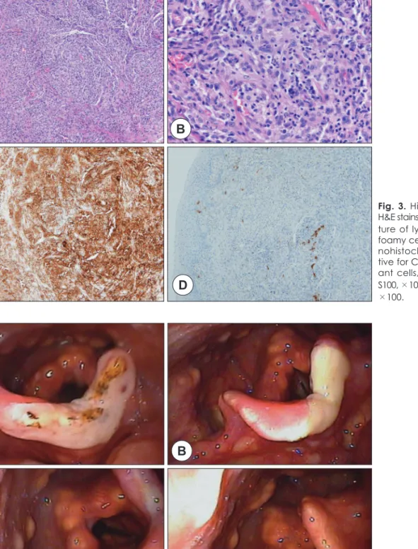
(3) 후두에 발생한 파종성 황색종. 절성 병변이 환자의 증상과 관련이 있을 것으로 생각되어 수. 하였다. 수술 후 시행한 조직검사에서 H&E 염색에서 림프구,. 술적 절제를 계획하였다. 현수후두경을 이용한 경구강적 접근. 포말 조직구 등의 침윤이 관찰 되었으며 CD68에 대하여 양. 법을 통해 후두개 종괴의 수술적 제거를 시행하였다. CO2 레. 성 소견, S-100에 대해서는 음성소견을 보여 파종성 황색종. 이저를 이용하여 종괴의 경계를 확인하며 조심스럽게 박리를. 으로 진단할 수 있었다(Fig. 3). 수술적 치료 시행 후 2주, 1달. 하였다. 종괴는 주변 조직에 유착 없이 비교적 쉽게 절제되었. 째 외래 내원하였으며 환자는 호흡곤란, 삼킴곤란 등의 증상. 으며 출혈이나 심한 후두개 부종은 발생하지 않았다.. 수술 후. 을 호소하지 않았고 후두내시경검사 상 합병증 및 재발 소견. 3일째 별다른 합병증 없이 퇴원하였고 외래 추적관찰 하였다.. 은 관찰되지 않았다. 수술 6개월 후, 후두내시경검사에서 결절. 내과적으로는 지질강하제(statin)를 복용하며 경과관찰 시행. 성 병변의 진행은 더이상 관찰되지 않았으며 호흡곤란 등의. A. B. C. D. A. B. C. Fig. 3. Histopathologic findings. H&E stains (A) ×100, (B) ×400, a mixture of lymphocytes, histiocytes, foamy cells and giant cells. Immunohistochemical stains, (C) positive for CD68 in histiocytes and giant cells, ×100, (D) negative for S100, ×100 C : CD68, ×100, D : S100, ×100.. Fig. 4. Post-operative findings, (A) 1day, mild swelling and crust on epiglottis, (B) 2weeks, mucosal healing state, (C) 1month, and (D) 6months, complete healing state, without complications.. D - 60 -.
(4) 박진수·이용우·이상혁·진성민. 혹은 기도 폐색이 예상되는 경우 크기 변화 및 증상에 대한 주. 증상 또한 호소하지 않았다(Fig. 4).. 의 깊은 경과 관찰이 필요하며 재발 여부와 상관 없이 수술적. 고. 찰. 절제가 필요할 것으로 생각된다. 본 증례에서는 병변에 대한 수술적 절제를 시행하였고 이. 파종성 황색종은 정상지방혈증의 비 유전성 점막 피부 질. 와 함께 항고지혈증제인 statin 제제를 복용하도록 하였으며. 환이다. 문헌 상 100예가 보고될 정도로 매우 드문 질환으로. 외래 경과 관찰을 하였다. 증상 및 폐쇄성 병변의 재발 소견. 60%에서 25세 이전에, 주로 남성에서 호발하는 것으로 알려. 은 관찰되지 않았으며, 결절성 병변의 진행도 관찰되지 않았. 7,8). 적황색 혹은 적갈색의 구진성 피부 병변이 주로 얼. 다. 후두내시경검사 상 성대의 움직임 저하 소견이 관찰되었. 굴, 신체 굴측부 등을 침범하며 40~60%에서 구인두, 후두 및. 는데, 파종성 황색종이 피열연골부를 침범하여 호흡곤란과. 결막, 각막 등을 침범한다. 주로 양성의 경과를 나타내나 침범. 성대의 가성마비가 발생한 증례가 보고된 바 있으며 심한 호. 한 병변의 해부학적 위치나 병변의 진행 속도에 따라 심각한. 흡곤란 및 기도 폐색으로 기관절개술 및 피열연골 내측절제. 져 있다.. 9). 합병증을 초래할 수 있다. 구인두 및 후두에 침범했을 경우. 술을 시행한 사례의 보고도 있었기에 이에 대해 보다 주의. 연하곤란 및 호흡곤란 등을 호소할 수 있으며 결막 및 각막. 깊은 경과관찰이 필요할 것으로 보인다.19). 침범 시 시야 및 시력에 장애가 생길 수 있다. 이외에도 드물. 파종성 황색종은 매우 드문 질환으로 증례수의 제한으로. 게 중추신경계 및 호흡기계를 침범하는 경우도 있으며 이는. 인해 아직까지 확실한 치료법이 없다. 기존의 문헌에 소개 된. 치명적인 결과를 야기하기도 한다.10,11). 치료 방법들이 다른 환자에게 효과가 있을 지 명확하게 밝혀. 파종성 황색종의 조절 및 치료를 위해 많은 전신적 치료제들. 지지 않았기에 치료 방향을 결정하는데 있어 어려움이 따른. 이 사용되어 왔으나 아직까지 확립된 치료법은 보고되지 않았. 다. 그러나 환자의 증상과 질환의 심각성을 고려하여 적절한. 다. 파종성 황색종의 전신적 치료로 대부분이 스테로이드 및. 국소적 치료와 전신적인 치료를 병행한다면 보다 안전하고 효. 면역억제제를 사용하였으나 치료에 반응을 보인 경우는 드물. 과적인 치료를 기대할 수 있을 것이다.. 다고 보고되고 있다. 그 밖의 치료제로 항고지혈증제 혹은 azathioprine과 cyclophosphamide의 병용요법, clofibrate와 항. 중심 단어 : 파종성 황색증ㆍ후두ㆍ비 랑게르한스 세포 조직구증.. 암치료의 병용요법이 효과적이었다는 보고가 있었으며,12) 특 히나 피부 병변의 장기적인 관해를 유지하는데 2-chlorodeoxyadenosine가 효과적이었다는 보고도 있었다.13) 최근 항고 지혈증제인 fenofibrate, statin, PPAR-γ를 병용하였을 때 병 변의 50% 이상의 호전을 보였다는 증례보고가 있었으나 3년 의 경과관찰 기간 동안 완전한 관해는 관찰되지 않았다.14) 파종성 황색종의 국소치료로 다양한 방법들이 이용되어 왔으며 다양한 결과를 나타내었다. 과거에는 인두, 후두, 기관 등에 침범한 병변의 조절을 위해 방사선 치료를 시도한 경우 도 있었으나 그 수가 적어 효과가 명확하게 밝혀지지 않았다. 병변이 증상과의 연관성이 명확하고 수술적 절제가 가능한 경우 수술적 절제는 비교적 확실한 효과의 치료법이 될 수 있 다.15,16) 수술적 절제가 어려운 경우, 특히 전신적으로 피부병변 이 퍼져 있는 경우 CO2 laser가 대안적인 치료 방법이 될 수 있었으며 이외에도 냉동치료, 병변 내 스테로이드 주입술, 전 기소작술, 박피술 등의 치료법이 시도된 경우가 있었다.17,18) 본 증례에서는 전형적인 황색 구진성 병변이 전신에 걸쳐 침 범하였고 구강 점막과 하인두 및 후두의 점막을 침범하였다. 특히 후두개 상부에서 2 cm 크기의 결절성 종괴가 관찰되었고 이로 인해 호흡곤란 및 삼킴 곤란 등의 증상이 나타나는 것 으로 생각되었다. 이와 같이 기도폐색 증상을 일으키는 경우,. REFERENCES 1) Montgomery H OA. Xanthomathous correlation of clinical, his-. tophathological and chemical studies of cutaneous xanthoma. Arch Derm Syphilol 1938(37):373-402. 2) Seaton ED, Pillai GJ, Chu AC. Treatment of xanthoma disseminatum with cyclophosphamide. The British Journal of Dermatology 2004;150(2):346-9. 3) Ansarin H, Berenji Ardestani H, Tabaie SM, Shayanfar N. Xanthoma disseminatum with tumor-like lesion on face. Case Reports in Dermatological Medicine 2014;2014:621798. 4) Kalz F, Hoffman MM, Lafrance A. Xanthoma disseminatum. Clinical and laboratory observations over a ten year period. Dermatologica 1970;140(3):129-41. 5) Caputo R, Veraldi S, Grimalt R, Gianotti R, Tosti A, Varotti C, et al. The various clinical patterns of xanthoma disseminatum. Considerations on seven cases and review of the literature. Dermatology 1995; 190(1):19-24. 6) Park M, Boone B, Devos S. Xanthoma disseminatum: case report and mini-review of the literature. Acta Dermatovenerologica Croatica: ADC 2014;22(2):150-4. 7) Natanzi N, Peng D, Ahdoot E, Ghatan S, Reinstandler A, Ram R. Xanthoma disseminatum in a pair of blind, deaf male twins. ISRN Dermatology 2011;2011:342909. 8) Kang TW KS. A case of xanthoma disseminatum presenting as pedunculating nodules and plaques. Korean J Dermatol 2007;45: 290-3. 9) Giller RH, Folberg R, Keech RV, Piette WW, Sato Y. Xanthoma disseminatum. An unusual histiocytosis syndrome. The American Journal of Pediatric Hematology/Oncology 1988;10(3):252-7. 10) Zak IT, Altinok D, Neilsen SS, Kish KK. Xanthoma disseminatum. - 61 -.
(5) 후두에 발생한 파종성 황색종 of the central nervous system and cranium. AJNR American Journal of Neuroradiology 2006;27(4):919-21. 11) Davies CW, Marren P, Juniper MC, Gray W, Wojnorowska F, Benson MK,et al. Xanthoma disseminatum with respiratory tract involvement and fatal outcome. Thorax 2000;55(2):170-2. 12) Mahajan VK, Sharma AL, Chauhan PS, Mehta KS, Sharma V, Sharma S, et al. Xanthoma disseminatum: a red herring xanthomatosis. Indian Journal of Dermatology, Venereology and Leprology 2013;79(2):253-4. 13) Khezri F, Gibson LE, Tefferi A. Xanthoma disseminatum: effective therapy with 2-chlorodeoxyadenosine in a case series. Archives of Dermatology 2011;147(4):459-64. 14) Kim WJ, Ko HC, Kim BS, Kim MB. Successful treatment of xanthoma disseminatum with combined lipid lowering agents. Annals of Dermatology 2012;24(3):380-2. 15) Caputo R, Veraldi S, Grimalt R, Fianotti R, Tosti A, Varotti C, et al.. The various clinical patterns of xanthoma disseminatum: considerations on seven cases and review of the literature. Dermatology 1995;190:19-24. 16) Hammond RR, Mackenzie IR. Xanthoma disseminatum with massive intracranial involvement. Clinical Neuropathology 1995;14(6): 314-21. 17) Kuligowski M, Gorkiewicz-Petkow A, Jablonska S. Xanthoma disseminatum. International Journal of Dermatology 1992;31(4):81-3. 18) Carpo BG, Grevelink SV, Brady S, Gellis S, Grevelink JM. Treatment of cutaneous lesions of xanthoma disseminatum with a CO2 laser. Dermatologic surgery: official publication for American Society for Dermatologic Surgery 1999;25(10):751-4. 19) Cantarella G, Neglia CB, Marzano AV, Ottaviani A. Bilateral laryngeal pseudoparalysis in xanthoma disseminatum treated by endoscopic laser medial arytenoidectomy. The Annals of Otology, Rhinology, and Laryngology 2001;110(3):263-7.. - 62 -.
(6)
수치

관련 문서
A Case of Chiasmal Optic Neuritis with Bitemporal Hemianopsia in Multiple Sclerosis.. Sung-Min Kim, M.D., Min-Jeong Kim, M.D., Jeong-In Cha, M.D., Jung-Joon Sung, M.D.,
A Study on Establishment of Civil UAV’s Flight Test Operation Procedures for Goheung Flight Test Aerodrome.. Ji-Sung Lim*, Dae-Jin Park*, Hyun-Woo
Sangheon Park, MD, Soo Min Hwang, MD, Min Woo Park, MD, Kwang-Yoon Jung, MD, PhD Department of Otolaryngology-Head and Neck Surgery, Korea University College of Medicine,
One Case Report of Chronic Motor Tic Disorder Patient with Extreme Headache and Insomnia.. Ja-Young Kim, Hyun-Sun Kang, Jin-Hwan Lee, Woo-Yong Sung,
A Case of Pneumatosis Cystoides Intestinalis in a Patient with Dermatomyositis.. Dong Jin Go, Jeong Seok Lee, Sang Hyun Joo, Jinyoung Moon, Jae Hyun Lee, Eun
A Case of Giant Cell Arteritis Accompanied by Acute Myeloid Leukemia.. Yong Hwang, Sang-Heon Lee, Sung-Chul Hong, Do-Young Kim, Yong-Jin Kim, Jung-Woong Seo,
Initial Hematochezia and Kidney Involvement are Important Prognostic Factors of Adult Onset Henoch-Schönlein Purpura in Korea.. Chang-Bum Bae, Jin-Woo Lee, Hyoun-Ah
Sang Hyun Lee, MD, Kook Jin Chun, MD, Dae Sung Lee, MD, Soo Yong Lee, MD, Jongmin Hwang, MD, Min Ku Chon, MD, Ki Won Hwang, MD, Jeong Su Kim, MD, Yong Huyn Park, MD, and June Hong
