Introduction
Cell damage caused by reactive oxygen species (ROS) and reactive nitrogen species (RNS) promotes degenerative diseases associated with aging [1]. ROS, which include free radicals such as super anion radicals (O
2-) and hydroxyl radicals (ㆍOH), and non-free radicals, such as hydrogen peroxide (H
2O
2), are physiological metabolites formed by oxygen metabolism [2, 3]. ROS are potent causes of biomolecular destruction and contribute to cancers, strokes, arteriosclerosis, and heart disease [4]. ROS are produced continuously during normal physiological function and are eliminated by an
endogenous antioxidant defense mechanism. The presence of antioxidant enzymes such as catalase, superoxide dismutase, and glutathione peroxidase is an evolutionary consequence of the threat that oxidative damage poses to cell and organism survival [5, 6].
A large number of synthetic and natural antioxidants have been identified. Butylated hydroxytoluene (BHT), butylated hydroxyanisol (BHA), and propyl gallate are synthetic antioxidants that are commonly used for food preservation. However, these antioxidants have also been suggested to be associated with disease and may have toxic effects [7]. Plants naturally produce antioxidants, such as ascorbic acid, α-tocopherol, chlorogenic acid,
* Corresponding author
Phone: +82-55-639-8630 Fax: +82-55-639-8509 E-mail: [email protected]
This is an open-access journal distributed under the terms of the Creative Commons Attribution Non-Commercial License
(http://creativecommons.org/licenses/by-nc/4.0/)
https://doi.org/10.15433/ksmb.2018.10.2.053 ISSN 2383-5400 (Online)
Evaluation of the Antioxidant and Antiproliferative Properties of a Hot-water Extract from Gulfweed, Sargassum fulvellum
So Jung Kim
1,2, Mingyeong Kang
1, Taek-Kyun Lee
1,*1
South Sea Environment Research Department, Korea Institute of Ocean Science and Technology, Geoje 53201, Republic of Korea
2
Gyeongbuk Institute for Marine Bio-Industry, Uljin 36315, Republic of Korea
(Received 12 November 2018, Revised 9 December 2018, Accepted 11 December 2018)
Abstract Sargassum fulvellum (gulfweed) is a widespread seaweed in the coastal areas of north- east Asia. In the present study, we identified the phenolic compounds present in aqueous and ethanolic extracts of S. fulvellum and evaluated their antioxidative properties and their abilities to block cell proliferation using in vitro assays: antioxidant activity was assessed by using a DPPH assay and superoxide anion scavenging activity, anti-tyrosinase activity, and anti-proliferative activ- ity were assessed using MTT and lactate dehydrogenase [LDH] assays in vascular smooth muscle cells. The hot-water (65°C) extract had a higher phenol content than the ethanolic extract. The hot-water extract showed a statistically significant increase in free radical scavenging activity and a greater ability to reduce proliferation of vascular smooth muscle cells stimulated with plate- let-derived growth factor-BB. Taken together, hot-water extracts of S. fulvellum may be an im- portant source of antioxidative and antiproliferative agents.
Keywords : S. fulvellum; phenolics; antioxidants; antiproliferation; vascular smooth muscle cell
and catechin. These natural antioxidants are often regarded with favor by consumers who are increasingly conscious of the nutritional value and safety of foods and ingredients [8]. Some dietary factors show in vivo antioxidant effects by functioning as metallo-enzyme cofactors, such as selenium (in glutathione peroxidase) and copper (in superoxide dismutases). In addition, some compounds in fruits and vegetables may promote health as they can act as powerful antioxidants [9]. For these reasons, there is increasing attention on natural antioxidants from plants and seaweeds.
The marine environment is a rich source of natural products because of the great variety of species [10, 11]. Seaweeds are used in many maritime countries as foods, for industrial applications, and to promote fertility [12]. Seaweeds contain essential minerals and various functional polysaccharides and proteins [13].
They are currently used as raw materials for foods, medicines, and cosmetics [7, 13]. All seaweeds are photosynthetic and oxygen-evolving organisms.
Although photosynthesis induces the formation of free radicals and other strong oxidants, seaweeds show little evidence of photodynamic damage indicating that their cells contain protective antioxidant mechanisms and/or compounds [9, 14]. Over the past several decades, many studies have tested algae or their extracts as potential sources of natural antioxidants [9, 15]. Previous studies have reported that seaweed extracts show a wide range of effect: protection against liver damage;
antiproliferative effects in HeLa cells; antibacterial properties, antiviral activities; anti-cancer activity;
anti-inflammatory activity; anti-hypoglycemic activity;
and neuroprotective activities [4, 16-21].
Sargassum fulvellum is a perennial brown alga that is widespread around the coast of Korea and provides a habitat for coastal marine animals to feed or to lay eggs. S. fulvellum is harvested for food and for isolation of alginic acid, which is widely used as a raw material in industry or as a fertilizer. Extracts of S. fulvellum have been reported to exhibit antioxidant, anti-cancer, and anti-inflammatory activities, and to suppress cell division in cancer cell lines and HeLa cells [22]. However, to
date, no studies have been conducted on the effect of seaweed extract on vascular cell proliferation.
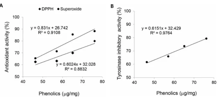
In this study, we obtained aqueous and ethanol extracts from gulfweed (S. fulvellum) and compared their bioactivities. Total phenol contents were determined, and the antioxidant and anti-tyrosinase activities were compared to several commercially available antioxidants (BHT, ascorbic acid, α-tocopherol, (+)-catechin, and epigallocatechin gallate [EGCG]). We examined the correlation between total phenol content and antioxidant or antiproliferative activities. Additionally, we screened proliferation of vascular smooth muscle cells after treatment with seaweed extracts.
Materials and Methods
Preparation of seaweed extract
Gulfweed (S. fulvellum) was collected from coastal regions of Geoje Island in Korea. The samples were washed and then dried at –40°C (2-3 days) using a freeze dryer (Ilshin Lab Co., PVTYFD 10A, Korea), and then macerated using a blender (Waring Blender, 51BL30, USA) as described previously [15]. Powdered samples (5 g) were mixed with 200 mL of 4 different solvents:
room-temperature distilled water (DW); 65°C distilled water (HW); 50% ethanol; or 100% ethanol. The mixtures were stirred at 250 rpm for 2 h and then centrifuged at 5000 rpm for 5 min. The supernatants were dried using a rotary evaporator. The dried samples were ground and filtered to obtain a fine powder, which was used for analyses of total phenol content and biological activity.
Determination of total phenol content
Total phenol content was determined using the Folin-Ciocalteu reagent according to the method of Capannesi and Palchetti [23]. Briefly, 0.5 mL of gulfweed extract (concentration 1 mg/mL) was mixed with 0.5 mL FCR for 30 sec and 1 mL 7.5% Na
2CO
3was added. The solution was then diluted with 8 mL of distilled water and left at 65°C for 20 min.
Absorbance of the blue reactant was measured at 765
nm using a UV spectrophotometer. Gallic acid was used
as a standard for quantitative determination of phenolic compounds. The analysis was performed in triplicate and the total phenol content was expressed as gallic acide quivalent (GAE).
DPPH-free radical scavenging activity
The DPPH assay measures basic antioxidant activity, and the results indicate the level of phenolic and flavonoid compounds present [24, 25]. Researchers commonly use this test to measure aromatic compounds, especially phenols, because it measures the free radical scavenging activity [26]. Free radical scavenging activity was determined using the modified DPPH (1,1-diphenyl-2-picrylhydrazyl) method of Lu and Foo [27]. A 0.1 mL aliquot of gulfweed extract (concentration of 1 mg/mL) was added to freshly prepared DPPH solution (100 mM DPPH in 80%
methanol). The mixed solution was left in the dark at 25°C for 10 min and absorbance was measured at 517 nm. The percent DPPH free radical scavenging capacity was calculated as: [(A
0-A
1)/A
0]×100, where A
0is the absorbance of a blank sample and A
1is the absorbance of the extract sample. The IC
50value was determined by plotting scavenging activity versus extract concentration. The experiment was performed in triplicate and free radical scavenging activity was expressed as percentage scavenged DPPH.
Superoxide anion scavenging activity
Superoxide anion scavenging activity of seaweed extracts was measured by the method of Oktay et al [28]. Superoxide anion (O
2-) is a potent free radical that is a precursor of more reactive ROS, such as singlet oxygen and hydroxyl radicals, which react with biological macromolecules to induce tissue damage and lipid peroxidation [5]. We used the superoxide anion scavenging (SAS) activity assay to measure the ability of superoxide anion radicals to be removed by the phenazine methosulfate/nicotinamide adenine dinucleotide (PMS/NADH) system, and measured the extent of nitroblue tetrazolium (NBT) reduction [28].
In brief, 1 mL nitroblue tetrazolium (NBT) solution
(156 μM) and 1 mL 468 μM NADPH solution were added to 0.1 mL seaweed extract (concentration 1 mg/mL), and solution was mixed. The reaction was started by adding 100 μL phenazine methosulfate (60 μM), and the combined mixture was incubated at 25°C for 5 min. Absorbance was measured at 560 nm and the percent scavenging activity was calculated as:
[(A
0-A
1)/A
0]×100, where A
0is the absorbance of a blank sample and A
1is the absorbance of the extract sample. The experiment was performed in triplicate and superoxide anion scavenging activity was expressed as percentage NBT diformazan produced.
Tyrosinase inhibitory activity
Anti-tyrosinase activity was assessed by the method of Kim et al [15]. In this assay, we determined anti-tyrosinase activity by measuring the absorbance of dopachrome at 475 nm, which forms from L-lysine or L-DOPA in the presence of antioxidants [29].
Mushroom tyrosinase (100 U/mL, 0.2 mL), 60 mM potassium phosphate buffer (pH 6.8, 0.2 mL) and 10 mM dihydroxyphenylalanine (DOPA, 0.4 mL) were mixed. Then, 0.2 mL of extract (concentration 1 mg/mL) was added, and absorbance was measured at 475 nm. The percent anti-tyrosinase activity was calculated as: (1-A
1/A
0)×100, where A
0is the absorbance of a blank sample and A
1is the absorbance of the extract. The IC
50was determined by plotting anti-tyrosinase activity versus extract concentration and is defined as the amount of extract necessary to decrease the initial dopachrome concentration by 50%.
The experiment was performed in triplicate and activity was expressed as percentage dopachrome produced.
Cell culture
Human vascular smooth muscle cells (VSMCs) were
purchased from the American type culture collection
(ATCC, USA) and maintained in a humid environment
(37°C) supplied with 5% CO
2. The cells were cultured in
Dulbecco's modified Eagle's medium (Life Technologies,
Invitrogen) supplemented with 10% fetal bovine serum
(Atlanta Biologicals, Atlanta, GA, USA), 1% L-glutamine
(29.2 mg/mL), penicillin G (10,000 U/mL), and streptomycin sulfate (10,000 µg/mL). Cells were plated at a density of 2 × 10
6cells/100 mm dishes at 70% confluency for platelet-derived growth factor-BB (PDGF-BB) treatment. Cultured VSMCs were used in the assay after treatment with PDGF-BB (10 ng/mL) for 24 h.
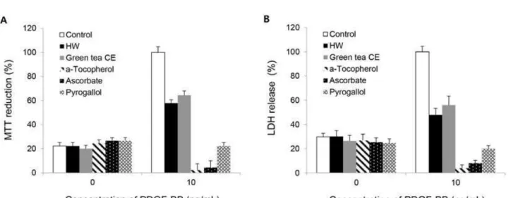
Cell proliferation assays
Cell proliferation was determined by the MTT and lactate dehydrogenase (LDH) release assays. VSMCs were serum-starved for 24 h, and then grown in the presence or absence of PDGF-BB (10 ng/mL) for 24 h. The cells were then exposed to 50 µM 4-hydroxynonenal (HNE) in Hank’s buffered saline solution (HBSS) for 30 min, which was replaced with DMEM containing 10% FBS. For the MTT assay, VSMCs were cultured with or without HW extracts in the presence or absence of PDGF-BB. After 24 h, the MTT assay was performed according to the method described by Calabro et al [30]. In brief, 0.5 mg/mL MTT solution was added to the treated cells and incubated at 37°C for 2 h. Then 200 mL of dimethyl sulfoxide (DMSO) was added and the mixture was gently shaken for 20 min to dissolve formazan crystals, the product of MTT reduction. Absorbance of extracted formazan crystal solution was measured by ELISA (550 nm). LDH release was evaluated as previously described by Perez et al [31]. After PDGF-BB treatment, the culture solution was collected, and the scraped cells were added to 100 μL PBS containing 0.1% Triton X-100 to prepare a cell lysate. After lysates and culture solution were centrifuged (5,000 rpm for 4 min at 37°C), absorbance was measured at 340 nm using a spectrophotometer. Cytotoxicity was then calculated by comparing the absorbance of the culture solution with the absorbance of the cell lysate.
Data analysis
All experiments were performed in triplicate, and results are presented as means ± SEs. Statistical analysis was performed using SPSS software, and the normality and homogeneity of the data were confirmed using
analysis of variance (ANOVA). The differences between the control and experimental groups were assessed using a one-way ANOVA (Tukey's multiple comparison test).
Statistical significance was set as p < 0.05.
Results and Discussion
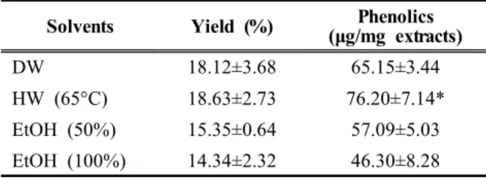
Total phenol contents of S. fulvellum extracts The phenol contents of S. fulvellum powder following were determined using four extraction solvents (Table 1). We quantitated the phenolic content using a calibration curve with gallic acid as the standard. Of the four samples tested, the HW extract had the highest phenol content (76.20 µg/mg), followed by the DW extract (65.15 µg/mg), 50% EtOH extract (57.09 µg/mg) and 100% EtOH extract (46.30 µg/mg).
Statistically significant differences in total phenol contents were found between HW extract and the other extracts. These results indicate that extraction using ethanol alone or ethanol with water led to lower total phenol yields than HW alone.
Solvents Yield (%) Phenolics
(μg/mg extracts)
DW 18.12±3.68 65.15±3.44
HW (65°C) 18.63±2.73 76.20±7.14*
EtOH (50%) 15.35±0.64 57.09±5.03
EtOH (100%) 14.34±2.32 46.30±8.28
Table 1. Total phenolics of S. fulvellum samples following extraction with different solvents.
Antioxidant and anti-tyrosinase activities of S. fulvellum extracts
The antioxidant activities of different extracts of S.
fulvellum were measured by DPPH radical scavenging,
superoxide anion scavenging (SAS), and anti-tyrosinase
activity (Table 2). DPPH radical scavenging activity was
lowest for the 100% ethanol extract and greatest for the
HW extract (62.49% vs. 79.84%, p < 0.05). The
activities of the DW and 50% ethanol extracts were
69.95% and 63.12%, respectively. The results of the
SAS assay paralleled those of the DPPH assay (Table 2):
Solvents DPPH (%) SAS (%) Anti-tyrosinase activity (%)
DW 69.95±4.41 85.42±7.32 73.56±6.22
HW (65°C) 79.84±5.53* 88.26±6.56* 79.24±7.64*
EtOH (50%) 63.16±6.84 71.26±9.26 65.87±5.26 EtOH (100%) 62.49±5.63 65.41±7.35 61.59±6.31 Table 2. Antioxidant and tyrosinase inhibitory activities of S.
fulvellum samples following extraction with different solvents.
the greatest SAS activity was found in the HW extract, and the lowest in the 100% ethanol extract (88.26% vs.
65.41%, p < 0.05). In addition, we measured the anti-tyrosinase activity (“whitening activity”) of the different extracts. Anti-tyrosinase activity was greatest in the HW extract, and lowest in the 100% ethanol extract (79.24% vs. 61.59%, p < 0.05).
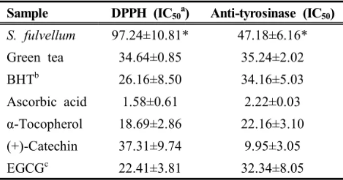
The DPPH radical scavenging and anti-tyrosinase activities of the HW extract was compared to those of a green tea extract and other antioxidants (BHT, ascorbic acid, α-tocopherol, (+)-catechin, and epigallocatechin gallate [EGCG]) by determining their IC
50concentrations (Table 3).
The IC
50values for DPPH radical scavenging and anti-tyrosinase activity were greatest for the S. fulvellum extract (97.24 ± 20.81 µg and 47.18 ± 6.16 µg, respectively.
In the other tested antioxidants, IC
50values were lowest for ascorbic acid (DPPH: 1.58 ± 0.61 µg; tyrosinase,
Sample DPPH (IC
50a) Anti-tyrosinase (IC
50) S. fulvellum 97.24±10.81* 47.18±6.16*
Green tea 34.64±0.85 35.24±2.02
BHT
b26.16±8.50 34.16±5.03
Ascorbic acid 1.58±0.61 2.22±0.03 α-Tocopherol 18.69±2.86 22.16±3.10
(+)-Catechin 37.31±9.74 9.95±3.05
EGCG
c22.41±3.81 32.34±8.05
a
= Concentration of extract required to quench 50% of free radical.
b
= Butylated hydroxytoluene
c