Corresponding Author: Seok Kil Zeon, M.D., Department Nuclear Medicine, Keimyung University School of Medicine 56 Dalseong-ro, Jung-gu, Daegu 700-712, Korea
Tel : +82-53-250-7132 E-mail : [email protected]
Lymphedema of the lower extremity is resulting from absence or obstruction of lymphatic vessels [1]. There are several etiologies such congenital absence, primary disorder of lymphatic vessels or lymph nodes by benign or malignant
neoplasm, obstruction following to infection/
inflammation of the lymphatic systems, post- phlebitic syndrome, surgically resection of the lymph node or lymphatic vessels at pelvic cavity and radiation therapy of the pelvic cavity [1-3]. In
Abstract
Lymphedema of the extremity is a chronic debilitating disease that is edema from chronic lymphatic insufficiency and frequently misdiagnosed, treated too late, or not treated at all. The effective therapy for lymphedema can be implemented, particularly after the disorder is properly diagnosed and characterized with lymphoscintigraphy. The lymphoscintigraphy is the most common and effective method to determine whether the limb swelling is due to lymphedema and follow-up study after proper management. For effective use of lymphoscintigraphy to plan therapy, it is necessary to understanding pathophysiology of lymphedema and the influence of technical factors such as selection of the radiopharmaceuticals, imaging times after injection, and patient activity after injection on the images. Several clinical cases of lymphoscintigraphy will be presented with depicting typical lymphoscintigraphic findings and their clinical applications.
Key Words :
Lymphoscintigraphy, Primary lymphedema, Secondary lymphedema Department Nuclear Medicine, Keimyung University School of Medicine,Daegu, Korea
Seok Kil Zeon, M.D.
Atlas: Impact of Lymphoscintigraphy in Evaluation of the
Lymphedema at Lower Extremity
from chronic leg discomfort with problems with walking, running, and fitting shoes. Advanced lymphedema of the leg may cause severe lifelong disability [1]. Genital lymphedema, secondary to therapy for pelvic cancer, could be involved.
Primary lymphedema is presented as congenital, prepuberty (lymphedema precox) and post- puberty onset (lymphedema tarda) [1-4].
Primary congenital lymphedema may result from genetic disorders and usually shows evidence at birth or so [2,3]. And some of the primary lymphdema is one of the main components of the several syndrome such as Milroy’s disease, Turner’s syndrome, Klippel-Trenaunay syndrome, Noonan’s s y n d r o m e, a n d M e i g e’s d i s e a s e. P r i m a r y lymphedema precox is developed before 35 years of age, and primary lymphedema tarda is developed after 35 years of age [1]. Usually primary lymphedema involves lower extremity [4].
On lymphoscintigraphy, primary lymphedema shows no definite visualization of lymphatic systems or cutoff the lymphatic vessels, and usually no evidence of the dermal back flow.
Acquired lymphedema is usually due to infection or inflammatory process of the groin or pubis making secondary lymphedema, much more prevalent than primary lymphedema. Postsurgical lymphedema (due to lymph node dissection) with or without radiation therapy in malignant disease of the pelvic cavity and postphlebitic syndrome are the most common causes of acquired, regional lymphatic insufficiency [1].
Regardless of etiology, lymphedema is presented usually as slowly progressive extremity edema. Initially, the edema is soft and pitting, but over the course of weeks to months the skin is thickened and the swelling becomes hard and non- pitting. Because of the non-functioning cutaneous lymphatics and impaired the local immune
are resulted in further injury to the tissue and worsening of edema. If lymphedema is untreated, it will progress to the point of chronic limb enlargement, followed by disfiguration of the limb associated with severe functional and psychologic impairment [1].
Diagnostic Imaging
Lymphedema can be extremely difficult to diagnose, especially in its early stages. Without a proper diagnosis, therapy is often delayed, allowing secondary fibrosis and lipid deposition to take place. Early diagnosis of these patients may allow specific implementation of preventive strategies to minimize the risk of lymphedema.
Lymphoscinti-graphy offers an objective and reliable approach to diagnose and characterize the severity of lymphedema [5,6].
On lymphoscintigraphy, there shows non- visualization or decreased number of the corresponding lymph nodes and mild to severe dermal backflow with delayed drainage or washout of the radiotracer from foot up to thigh and pelvis [5]. Lymphoscintigraphy is essential for diagnosis and follow-up examination. Computed Tomogram (CT) and Magnetic Resonance Image (MRI) could be also used as diagnostic imaging tool, however these are more suitable for rule-out other etiology of the lower extremity swelling such as deep vein thrombosis [5].
Lymphoscintigraphy
The protocol for lymphoscintigraphy is not
standardized and variable between institutions.
The distinctive points are kinds of radiotracer, type and site of injection, use of dynamic and/or static acquisitions, and acquisition times itself. 99mTc- antimony sulfide colloid (particle size: 2-15 nm),
99mTc-sulfur colloid (particle size: 500-2,000 nm),
99mTc-albumin microcolloid (particle size: < 80 nm), and 9mTc-phytate (particle size: 8-12 nm) have become the primary agents for clinical use [7].
There are several references about their characteristics and disadvantages of each radiotracer. The choice of the radiotracer is dependent on institution and country of available radioisotope and pharmaceuticals. Because 99mTc- phytate is commercially available and more easily to handle in Korea, we used this radiotracer routinely. Ordinarily, radionuclide lower extremity lymphoscintigraphy is performed by intradermal injection of radiotracer into 1st and 2nd pedal interdigital webs of both feet [5]. Usually anterior scans of the feet, calf, thigh and pelvis are
acquisitioned on 5 minutes after injection, and anterior scans of whole body on 1/2, 1, 2 and 4 h o u r s a f t e r i n j e c t i o n. A l t h o u g h s t r e s s lymphoscintigraphy (walking, standing, or limb massage) is recommended for its enhanced sensitivity and for its utility in the quantification of lymphatic flow, this approach is not universally adopted [8].
Case Presentation
This article aims to review the typical cases of the lymphoscintigraphy of the lower limb lymphedema with the clinical presentations as follows.
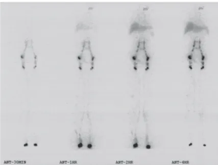
Fig. 1 is a typical case of normal ymphoscinti- graphy, as a reference case.
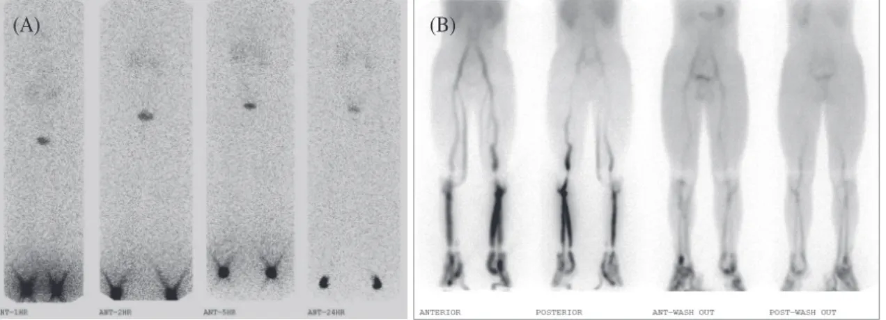
Fig. 2 is a case of primary lymphdema precox, and Fig. 3 is an example of primary lymphedema tarda.
Fig. 1. 62-year-old female patient suffered from lower extremity swelling. Radionuclide lymphoscintigraphy shows no evidence of abnormality in lymphatic system of lymphatic vessels and lymph nodes of bilateral inguinal, external iliac, common iliac and abdominal para-aortic chains.
Fig. 2. 18-y-o male referred from both feet swelling after prolonged standing or prolonged sitting. (A) Radionuclide lower extremity lymphoscintigraphy reveals non-visualization of lymphatic drainage of radiotracer in both lower extremities and non-visualization of both inguinal, bilateral iliac and para-aortic lymph nodes until 24-h delayed serial image. (B) Radionuclide bipedal venogram of both lower extremities by Tc-99m O4 injection with and without tourniquet compression reveals well visualization of superficial and deep venous systems of both lower extremities. There shows symmetric visualization of bilateral iliac veins and bilateral inferior vena cava (a normal variation). This finding is compatible with primary lymphedema precox.
(A) (B)
Fig. 3. 48-y-o female suffered from both leg swelling on long waliking for 1 year. There was no specific surgical intervention on pelvis. The color Doppler examination revealed good venous flow at both groins and both popliteal fossae (not shown here). Radionuclide lower extremity lymphoscintigraphy reveals markedly decreased in number of bilateral inguinal lymph nodes and nearly non-visualization of bilateral iliac lymph nodes, although relatively well lymphatic drainage of radiotracer in both lower extremities up to pelvic cavity. There shows no definite dermal backflow. This finding is compatible with primary lymphedema tarda.
Fig. 4 and 5 are the case of lymphedema involving pelvis and lower abdomen with collateral circulation.
Fig. 6 is a case of lymphedema involving pubis and bilateral groins with progress on follow-up lymphoscintigraphy.
Fig. 7 and 8 are the case of lymphedema at bilateral groins.
Fig. 9 shows the case of lymphedema at bilateral groins with progress on follow-up lymphoscintigraphy.
Fig. 10 shows the example of lymphedema
involving whole left lower extremity with poor prognosis on follow-up lymphoscintigraphy.
Fig. 11 shows the example of lymphedema involving whole left lower extremity with good prognosis on follow-up lymphoscintigraphy.
Fig. 12 and 13 are the example of lymphedema involving left lower leg, and Fig. 14 is a case involving right lower leg.
Fig. 15 shows a unique case of lymphocele at right medial thigh following to local burn scar.
Fig. 4. 52 year old female complaints both lower limb swelling for several months. Five years ago, cancer chemotherapy with radiation therapy was done for cancer of the uterine cervix. The CT angiography (A,B) shows significant subcutaneous soft tissue density at right abdominal wall with moderately thickened dermis, and the middle of the left thigh shows the subcutaneous soft tissue density with mild swelling. The lymphoscintigrahy (C) shows extensive dermal back flow at groins, pubic area, pelvis and lower abdominal wall with well visualization of multiple subcutaneous lymph nodes. There shows no definite visualization of bilateral iliac lymph nodes although well visualized bilateral inguinal lymph nodes. This finding shows secondary lymphedema of pelvis and abdominal wall with profuse superficial collateral pathway.
(C)
(A) (B)
Fig. 5. 58-y-o female referred for left lower leg edema for 8 years after surgical therapy, chemotherapy and radiation treatment of the stage IIb cancer of the uterine cervix. Lymphoscintigraphy shows marked dermal backflow at pubic area up to 4 hours delayed scan with collateral circulation through profuse and focal lymphovascular dilatation at right lower abdominal wall and bilateral lateral abdominal walls. There reveals non- visualization of bilateral iliac lymph nodes by surgical resection and radiation therapy although well visualization of bilateral inguinal lymph nodes.
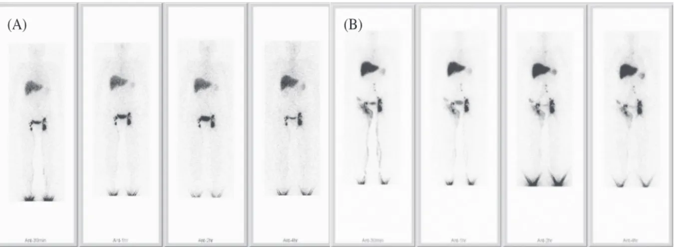
Fig. 6. 46-y-o female suffered from right leg edema for 1 month after surgical tretment of the stage Ib2 cancer of the uterine cervix with chemotherapy and radiation therapy. (A) Lymphoscintigraphy(Nov-2006) shows no definite abnormality in drainage of the radiotracer on both lower extermities up to relatively well visualized bilateral inguinal lymph nodes, but there shows non-visualization of bilateral iliac lymph nodes following to surgical resection and radiation therapy. (B) Follow-up lymphoscintigraphy (May-2011) after 6 years shows extensive dermal backflow at left groin and left sided plevic wall with collateral circulation through left lateral wall, and there shows moderate dermal back flow of the right groin and right sided pelvic wall. The delayed scan of 4 hours reveals relative improvement of the dermal backflow. There shows markedly decreased number of bilateral inguinal lymph nodes, due to delayed effect of ratiation therapy or malignant metastases (not confirmed).
(A) (B)
Fig. 7. 33-y-o female suffered from right leg swelling for 5 months, which was developed 3 months after radical hysterectomy with cancer chemotherapy and radiation therapy. On physical examination, there was stage 2 (pitting edema and hotness) lymphedema of the right lower limb, and no abnormality in left lower limb.
Lymphoscintigraphy shows markedly and profuse dermal backflow at both groins (RIGHT>LEFT) and pubis, with prolonged visualization of enlarged bilateral inguinal lymph nodes and faint collateral lymphatic vessel on right sided pelvic wall and right lower abdominal wall until 5-hours scan. There shows non-visualization of bilateral iliac lymph nodes by surgically absence and post-radiation therapy. This finding is compatible with secondary lymphedema.
Fig. 8. 45-y-o female complaints both leg swelling and heat sensation. Five months ago, cytoreductive surgery and cancer chemotherapy were done for ovarian cancer of stage IIIc. Lymphoscintigraphy shows mild swelling of both lower limbs (best seen in ANT-4 hr scan), with relatively delayed washout of the radiotracer from both lower legs until 5 hours after, and absence of right inguinal lymph nodes and bilateral iliac lymph nodes at pelvic cavity although well visualization of abdominal paraaortic lymph nodes and lymphatic vessels. CT angiogram of both lower extremities shows no abnormality in arterial and venous systems (not shows here). This finding is compatible with early stage of secondary lymphedema.
Fig. 9. 53-year-old female suffered from intermittent right lower extremity swelling for 1 year. One year ago, there was a clinical history of endometrial cancer and radical hysterectomy, followed by cancer chemotherapy and radiation therapy. (A) Lymphoscintigraphy shows well visualization of lymphatic channels of both lower limbs with well washout, although non-visualization of bilateral iliac lymph nodes. (B) Follow-up lymphoscintigraphy 18 months later reveals profuse dermal back flow at right groin with a small collateral lymphatic channel at right lateral pelvic wall. There shows well drainage of the lymphatic channels of left lower limb, and faint visualization of left iliac lymph nodes and abdominal paraaortic lymph nodes which were not seen in previous examination, representing regeneration of the left iliac lymph nodes. This finding is compatible with secondary lymphedema of right lower limb with progression.
Fig. 10. 57-y-o female suffered from left leg swelling for 1 month. Two years ago, total radical hysterectomy (June- 10/2009) with cancer chemotherapy and radiation therapy for cancer of the uterine cervix. There was skin color change with redness and positive Stemmer sign. (A) Lymphoscintigraphy (July-7/2011) shows moderate dermal backflow from left lower leg to left thigh and non-visualization of left inguinal lymph nodes. There shows surgically absent bilateral iliac lymph nodes. This finding is compatible with secondary lymphedema. (B) Follow- up lymphoscintigraphy (Sept-5/2012) shows progression of the secondary lymphedema of whole left lower limb.
(A)
(A)
(B)
(B)
Fig. 11. 54-y-o female suffered from intermittent left leg swelling for several months, and in 2000 total abdominal hysterectomy was done for cancer of the uterine cervix. On physical examination, stage 2 with positive Stemmer sign and no skin color change of the left lower limb is noted. (A) Initial lymphoscintigraphy shows dermal backflow at left thigh with non-visualization of left inguinal lymph nodes, and non-visualization of bilateral pelvic lymph nodes (surgical resection). This finding is compatible with secondary lymphedema. (B) The follow-up lymphoscintigraphy after 20 months shows gradual visualization of left sided pelvic lymph nodes after conservative therapy such as the vigorous massage and the elastic stocking. This finding shows good prognosis.
(A) (B)
Fig. 12. 55-y-old female suffered from left calf tenderness and progressive left leg swelling. Ten years ago, radical hysterectomy was done for cancer of the uterine cervix Ib1, and 1 year after left leg swelling was developed.
Lymphoscintigraphy shows extensive dermal backflow at left lower leg and non-visualization of left inguinal lymph nodes. There shows surgically absent bilateral iliac lymph nodes. This finding is compatible with secondary lymphedema.
Fig. 13. 69-y-o female suffered from left ankle swelling for 4 months and aggravated. Eighteen years ago, radical hysterectomy and radiation therapy were done for cancer of the uterine cervix. CT angiogram of lower extremity shows marked dermal thickening and swelling with subcutaneous increased density and significantly irregular strands (A: thigh, B: below knee). (C) Radionuclide lower extremity lymphoscintigraphy reveals non-visualization of left inguinal lymph nodes and bilateral iliac lymph nodes, and marked dermal backflow of left lower leg with swelling of left lower extremity up thigh. It shows well lymphatic drainage of radiotracer in right lower extremity up to right groin and well visualization of right inguinal lymph nodes. This finding is compatible with secondary lymphedema, following to surgical resection of bilateral iliac lymph nodes and post-radiation therapy change of left inguinal lymph nodes.
(C)
(A) (B)
Fig. 14. 51-y-o female suffered from fever and chillness with right lower extremity swelling 1 week after cholecystectomy of chronic calculus cholecystitis. Eighteen years ago, uncertain surgery for cancer of the uterine cervix with external radiation therapy was done, and intermittent swelling of the right lower extremity was developed 1 year after. Radionuclide lymphoscintigraphy after treatment of lymphangitis of the right lower extremity with antibiotics reveals extensive dermal back flow at right lower leg, with non-visualization of drainage of radiotracer in the right upper extremity up to right groin, and non-visualization of right inguinal lymph nodes, bilateral external iliac and bilateral common iliac nodes. There shows well drainage of the radiotracer and well visualization of the left inguinal lymph nodes. This finding is compatible with secondary lymphedema and acute onset of lymphangitis following to surgical resection of the pelvic lymph nodes and radiation therapy.
Fig. 15. 67-y-o male suffered from fluctuation and progressive enlargement of the burn scar at right medial thigh, 1 month after contact burn. (A) MRI of the right medial thigh shows suggestive of seroma. (B) Lymphoscintigraphy shows a large, well-defined, elongated, radioactivity accumulation mass at right medial thigh, compatible with lymphocele. The draining catheter with Hemovac shows considerable radioactivity from lymphocele.
(A) (B)
Lymphatic imaging of lymphoscintigraphy can play a pivotal role in defining the etiology of extremity swelling and in predicting the success of the ordinary therapies. It will be important to d e v e l o p s t a n d a r d i z e d p r o c e d u r e s a n d r a d i o p h a r m a c e u t i c a l s t o p e r f o r m t h e s e examinations and standardized criteria to interpret the results.
Acknowledgement
Author wants cordially to thank to Professor a n d D r. S o-y o u n g L e e, D e p a r t m e n t o f Rehabilitation Medicine for generous sharing the cases, and Dr. Sung Hoon Kim and Dr. Il Jo, Department of Nuclear Medicine for great assistances.
References
1. Szuba A, Shin WS, Strauss HW, Rockson S. The Third Circulation: radionuclide lymphoscintigraphy in the evaluation of lymphedema. J Nucl Med 2003;44:43-57.
Boccardof F, et al. Lymphoscintigraphic evaluation of congenital lymphedema of the newborn. Clin Nucl Med 2002;27:383-4.
3. Shinawi M. Lymphedema of the lower extremity: Is it genetic or nongenetic? Clin Pediatr 2007;46:835-41.
4. Williams WH, Witte CL, Witte MH, George CM.
Radionuclide lymphangioscintigraphy in the evaluation of peripheral lymphedema. Clin Nucl Med 2000;25:451- 64.
5. Choi JY, Hwang JH, Park JM, Lee KH, Kim SE, Kim DI. Risk assessment of dermatolymphangioadenitis by lymphoscintigraphy in patients with lower extremity lymphedema. Korean J Nucl Med 1999;33:143-51.
6. Luongo JA, Scalcione LR, Katz DS, Yung EY.
Progression of clinically stable lymphedema on lymphoscintigraphy. Clin Nucl Med 2009;34:585-8.
7. Alavi A, Staum MM, Shesol BF, Bloch PH. Technetium 99m stannous phytate as an imaging agent for lymph nodes. J Nucl Med 1978;19:422-6.
8. Tartaglionea G. Pagan M. Morese R, Cappellini GA, Zappala AR, Sebastiani C, et al. Intradermal lymphoscintigraphy at rest and after exercise: a new technique for the functional assessment of the lymphatic system in patients with lymphoedema. Nucl Med Commun 2010;31:547-51.