INTRODUCTION
Ingestion of acid or alkaline caustic substances may cause serious injuries in the esophagus and stomach.
1The degree of injury is determined by the nature of the substance (the de- gree to which it could cause corrosion), the amount con- sumed or its concentration and state (solid or liquid), and the time of contact with the gastrointestinal (GI) mucosa. In the United States, >5,000 cases of ingestion of caustic substances are reported annually. Although most cases occur in pediatric patients, some cases involve adults who attempt suicide, psy- chiatric patients, and alcoholic patients. More severe injuries to the esophagus and stomach occur if large quantities of sub- stances are ingested, particularly in persons who attempt sui- cide.
2However, worldwide estimates report that about 80% of cases are in pediatric patients.
3Although precise data are scant, in Korea, 60% of cases were caused by ingesting caustic sub- stances with the intention of committing suicide, and another 40% of cases were accidental.
4,5Most of the cases involving the intent to commit suicide occur especially among young per-
sons in their teens and 20s.
4,5Also, many of the reported pa- tients who ingested caustic substances have accompanying psychiatric disorders including depression, schizophrenia, ad- justment problems, and personality disorders.
4,5Although the numbers have decreased compared with in the past, cases of patients who ingest caustic substances and visit the emergency room are not rare. Therefore, in this article, I will discuss the etiologic causative agents, injury mechanism, and clinical ch- aracteristics, as well as the endoscopic evaluation of the degree of injury and proper management of the patient, in GI caus- tic injury. As the focus of this discussion is the evaluation and management of patients under emergency settings, the discus- sion about chronic complications has been omitted.
SUBSTANCES CAUSING CAUSTIC INJURY
In pediatric patients younger than 5 years, consuming caus- tic substances occurs accidentally; however, in teens and ad- ults, it is mostly intentional with the goal of committing suicide.
2Although many kinds of substances cause caustic injury,
6,7the most common agent is a strong alkaline substance such as sodium hydroxide (NaOH) or potassium hydroxide (KOH), which usually includes disinfectants used in the home or laundry facilities, and discoid batteries.
8The term “lye” refers to the liquid obtained from the leaching of ashes, including NaOH or KOH. Highly acidic substances, such as hydrochlo- ric acid, sulfuric acid, and phosphoric acid, are used frequently to remove rust in bathrooms or swimming pools and may be
Evaluation and Management of Caustic Injuries from Ingestion of Acid or Alkaline Substances
Kyung Sik Park
Department of Internal Medicine, Keimyung University School of Medicine, Daegu, Korea
Although the numbers have decreased compared with in the past, cases of patients who ingest caustic substances and visit the emergency room are not rare. However, well-summarized data about caustic injuries are insufficient. Therefore, in this article, I will discuss the etio- logic causative agents, injury mechanism, and clinical characteristics, as well as the endoscopic evaluation of the degree of injury and proper management of the patient, in gastrointestinal caustic injury.
Key Words: Caustic injury; Acids; Alkalies Open Access
Received: March 21, 2014 Revised: April 13, 2014 Accepted: April 14, 2014
Correspondence: Kyung Sik Park
Department of Internal Medicine, Keimyung University Dongsan Medical Center, Keimyung University School of Medicine, 56 Dalseong-ro, Jung-gu, Daegu 700-712, Korea
Tel: +82-53-250-7088, Fax: +82-53-250-7088, E-mail: [email protected]
cc This is an Open Access article distributed under the terms of the Creative Commons Attribution Non-Commercial License (http://creativecommons.org/
licenses/by-nc/3.0) which permits unrestricted non-commercial use, distribution, and reproduction in any medium, provided the original work is properly cited.
included in car batteries. These acidic materials are used less frequently than alkaline substances as a tool for suicide be- cause they can induce severe pain.
1In Korea, ingestion of alkaline substances was more com- mon in the past; however, recently, cases of ingestion of acidic material have been increasing. This is thought to be due to a rapid decrease in the use of lye owing to the development of synthetic detergents, and at the same time, a relative increase in the use of acetic acid, which can be purchased easily.
4,5,9A solution of 5% sodium hypochlorite, which is used as bleach, is commonly known as “Rox.” This agent is frequently reported to be ingested, but it rarely injures the esophagus or stomach.
10Button-type batteries contain highly alkaline sub- stances, and if swallowed, serious tissue injury secondary to local current or pressure necrosis may occur. When these bat- teries are trapped in the esophagus, burns may occur within 4 hours and perforation may occur within 6 hours.
8Therefore, in those cases, the batteries should be removed through emer- gent endoscopy.
PATHOPHYSIOLOGY
Ingestion of alkaline substances is known to mainly injure the esophagus rather than the stomach or duodenum, where- as acidic materials injure mainly the stomach rather than the esophagus.
1,11However, recent reports indicating that exten- sive esophageal injury or even perforation is not uncommon after ingestion of acids are putting this traditional notion into question.
12In addition, both acidic and alkaline substances can injure the larynx, trachea, and bronchi.
8Ingestion of alkaline substances leads to liquefaction ne- crosis due to bonding with tissue protein (Fig. 1).
1,8Therefore, it causes rapid injuries outside the esophagus toward the me- diastinal wall. These responses continue until the alkaline substance is neutralized by the tissue fluids. In addition, alka- line fluid has a stronger surface tension and stays in the tissue for a longer period, thereby worsening the injury. Within the stomach, injuries are limited by the partially neutralizing me- chanism of gastric acid.
13In the case of extensive injury to the intestinal wall, complications such as perforation, mediasti- nitis, and peritonitis may occur and result in death. Liquid materials, rather than solid batteries, result in more extensive injuries.
8Liquefaction necrosis occurs for 3 to 4 days and causes intravascular thrombus and mucosal inflammation, in addition to causing local or extensive putrefaction and ul- ceration. Over a period of 2 weeks, the esophageal wall is thinned with tissue putrefaction, granulation, and fibrosis;
the process of reepithelialization takes 1 to 3 months. There- fore, endoscopic procedures should be avoided from 5 to 15 days after the ingestion of alkaline substances.
14Stricture for-
mation, as a chronic complication, is ultimately affected by the depth of the esophageal injury and the degree of collagen ac- cumulation. Therefore, the subsequent possibility of chronic complications or death increases in second- or third-degree injuries.
13Ingestion of acids usually causes superficial coagulation necrosis, in which formation of thrombi within the vessels and bundling of connective tissue lead to the formation of scar tissue. Therefore, very deep injuries that extend through all layers of the GI wall do not commonly develop after the in- gestion of acidic substances.
15Upon swallowing, acids cause severe oropharyngeal pain;
thus, they are usually consumed in small volumes compared with the alkaline substances. Acidic substances have less sur- face tension; therefore, they pass rapidly through the esopha- gus, resulting in a less frequency and a lower degree of esoph- ageal injuries (Fig. 2A).
1,8The ingested acidic substance flows rapidly to the pylorus through the lesser curvature of the stomach, causing widespread injury in the antrum (Fig. 2B).
If there is some amount of food inside the stomach, injury can be prevented because of a neutralization effect. Despite this, if large quantities are consumed, esophageal or gastric perfo- ration may occur.
Although, as stated above, caustic injury caused by alkaline
substances is more severe, a comparative study revealed that
the prognosis was less favorable for those who swallowed a
strong acid.
16In this study, the acid ingestion group showed a
high frequency of severe complications such as perforation or
death, a high degree of mucosal injury, and a high frequency
of intensive care unit (ICU) admission. This is thought to be
Fig. 1. Diffuse liquefaction necrosis of the entire esophagus is noted after the ingestion of alkaline substances.because the prognosis of caustic injury is influenced by the quantity ingested and the types of complication after inges- tion rather than by the type of the ingested substance (acid or alkaline).
GRADING OF THE CAUSTIC INJURY
The degree of injury from caustic substances can be cate- gorized similarly to that of skin burns.
14First-degree injuries are confined to the mucosal surface, and can show diffuse or localized erythema, edema, and bleeding. Scar formation does not ultimately occur. Second-degree injuries involve the mu- cosa and submucosa. Endoscopic examination can show vari- ous findings such as exudates or blister formation. Ultimately, scar changes can develop through the formation of granula- tion and fibrosis. Third-degree injuries include the entire wall layer, and are characterized by deep penetrating ulcers, black discoloration, or perforation of the intestinal wall.
CLINICAL PRESENTATIONS
The clinical presentations of caustic injuries are very di- verse, and initial symptoms do not show deep correlations with the ultimate degree of the injury.
17Crystal or other solid materials easily adhere to the oral mucosa; therefore, they cause more severe injuries to the pharynx and upper airway rather than to the esophagus or stomach. In contrast, liquids can easily pass through the esophagus and stomach; therefore, they can cause extensive injury. The degree of injury is propor- tional to the mortality. Furthermore, the morbidity of chron- ic complication is also related to the degree of injury.
18,19De- pending on the injured area, pain may arise in the oropharynx, retrosternum, or epigastrium. Dysphagia, odynophagia, and excessive salivation may also occur. Continuous severe retro-
sternal or back pain may indicate esophageal perforation or mediastinitis. Perforation may occur up to 2 weeks after in- gestion; therefore, strict monitoring is essential.
3In addition, depending on the extent of injury, vomiting, hematemesis, abdominal tenderness, and rebound tenderness may suddenly occur. Although rare, vocal hoarseness, wheezing, and short- ness of breath may also occur upon injury to the larynx and epiglottis (Fig. 3). Fever, palpitations, and shock indicate ex- tensive damages.
DIAGNOSIS AND EVALUATION OF THE EXTENT OF INJURY
Laboratory tests
Although the results of laboratory tests are not well corre-
Fig. 2. Caustic injury after the ingestion of acid material. Mild esophageal injury (A) is noted compared with widespread severe injury (B) in the stomach.A B
Fig. 3. Injury to the larynx and epiglottis after the ingestion of al- kaline material. Vocal hoarseness, wheezing, and shortness of breath may occur upon injury to these areas.
lated with the degree of injury, a study suggests that white blood cell counts >20,000 cells/mm
3, increase in the C-reac- tive protein level, old age, and the presence of esophageal ul- cers are predictive factors that are correlated with mortality.
20Some reports state that an arterial pH <7.22 may indicate eso- phageal injury requiring surgical management.
21Usually, lab- oratory results contribute toward setting the direction of mon- itoring and management of the patient, rather than predicting the mortality.
22Endoscopy
Some reports suggest that nonsymptomatic pediatric pa- tients who have ingested low-potency substances do not re- quire endoscopy. However, for adult patients who have in- gested caustic substances with the intent to commit suicide, most of those substances are very potent, and therefore, emer- gent endoscopy is recommended for all patients.
23As an intact-looking oral cavity or pharynx cannot be used to exclude the possibility of esophageal or gastric injury, up- per GI endoscopy should be performed within 24 hours of ingestion of caustic substances; however, several reports in- dicate that it can be safely done up to 96 hours after inges- tion.
16,24Upper GI endoscopy can be used to evaluate esoph- ageal and gastric injuries, as well as to predict prognosis and establish a management plan.
16,25However, it is contraindi-
cated in several patients, such as hemodynamically unstable patients, those suspected to have a perforation, those in severe respiratory distress, and patients with severe laryngo-pha- ryngeal edema or necrosis. For patients whose injury is fo- cused around the lips and oral cavity, esophageal or gastric injury usually does not exceed the first-degree grading.
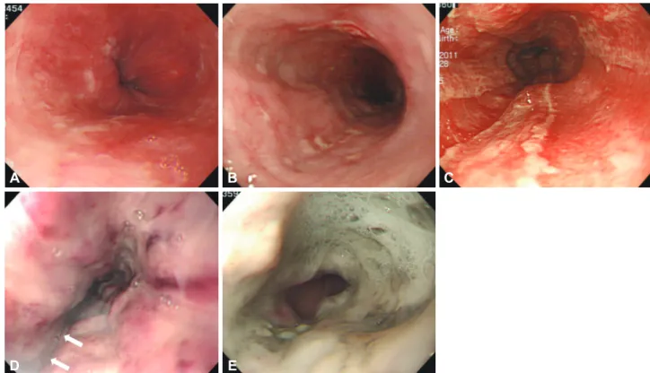
26To predict the treatment outcome and prognosis of patients with caustic injuries, endoscopic categorization of 81 patients was performed, and the subsequent categorization is widely used.
14Grade 0 indicates a normal mucosa; grade 1 indicates only slight swelling and redness of the mucosa (Fig. 4A); grade 2A indicates the presence of superficial ulcers, bleeding, and exudates (Fig. 4B); grade 2B indicates local or encircling deep ulceration (Fig. 4C); grade 3A indicates focal necrosis (Fig.
4D); and grade 3B indicates extensive necrosis (Fig. 4E). Al- though further studies are needed to evaluate the validity of this endoscopic categorization, most patients with grade 1 or 2A injuries have good prognoses without sudden deaths, and they do not develop outlet obstruction or stricture of the eso- phagus. Approximately 70% to 100% of patients with grade 2B and 3A injuries develop stricture. For patients with grade 3B injuries, a mortality of 65% has been reported, and in the majority of cases, esophagectomy and colonic or jejunal re- placement surgeries are required. However, several reports have indicated that 12% of gastrectomies and 15% of esopha-
A
D
B
E
C
Fig. 4. Endoscopic grading of the caustic gastrointestinal injury. (A) Grade 1 indicates only slight swelling and redness of the mucosa. (B) Grade 2A indicates the presence of superficial ulcers, bleeding, and exudates. (C) Grade 2B indicates local or encircling deep ulceration.
(D) Grade 3A indicates focal necrosis. White arrows indicate focal necrosis. (E) Grade 3B indicates extensive necrosis.
gectomies are unnecessary; therefore, a more accurate stan- dard is required.
27Endoscopic ultrasound
Miniprobe endoscopic ultrasound (EUS) can be used safe- ly, and compared with conventional endoscopy, no differences have been reported for their use in predicting the develop- ment of early complications.
28However, a study had indicat- ed that no strictures form later if the muscle layer is intact in EUS.
29In addition, several studies with radial EUS indicated that if the proper muscle layer is included, the treatment re- sponse to balloon dilatation decreases, and subsequent repeat- ed procedures are required; however, additional studies are needed to investigate the role of EUS in evaluating caustic eso- phageal injuries.
Radiologic examinations
A simple chest X-ray may be done to observe if there is a gas shadow in the mediastinum or under the diaphragm, indicat- ing esophageal or gastric perforation, respectively. For confir- mation, esophagography or upper GI series with a water-sol- uble contrast medium may be attempted carefully.
The diagnostic efficacy of computed tomography (CT) is slightly higher than endoscopy in terms of assessing the depth and boundary of esophageal or gastric injury, and it is effec- tive in diagnosing impending perforation. In a retrospective analysis of 49 patients who underwent CT, a scoring system for the degree of esophageal causticity and injury of periph- eral tissues was attempted, and in terms of predicting the de- gree of stricture, CT showed better results than endoscopy.
30MANAGEMENT OF PATIENTS
No randomized trial has compared various models for the management of caustic esophageal injury in humans. Instead, most of the currently used management procedures are based on animal experiments.
General management
If a person is suspected to have ingested large quantities of acidic or alkaline substances according to history taking, phys- ical examination, and upper endoscopy (higher than grade 2B), admission to a medical or surgical ICU is mandatory. By using this management approach, the development of seri- ous complications, if any, can be rapidly treated, and the for- mation of strictures after recovery can be reduced. However, clinical signs may vary between patients; therefore, strict in- dividual evaluation is necessary. It should be considered that the presence of a symptom or sign alone cannot be used to predict the degree of injury.
Endoscopy is not necessary if there are no symptoms and if unintentional ingestion of small volumes of acid or alkaline substances can be confirmed on the basis of history taking.
The patient can be followed at the outpatient clinic after dis- charge.
8Other patients should be admitted and kept nil per os, and chest and abdominal X-ray scans should be taken to assess for perforation. In addition, an intravenous (IV) line must be kept for fluid resuscitation in patients with hypotension. To prevent stress ulcers and additional damage to the esophagus from the regurgitated gastric acid, IV proton pump inhibi- tors can be administered.
31If patients experience pain, it should be controlled with adequate administration of nar- cotic anesthetics. Perforation, mediastinitis, and peritonitis are indications for emergent surgery. For the treatment of patients with injuries higher than grade 3 that are observed by using endoscopy or those suspected to have esophageal per- foration, broad-spectrum IV antibiotics such as third-genera- tion cephalosporins should be administered. In patients with respiratory difficulties, laryngoscopic observation is required to evaluate for the need for a tracheostomy. In patients with accompanying oropharyngeal injury, careful management considering airway obstruction is necessary. If there is swelling of the larynx or epiglottis, the airway should be maintained by performing tracheotomy rather than tracheal intubation.
Several methods for the management of caustic esophageal injuries must be avoided before their evaluation, which in- clude administering drugs that may induce vomiting. This is because such methods may reexpose the esophagus to the caustic substances residing in the stomach. Neutralizers must also be avoided because the heat generated from the neutral- ization reaction may worsen the tissue injury. Furthermore, nasogastric tubes must also be avoided, as they may induce vomiting and reexpose the esophagus to the caustic substanc- es; the pressure generated during vomiting may also increase the risk of perforation.
Upper GI endoscopy
As described above, upper GI endoscopy to evaluate the degree of injury must be performed within 24 hours. Patients with grade 0 injuries can be discharged immediately, and those with grade 1 or 2A injuries do not require specific treatment.
Patients can consume liquids, and advance to regular food
within 24 to 48 hours. Patients with grade 2B or 3 injuries can
be given liquids through a nasogastric tube 24 hours after the
ingestion of caustic substances, and may drink water if they
are able to swallow saliva after 48 hours. Patients with grade 3
injuries must be carefully observed for perforation symptoms
for at least 1 to 2 weeks after the ingestion.
Surgery
Esophagectomy is required for patients with severe stric- ture;
32however, this may result in negative long-term conse- quences concerning the survival rate or functional capacity.
27Emergent surgery is required for patients with perforation, mediastinitis, and peritonitis. If performed by an experi- enced surgeon, minimally invasive thoracoscopic and laparo- scopic procedures result in better outcomes than conventional methods.
33The most important factors to ensure successful recovery include vascular supply and low tension of the anas- tomotic site. In patients with damage to both the esophagus and stomach, the colon is usually used as a source of replace- ment tissue. On the other hand, in patients with damage to only the esophagus, the stomach is pulled up toward the me- diastinum to replace the esophagus.
Prevention of strictures
The use of corticosteroids to prevent the formation of stric- tures is controversial. Usually, it is not recommended because corticosteroids increase the adverse effects without actually preventing stricture formation, as stricture formation is de- termined by the initial depth of the injury.
34,35Grade 3 injuries especially are not affected by the use of corticosteroids. Intra- lesional injection of triamcinolone has been attempted; how- ever, there are no clear data on the effectiveness, appropriate dosage, and frequency of administration that is required to prevent stricture formation.
36There is an old study that has shown that antibiotic use significantly helps in preventing strictures.
37However, the ef- ficacy has not been proven in patients without infection. Cur- rently, the use of antibiotics is not recommended for prevent- ing strictures in patients who are not being treated with cor- ticosteroids.
Nasogastric tube insertion can cause infection, acid reflux, and long strictures; therefore, its unique use is not currently recommended. However, a report suggested that nasogastric tube insertion can be used to provide enteral nutrition; there- fore, it can be used selectively.
38Several reports stated that intramucosal injection of mito- mycin-C, a chemotherapeutic agent with DNA cross-linking activity, was helpful to prevent strictures;
39,40however, patients should be observed carefully because systemic absorptions can cause serious adverse effects. A recent meta-analysis indi- cated positive long-term results; however, additional prospec- tive studies are needed to determine the appropriate concen- tration, administration period, and frequency of adminis- tration,
39as theoretically, malignant tumors can develop. Th- erefore, this therapeutic method should be used with caution.
Several reports have shown the usefulness of a specially de- signed stent (silicone rubber,
41Polyflex stent;
42Boston Scien-
tific, Natick, MA, USA) for preventing stricture formation.
However, their low efficacy (<50%) and high expulsion rate (>25%) were problematic. Recently, researchers of a study on the use of a polytetrafluoroethylene stent reported an efficacy of 72% during a period of 9 to 14 months, and researchers of another study on a biodegradable stent reported a 45% effica- cy during 53 months, indicating the development of various stent models with varying efficacies. Bougie dilatation has been recommended; however, its efficacy is unclear and ad- ditional studies are required.
Apart from the treatment models described above, antioxi- dants (such as 5-fluorouracil and vitamin E), phosphatidyl- choline, octreotide, and interferon-α-2b are being studied for their utility in preventing stricture formation in animal mod- els; however, they are not yet at a stage where they can be used to treat humans. More time may be needed before antioxi- dants could be used for treatment.
Once a stricture develops, balloon dilatation can be attempt- ed carefully.
43,44PROGNOSIS
The most important prognostic factors include the degree of tissue injury and the underlying condition of the patient.
Most deaths occur because of complications such as medias- tinitis and peritonitis; therefore, strict management in the ini- tial stage is crucial to avoid the occurrence of complications.
This article does not address the complications of tissue inju- ry; however, the most representative chronic complications include stricture, squamous cell carcinoma, and a decrease in lower esophageal sphincter pressure, which leads to reflux esophagitis, esophageal motility disorder, intractable pain, gastric outlet obstruction, acidity, and protein losing enteropa- thy. Acid reflux may be an aggravating factor that cause refrac- tory stricture of the esophagus; therefore, regular observation and aggressive anti-acid therapy is necessary in patients with corrosive esophagitis.
28CONCLUSIONS
In this article, I have reviewed the etiologic causes of caustic GI injury, mechanisms of injury, clinical signs, endoscopic ev- aluation, and management of patients who have ingested caustic substances. The degree of caustic tissue injury is deter- mined by the nature of the swallowed substance and time the substance spent in contact with the mucosa.
In most adult patients, it may be beneficial to perform an
upper GI endoscopy within 24 hours to evaluate the degree
of tissue injury. It can help determine the treatment options
and to predict prognosis. However, endoscopy is usually con-
traindicated in patients with hemodynamic instability, possi- ble perforation, severe respiratory insufficiency, and severe swelling of the larynx or oropharynx. Patients with a moder- ate to severe level of injury should be admitted to the ICU while strictly monitoring for any life-threatening complica- tions. In addition, if complication such as mediastinitis, peri- tonitis, and other signs indicative of perforations is observed, emergent surgery is required.
Conflicts of Interest
The author has no financial conflicts of interest.
REFERENCES
1. Goldman LP, Weigert JM. Corrosive substance ingestion: a review. Am J Gastroenterol 1984;79:85-90.
2. Riffat F, Cheng A. Pediatric caustic ingestion: 50 consecutive cases and a review of the literature. Dis Esophagus 2009;22:89-94.
3. Gumaste VV, Dave PB. Ingestion of corrosive substances by adults.
Am J Gastroenterol 1992;87:1-5.
4. Yeom HJ, Shim KN, Kim SE, et al. Clinical characteristics and predis- posing factors for complication of caustic injury of the upper digestive tract. Korean J Med 2006;70:371-377.
5. Yoon KW, Park MH, Park GS, et al. A clinical study on the upper gas- trointestinal tract injury caused by corrosive agent. Korean J Gastrointest Endosc 2001;23:82-87.
6. Jung IS, Kim JS. A case of corrosive gastritis caused by salt-fermented Northern Sand Lance. Korean J Gastrointest Endosc 2011;42:366-368.
7. Joo HR, Park J, Kim TGS, E H, et al. A case of corrosive esophagogas- tritis after copper sulfate ingestion. Korean J Gastrointest Endosc 2011;
43:30-32.
8. Contini S, Scarpignato C. Caustic injury of the upper gastrointestinal tract: a comprehensive review. World J Gastroenterol 2013;19:3918-3930.
9. Sohn YT, Kim JG, Song DW, Lee SS. Clinical study of corrosive esopha- gitis. Korean J Otolaryngol-Head Neck Surg 1992;35:346-353.
10. Wasserman RL, Ginsburg CM. Caustic substance injuries. J Pediatr 1985;107:169-174.
11. Kim HG, Han KH, Lee SI, et al. A case of corrosive gastritis caused by hydrochloric acid. Korean J Gastrointest Endosc 1988;8:19-23.
12. Arévalo-Silva C, Eliashar R, Wohlgelernter J, Elidan J, Gross M. Inges- tion of caustic substances: a 15-year experience. Laryngoscope 2006;116:
1422-1426.
13. Zargar SA, Kochhar R, Nagi B, Mehta S, Mehta SK. Ingestion of strong corrosive alkalis: spectrum of injury to upper gastrointestinal tract and natural history. Am J Gastroenterol 1992;87:337-341.
14. Zargar SA, Kochhar R, Mehta S, Mehta SK. The role of fiberoptic en- doscopy in the management of corrosive ingestion and modified en- doscopic classification of burns. Gastrointest Endosc 1991;37:165-169.
15. Fisher RA, Eckhauser ML, Radivoyevitch M. Acid ingestion in an ex- perimental model. Surg Gynecol Obstet 1985;161:91-99.
16. Poley JW, Steyerberg EW, Kuipers EJ, et al. Ingestion of acid and alka- line agents: outcome and prognostic value of early upper endoscopy.
Gastrointest Endosc 2004;60:372-377.
17. Gaudreault P, Parent M, McGuigan MA, Chicoine L, Lovejoy FH Jr.
Predictability of esophageal injury from signs and symptoms: a study of caustic ingestion in 378 children. Pediatrics 1983;71:767-770.
18. Salzman M, O’Malley RN. Updates on the evaluation and management of caustic exposures. Emerg Med Clin North Am 2007;25:459-476.
19. Hoffman RS, Howland MA, Kamerow HN, Goldfrank LR. Compari- son of titratable acid/alkaline reserve and pH in potentially caustic household products. J Toxicol Clin Toxicol 1989;27:241-246.
20. Rigo GP, Camellini L, Azzolini F, et al. What is the utility of selected clinical and endoscopic parameters in predicting the risk of death after
caustic ingestion? Endoscopy 2002;34:304-310.
21. Cheng YJ, Kao EL. Arterial blood gas analysis in acute caustic ingestion injuries. Surg Today 2003;33:483-485.
22. Katzka DA. Caustic injury to the esophagus. Curr Treat Options Gas- troenterol 2001;4:59-66.
23. Celik B, Nadir A, Sahin E, Kaptanoglu M. Is esophagoscopy necessary for corrosive ingestion in adults? Dis Esophagus 2009;22:638-641.
24. Previtera C, Giusti F, Guglielmi M. Predictive value of visible lesions (cheeks, lips, oropharynx) in suspected caustic ingestion: may endos- copy reasonably be omitted in completely negative pediatric patients?
Pediatr Emerg Care 1990;6:176-178.
25. Cabral C, Chirica M, de Chaisemartin C, et al. Caustic injuries of the upper digestive tract: a population observational study. Surg Endosc 2012;26:214-221.
26. Aronow SP, Aronow HD, Blanchard T, Czinn S, Chelimsky G. Hair re- laxers: a benign caustic ingestion? J Pediatr Gastroenterol Nutr 2003;36:
120-125.
27. Chirica M, Resche-Rigon M, Bongrand NM, et al. Surgery for caustic injuries of the upper gastrointestinal tract. Ann Surg 2012;256:994-1001.
28. Chiu HM, Lin JT, Huang SP, Chen CH, Yang CS, Wang HP. Prediction of bleeding and stricture formation after corrosive ingestion by EUS concurrent with upper endoscopy. Gastrointest Endosc 2004;60:827-833.
29. Kamijo Y, Kondo I, Kokuto M, Kataoka Y, Soma K. Miniprobe ultraso- nography for determining prognosis in corrosive esophagitis. Am J Gas- troenterol 2004;99:851-854.
30. Ryu HH, Jeung KW, Lee BK, et al. Caustic injury: can CT grading sys- tem enable prediction of esophageal stricture? Clin Toxicol (Phila) 2010;
48:137-142.
31. Cakal B, Akbal E, Köklü S, Babali A, Koçak E, Taş A. Acute therapy with intravenous omeprazole on caustic esophageal injury: a prospective case series. Dis Esophagus 2013;26:22-26.
32. Bothereau H, Munoz-Bongrand N, Lambert B, Montemagno S, Cattan P, Sarfati E. Esophageal reconstruction after caustic injury: is there still a place for right coloplasty? Am J Surg 2007;193:660-664.
33. Zhou JH, Jiang YG, Wang RW, et al. Management of corrosive esopha- geal burns in 149 cases. J Thorac Cardiovasc Surg 2005;130:449-455.
34. Fulton JA, Hoffman RS. Steroids in second degree caustic burns of the esophagus: a systematic pooled analysis of fifty years of human data:
1956-2006. Clin Toxicol (Phila) 2007;45:402-408.
35. Anderson KD, Rouse TM, Randolph JG. A controlled trial of corticoste- roids in children with corrosive injury of the esophagus. N Engl J Med 1990;323:637-640.
36. Siersema PD, de Wijkerslooth LR. Dilation of refractory benign esopha- geal strictures. Gastrointest Endosc 2009;70:1000-1012.
37. Krey H. On the treatment of corrosive lesions in the oesophagus; an ex- perimental study. Acta Otolaryngol Suppl 1952;102:1-49.
38. Kochhar R, Poornachandra KS, Puri P, et al. Comparative evaluation of nasoenteral feeding and jejunostomy feeding in acute corrosive in- jury: a retrospective analysis. Gastrointest Endosc 2009;70:874-880.
39. Berger M, Ure B, Lacher M. Mitomycin C in the therapy of recurrent esophageal strictures: hype or hope? Eur J Pediatr Surg 2012;22:109-116.
40. Uhlen S, Fayoux P, Vachin F, et al. Mitomycin C: an alternative conser- vative treatment for refractory esophageal stricture in children? En- doscopy 2006;38:404-407.
41. De Peppo F, Zaccara A, Dall’Oglio L, et al. Stenting for caustic stric- tures: esophageal replacement replaced. J Pediatr Surg 1998;33:54-57.
42. Broto J, Asensio M, Vernet JM. Results of a new technique in the treat- ment of severe esophageal stenosis in children: poliflex stents. J Pediatr Gastroenterol Nutr 2003;37:203-206.
43. Lee SI, Shn SH, Park IS, Choi HJ. Dilatation of severe corrosive esoph- ageal stricture guided by right coronary artery catheter. Korean J Gas- trointest Endosc 1991;11:77-80.
44. Lee MS, Kim JW, Kim JH, Cho SW, Shim CS. Non-operative dilatation of corrosive esophageal and gastric angular stricture: a case report. Ko- rean J Gastrointest Endosc 1989;9:151-155.