444
CASE REPORT
A 75-year-old male, a resident of a rural area and a farmer by occupation, visited our outpatient clinic with the symptoms of poor cognition and memory decline over 2 weeks. He denied any history of fever, headache, blurred vision, vomiting or seizure.
He was afebrile and his vital signs were stable. There were no lab- oratory abnormalities including leukocytosis or C-reactive pro- tein rising. Upon the neurologic examination, he was conscious and there were no neurologic deficits except intermittent expres- sive dysphasia and disorientation. Brain magnetic resonance im- aging (MRI) was performed because of suspicion of some type of dementia. It showed a 20 mm sized nodular enhancing mass INTRODUCTION
Fungal brain abscesses are well known to be associated with the immunocompromised state. However, cerebral phaeohypho- mycosis (CP) caused by darkly pigmented fungi appears to be a common exception to this rule because about one-half of this fungal infection occurred in patients with no underlying disease or risk factors. CP is a very rare cause of brain abscess, but is often a fatal disease regardless of immune status
14,17,23).
The authors illustrate a 75-year-old, immunocompetent male patient who had a single brain abscess from dematiaceous fungi.
To the authors’ knowledge, this is the first case of CP in Korea.
Cerebral Phaeohyphomycosis : A Rare Cause of Brain Abscess
Na-Young Jung, M.D., Ealmaan Kim, M.D., Ph.D.
Department of Neurosurgery, Dongsan Medical Center, Keimyung University School of Medicine, Daegu, Korea
Cerebral phaeohyphomycosis (CP) is a very rare but serious form of central nervous system fungal infection that is caused by dematiaceous fungi. It is commonly associated with poor prognosis irrespective of the immune status of the patient. In this study, the authors describe the first case of CP in Korea that occurred in a 75-year-old man without immunodeficiency and showed favorable outcome after surgical excision and antifungal thera- py. In addition, the authors herein review the literature regarding characteristics of this rare clinical entity with previously reported cases.
Key Words : Brain abscess · Cerebral phaeohyphomycosis · Fungal infection · Treatment.
Case Report
•Received : August 7, 2014 •Revised : October 1, 2014 •Accepted : October 22, 2014
•Address for reprints : Ealmaan Kim, M.D., Ph.D.
Department of Neurosurgery, Dongsan Medical Center, Keimyung University School of Medicine, 56 Dalseong-ro, Jung-gu, Daegu 700-712, Korea Tel : +82-53-250-7823, Fax : +82-53-250-7356, E-mail : [email protected]
•This is an Open Access article distributed under the terms of the Creative Commons Attribution Non-Commercial License (http://creativecommons.org/licenses/by-nc/3.0) which permits unrestricted non-commercial use, distribution, and reproduction in any medium, provided the original work is properly cited.
J Korean Neurosurg Soc 56 (5) : 444-447, 2014
http://dx.doi.org/10.3340/jkns.2014.56.5.444
Copyright © 2014 The Korean Neurosurgical Society Print ISSN 2005-3711 On-line ISSN 1598-7876
www.jkns.or.kr
A B C D
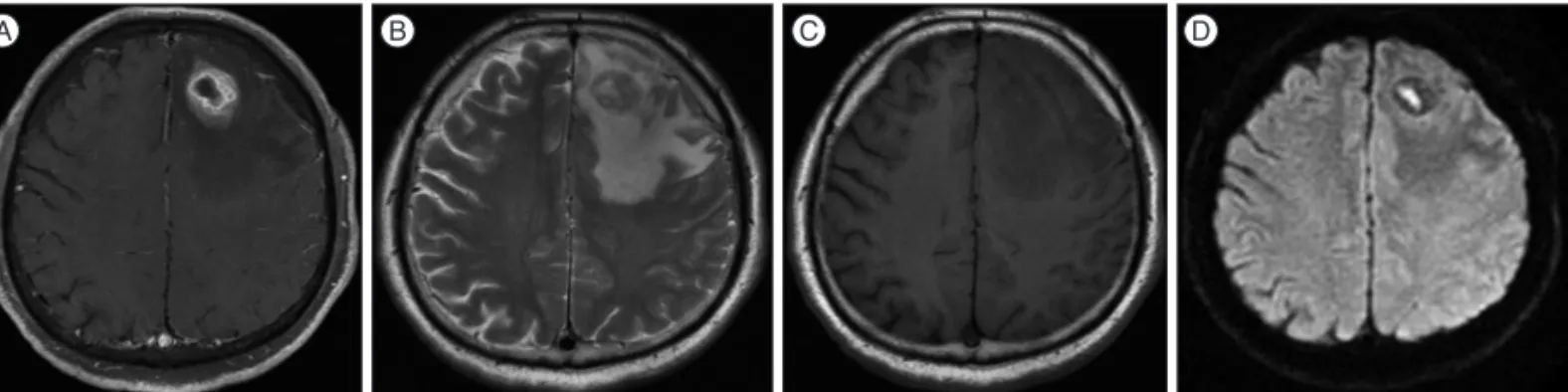
Fig. 1. Brain magnetic resonance imaging (MRI) shows heterogenous enhancing nodular lesion in left frontal lobe with adjacent edema (A). The mass is slightly hyperintense on T2-weighted image (B) and isointense on T1-weighted image (C). The cavity in central portion is brightly shown on diffu- sion-weighted image (D).
445
Cerebral Phaeohyphomycosis | NY Jung and E Kim
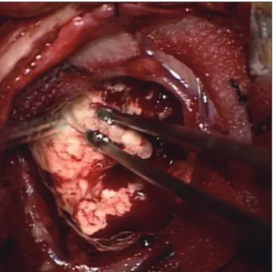
with peritumoral edema in the left frontal lobe. The mass had a central cystic portion with diffusion restriction (Fig. 1). High- grade glioma or metastatic tumor was initially presumed based on his age and progressive symptoms. Excisional biopsy was performed for tissue diagnosis. The lesion appeared to be white and took the form of a relatively hard mass with a clear boundary, permitting radical excision of the mass (Fig. 2). Pathological ex- amination revealed multiple necrotizing granulomas with brown pigmented fungal hyphae. Septated hyphae and melanin pigments were confirmed at Fontana-Masson stain consistent with CP (Fig. 3).
The patient was started on intravenous amphotericin B at a dose of 68 mg daily. After 10 days, he was switched to 270 mg of intravenous voriconazole twice a day because of the elevation of serum creatinine. He took the injection for 8 weeks, followed by oral voriconazole 200 mg twice a day for 2 months. A follow-up brain MRI 3 weeks after surgical excision demonstrated a sig- nificant resolution of the edema. Ongoing resolution of the lesion was found on the latest follow-up MRI (Fig. 4). He showed dra- matic improvement in his symptoms including disorientation and memory disturbance after completion of surgery and antifun-
gal therapy.
DISCUSSION
CP is a rare infection caused by darkly pigmented fungi, namely dematiaceous fungi
23). Dematiaceous fungi represent a group of filamentous molds that contain melanin pigment in their cell walls
3,6,14,17). Cladophialophora bantiana is the most frequently isolated species. Rhinocladiella mackenziei (formerly Ramichlo-
ridium mackenziei) is the second most common cause of CP,which is exclusively endemic in the Middle East area
17,22). Most agents are found in soil. Because of this, occupational predispo- sition has been reported in agricultural workers, especially farm- ers due to risk of soil exposure
17,23). CP commonly occurs in the second and third decades of life with male predominance, except
Rhinocladiella mackenziei which affects adults with a median ageof 62 years without male predominance
3,13,23).
The most unique characteristic of CP is its occurrence irrele- vant to the immune status of the host
15,17,19,23). Even though im- munodeficiency may play a role as a risk factor, there are many reports of this infection in immunocompetent individuals simi- lar to the patient in this report
12,15,17,19,25). The portal entry to brain
Fig. 2. Intraoperative photography demonstrates a white-yellowish and hard mass with well-defined capsule.
A B C D
Fig. 3. Gross preparation shows white, well-demarcated, round masses in brain parenchyma (A). Hematoxylin and eosin stain reveals necrotizing granulomas (red stared, ×100) and inflammatory infiltrates (yellow stared, ×100) (B), as well as brown colored septated hyphae (×1000) (C). Black col- ored melanin pigments are present in branched fungal hyphae on Fontana-Masson stain (×400) (D).
Fig. 4. Magnetic resonance imaging 1 year after surgery depicts complete resolution of abscess and edema in the frontal lobe.
446 J Korean Neurosurg Soc 56 | November 2014
is unclear, although several possible routes have been suggested, such as hematogenous dissemination of inhaled spores or acci- dental skin inoculation as well as direct extension from adjacent paranasal sinuses or ears
2,6,10,14,15,19,22,23). The authors were unable to ascertain the route of infection in the present case. Pathogen- esis of CP is associated with the presence of melanin as a virulence factor that provides advantages in evading host defense and cross- ing the blood-brain barrier by binding to hydrolytic enzyme
14,16,17). Clinical spectrum of phaeohyphomycosis was listed as a vari- able, ranging from solitary subcutaneous nodules to a life-threat- ening infection
5,11,16-18). In the central nervous system (CNS) mani- festation, brain abscess is a classic clinical presentation
3,23). Patients can also present meningitis, encephalitis, myelitis or arachnoid-
itis
17,19). Hemiparesis and headache are the most common symp-
toms followed by various clinical manifestations
17). About 70–
80% of cases typically manifest as a single brain abscess particularly on the frontal lobe (52%) like in our case, while multiple brain ab- scesses can be seen in immunocompromised patients
3,6,17,19).
The diagnosis of CP can be difficult because dematiaceous fungi are often considered contaminants when identified in cul- ture. Furthermore, the pathogen can not always be cultured and isolated from the serum or cerebrospinal fluid (CSF)
12,21,24). No mo- lecular techniques are available to speedily identify these fungi even to the genus level
17). Therefore, diagnosis is made by surgi- cal biopsy. Only the tissue examination can be useful to identify irregularly swollen hyphae with yeast-like structure and to con- firm the presence of dematiaceous hyphae in melanin-specific Fontana-Masson stain
14,17). Unfortunately in this case, fungus was not identified in the culture of surgical specimen, therefore, the species that causes CP could not be detected. Meanwhile, the brain MRI reveals a ring-enhancing lesion with a low-attenuation core, suggesting the presence of necrosis or pus
10,19). In cases where high- grade glioma or metastasis is mimicked by irregular and variably contrast-enhancing lesions, magnetic resonance spectroscopy may be used to differentiate the entities
6,7,19). Imaging findings of this patient were more suggestive of a glioma than an abscess, because nodular heterogeneity on contrast injection mimicked the images seen in high-grade tumors. Consequently, surgical biopsy is essential for the diagnosis of CP.
Because of the rarity of the cases, there is no standard treat- ment guidelines for CP. A combination of surgical and medical treatments is generally recommended
10,22). Complete excision of brain lesions may provide better results than simple aspiration unless the lesion is multiple or is located within the eloquent area of the brain
3,17). Antifungal agents are generally used in combina- tion of amphotericin B, 5-flucytosine and itraconazole because it is associated with improved survival rates
3,14,17,20). Voriconazole can be used as alternative to itraconazole because of its good pen- etration into both CSF and brain tissue
17,22). Duration of taking the medications is still unknown because most reported patients expired during treatment except a few survivors who received voriconazole for about 12 months
8,19). In addition, posaconazole may be a potent drug when pathogen is Rhinocladiella macken-
ziei1,3,8)
. In this case, amphotericin B was replaced by voricon-
azole because of serum creatinine elevation. Amphotericin B has fatal side effects such as nephrotoxicity, therefore, close ob- servation on kidney function is needed.
The prognosis of CP is poor. Mortality rate approaches 100%
in untreated patients, while that of treated cases as high as 65%
to 73% despite the aggressive treatment
6,9,10,17,19,23). Interestingly, mortality rate did not differ significantly between immunocom- promised and immunocompetent patients (75% vs. 71%)
12,17). Multiple brain abscesses are associated with worse prognosis than solitary lesion
4,23). Fortunately, the patient reported here had a good response to surgery and chemotherapy and showed fine recovery without any sequela. Solitary lesion and the good gener- al condition of the patient, together with an aggressive therapeu- tic approach, are therefore inferred to contribute to a favorable outcome. Further studies are necessary to find more potentially useful antifungal regimen for these refractory infections and to investigate more detailed pathophysiology and prognostic factors to increase the survival rate.
CONCLUSION
CP is rare disease, but challenging one with high mortality rate, particularly when the CNS is affected. As shown in this report, complete resection and adequate antifungal therapy are the most recommended modality for patients with CP-related abscess to this time.
References
1. Badali H, de Hoog GS, Curfs-Breuker I, Meis JF : In vitro activities of antifungal drugs against Rhinocladiella mackenziei, an agent of fatal brain infection. J Antimicrob Chemother 65 : 175-177, 2010
2. Brandt ME, Warnock DW : Epidemiology, clinical manifestations, and therapy of infections caused by dematiaceous fungi. J Chemother 15 Suppl 2 : 36-47, 2003
3. Cristini A, Garcia-Hermoso D, Celard M, Albrand G, Lortholary O : Ce- rebral phaeohyphomycosis caused by Rhinocladiella mackenziei in a woman native to Afghanistan. J Clin Microbiol 48 : 3451-3454, 2010 4. Dixon DM, Walsh TJ, Merz WG, McGinnis MR : Infections due to Xylo-
hypha bantiana (Cladosporium trichoides). Rev Infect Dis 11 : 515-525, 1989
5. Fader RC, McGinnis MR : Infections caused by dematiaceous fungi : chromoblastomycosis and phaeohyphomycosis. Infect Dis Clin North Am 2 : 925-938, 1988
6. Gongidi P, Sarkar D, Behling E, Brody J : Cerebral phaeohyphomycosis in a patient with neurosarcoidosis on chronic steroid therapy secondary to recreational marijuana usage. Case Rep Radiol 2013 : 191375, 2013 7. Hauck EF, McGinnis M, Nauta HJ : Cerebral phaeohyphomycosis mim-
ics high-grade astrocytoma. J Clin Neurosci 15 : 1061-1066, 2008 8. Jabeen K, Farooqi J, Zafar A, Jamil B, Mahmood SF, Ali F, et al. : Rhinoc-
ladiella mackenziei as an emerging cause of cerebral phaeohyphomycosis in Pakistan : a case series. Clin Infect Dis 52 : 213-217, 2011
9. Kantarcioglu AS, de Hoog GS : Infections of the central nervous system by melanized fungi : a review of cases presented between 1999 and 2004.
Mycoses 47 : 4-13, 2004
10. Li DM, de Hoog GS : Cerebral phaeohyphomycosis--a cure at what lengths?
Lancet Infect Dis 9 : 376-383, 2009
447
Cerebral Phaeohyphomycosis | NY Jung and E Kim
11. Matsumoto T, Ajello L, Matsuda T, Szaniszlo PJ, Walsh TJ : Developments in hyalohyphomycosis and phaeohyphomycosis. J Med Vet Mycol 32 Suppl 1 : 329-349, 1994
12. Ochiai H, Kawano H, Minato S, Yoneyama T, Shimao Y : Cerebral phaeo- hyphomycosis : case report. Neuropathology 32 : 202-206, 2012 13. Palaoglu S, Sav A, Basak T, Yalcinlar Y, Scheithauer BW : Cerebral phaeo-
hyphomycosis. Neurosurgery 33 : 894-897, 1993
14. Ravisankar S, Chander RV : Cerebral pheohyphomycosis : report of a rare case with review of literature. Neurol India 61 : 526-528, 2013 15. Revankar SG : Dematiaceous fungi. Mycoses 50 : 91-101, 2007 16. Revankar SG, Patterson JE, Sutton DA, Pullen R, Rinaldi MG : Dissemi-
nated phaeohyphomycosis : review of an emerging mycosis. Clin Infect Dis 34 : 467-476, 2002
17. Revankar SG, Sutton DA, Rinaldi MG : Primary central nervous system phaeohyphomycosis : a review of 101 cases. Clin Infect Dis 38 : 206-216, 18. Rinaldi MG : Phaeohyphomycosis. Dermatol Clin 14 : 147-153, 19962004 19. Rosow L, Jiang JX, Deuel T, Lechpammer M, Zamani AA, Milner DA,
et al. : Cerebral phaeohyphomycosis caused by Bipolaris spicifera after heart transplantation. Transpl Infect Dis 13 : 419-423, 2011
20. Sharkey PK, Graybill JR, Rinaldi MG, Stevens DA, Tucker RM, Peterie JD, et al. : Itraconazole treatment of phaeohyphomycosis. J Am Acad Derma- tol 23 (3 Pt 2) : 577-586, 1990
21. Shimosaka S, Waga S : Cerebral chromoblastomycosis complicated by meningitis and multiple fungal aneurysms after resection of a granuloma.
Case report. J Neurosurg 59 : 158-161, 1983
22. Sood S, Vaid VK, Sharma M, Bhartiya H : Cerebral phaeohyphomycosis by Exophiala dermatitidis. Indian J Med Microbiol 32 : 188-190, 2014 23. Suri P, Chhina DK, Kaushal V, Kaushal RK, Singh J : Cerebral phaeohy-
phomycosis due to cladophialophora bantiana - a case report and re- view of literature from India. J Clin Diagn Res 8 : DD01-DD05, 2014 24. Takei H, Goodman JC, Powell SZ : Cerebral phaeohyphomycosis
caused by ladophialophora bantiana and Fonsecaea monophora : report of three cases. Clin Neuropathol 26 : 21-27, 2007
25. Watson KC : Cerebral chromoblastomycosis. J Pathol Bacteriol 84 : 233-237, 1962