Roles of Prostatic Acid Phosphatase in Prostate Cancer
Hoon Young Kong
1, Hak Jong Lee
2and Jonghoe Byun
1*
1
Department of Molecular Biology, Institute of Nanosensor and Biotechnology, Dankook University, Yongin-si, Gyeonggi-do 448-701, Korea
2
Department of Radiology, Seoul National University College of Medicine and Seoul National University Bundang Hospital, Seongnam, Gyeonggi-do 463-707, Korea
Received May 24, 2011 /Accepted June 4, 2011
Prostatic acid phosphatase (PAP) is one of the widely used biomarkers in the diagnosis of prostate cancer. It was initially identified in 1935 and is the most abundant phosphatase in the human prostate.
PAP is a prostate-specific enzyme that is synthesized in prostate epithelial cells. It belongs to the acid phosphatase group that shows enzymatic activity in acidic conditions. PAP is abundant in prostatic fluid and is thought to have a role in fertilization and oligospermia. It also has a potential role in reducing chronic pain. But one of the most apparent functions of PAP is the dephosphorylation of macromolecules such as HER-2 and PI3P that are involved in the ERK1/2 and MAPK pathways, which in turn leads to inhibition of cell growth and tumorigenesis. Currently, clinical trials using PAP DNA vaccine are underway and FDA-approved immunotherapy using PAP is commercially available.
Despite these clinically important aspects, molecular mechanisms underlying PAP regulation are not fully understood. The promoter region of PAP was reported to be regulated by NF-κB, TNF-α, IL-1, androgen and androgen receptors. Here, the features of PAP gene and protein structures together with the function, regulation and roles of PAP in prostate cancer are discussed.
Key words : Prostatic acid phosphatase (PAP), prostate cancer, marker, tumor suppression
*Corresponding author
*Tel:+82-31-8005-3194, Fax:+82-31-8021-7201
*E-mail : [email protected]
서 론
Phosphatase는 kinase와 함께 단백질의 인산화/탈 인산화 를 균형적으로 조절하여 다양한 cellular process에 관여한다.
이러한 인산화/탈 인산화 과정은 signal transduction과정에 서 중요한 역할을 담당하게 되는데, 비정상적인 kinase에 의한 hyperphosphorylation은 tumorigenesis 과정 동안 세포 성장 신호를 지속적으로 유지시키는 역할을 하게 된다. 한편 비정 상적인 phosphatase는 단백질의 탈 인산화를 저해하여 세포 성장신호의 억제를 불가능하게 하는 결과를 가져온다. 현재까 지 kinase에 의한 단백질의 hyperphosphorylation으로 발생 하는 암에 관한 연구는 많이 진행되어 왔지만, phosphatase에 의한 세포 성장신호의 억제를 통한 암의 치료에 관해서는 많 이 연구가 진행되어 있지 않다.
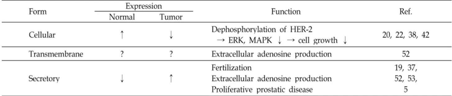
Prostatic acid phosphatase (PAP)는 1935년 전립선에서 처 음 발견되었는데[17], Table 1에서 보듯이 secretory form, transmembrane form, 그리고 cellular form의 세가지 형태가 존재한다[5,13,30]. 전립선 암의 진행 정도에 비례하여 secre- tory PAP의 발현이 증가하고 이 secretory PAP가 분열하는 세포막에서 탈 인산화 작용을 한다는 사실이 알려졌다[5,13].
PAP는 PSA와 같은 다른 전립선 암 표지자들보다 형태학적
특징과 높은 상관관계를 지니고 있기 때문에[9], 이를 바탕으 로 PAP는 과거 수십년간 전립선 암의 표지자(marker)로서 혈 청검사와 조직검사에 널리 사용되어 왔다. 이런 표지자로서의 기능과는 다른 측면으로 전립선이 암이 되는 과정에서 cel- lular PAP 발현의 감소로 인해 탈 인산화가 제대로 일어나지 않아 세포 성장 신호가 지속적으로 유지되는 현상도 밝혀졌다 [20,22,23,38,42]. 즉, cellular PAP 발현의 감소가 전립선 암 발 생 원인중의 하나가 되고, 이 경우 PAP는 tumor supressor의 역할을 하는 것으로 추정된다[42](Table 1). PAP의 전립선 특 이적 발현의 조절 기작 관련해서도 PAP promoter 부위에서의 cis-acting element들이 몇몇 보고된 바 있지만[27,29,33,34,50], 이들 외에 다른 조절 기작들이 존재할 것으로 여겨지고 있다.
이러한 PAP 발현의 증가 및 감소가 갖는 의미와 전립선에서 여러 형태의 PAP들이 갖는 역할에 대해서는 아직까지 많은 부분들이 알려져 있지 않다. PAP의 탈 인산화 능력은 전립선 세포에서의 신호전달 과정에 중요한 의미를 지니는 바, 이에 본 총설에서는 현재까지 밝혀진 PAP의 유전자 및 단백질의 구조와 기능, PAP 발현의 조절 기작과 전립선 암 발생에 갖는 의미 등을 고찰하였다.
재료 및 방법
Phosphatase의 중요성
단백질의 인산화/탈 인산화 과정은 대사과정, 세포주기,
DNA 복제, 전사, 번역, 신호전달 등 무수히 많은 세포 과정들
Table 1. Comparison of three forms of PAP in human prostate cells
Form NormalExpressionTumor Function Ref.
Cellular ↑ ↓ Dephosphorylation of HER-2
→ ERK, MAPK ↓ → cell growth ↓ 20, 22, 38, 42
Transmembrane ? ? Extracellular adenosine production 52
Secretory ↓ ↑ Fertilization
Extracellular adenosine production Proliferative prostatic disease
19, 37, 52, 53,
5
Fig. 1. Processing and modification of transmembrane form of hPAP. Human PAP is initially translated as an immature form.
After cleavage of signal peptide, hPAP becomes a mature form, where three N-linked glycosylation sites exist; two high mannose-type carbohydration sites (N62 and N301) and one sialylation site (N188). His12 is an essential active site residue that provides a nucleophile function. Other sites important for enzyme activity are marked in the mature form.
을 조절하는 중요한 기작이다. 탈 인산화에 관여하는 효소인 phosphatase는 크게 non-specific phosphatase와 protein phosphatase 그룹으로 나는데, non-specific phosphatase는 효 소의 적정 환경에 따라서 alkaline phosphatase와 acid phos- phatase로, 기질의 종류에 따라 tyrosine-specific phosphatase, serine/threonine-specific phosphatase, dual specificity phos- phatase, lipid phosphatase 등으로 나뉜다. 정상적인 세포에 서는 phosphatase와 kinase가 서로 길항적으로 작용하여 인산 화/탈 인산화가 평형상태를 이루고 있는데, 이 평형상태가 파 괴되면 다양한 질병으로 이어질 수 있다. 특히, 암의 발생과 진행에 있어서 hyperphosphorylation의 중요성이 강조되어온 바, 비정상적으로 인산화를 유도하는 kinase에 관한 연구가 그동안 활발히 이루어져 왔다. 그러나, 최근 암화 과정에서 kinase의 반대 역할을 담당하는 phosphatase도 중요한 역할을
담당할 수 있음이 부각되고 있어 이에 대한 연구들이 점차 많이 진행될 것으로 예상된다.
Prostatic acid phosphatase 유전자와 단백질의 구조 Human prostatic acid phosphatase (PAP)는 전립샘의 상피 세포(epithelial cells)에서 합성되어 전립선액으로 분비되는 glycoprotein이다[14,43,44]. PAP는 1935년에 전립선에서 처음 으로 밝혀져[17], 지난 수십 년간 관련 연구가 진행되어 왔지만 아직 명확한 생리적 기능은 밝혀지지 않은 효소이다. Human PAP 유전자는 3q21~23에 위치하고 있으며[47], cDNA는 354 개의 아미노산을 지정한다. PAP는 32개의 잔기로 이루어진 signal peptide를 N-terminal에 갖는 41 kDa의 단백질이며 (Fig. 1), 산성 환경에서 탈 인산화 활성을 갖는 효소이다[48].
처음에는 10개의 exon을 가지는 세포 내 단백질(cellular PAP)
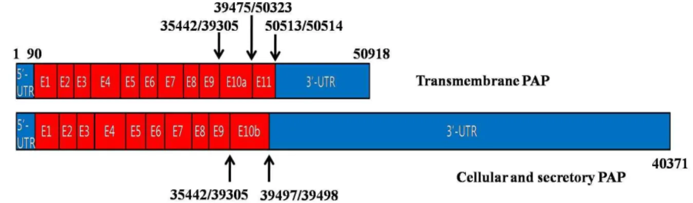
Fig. 2. Schematic diagram of hPAP transcripts. Two types of hPAP transcripts are generated by alternative splicing. Transmembrane PAP has exon 10a and 11, whereas cellular and secretory PAPs have exon 10b instead of exon 10a and 11. The 3’-UTR of transmembrane PAP is shorter than those of cellular and secretory PAPs. Cellular PAP and secretory PAP are identical but they differ in their cellular location. The number indicates relative nucleotide numbers from the start site of 5’-UTR, which was set at 1. Introns were omitted for easier comparison of exon structures.
로 알려졌지만[48], secretory PAP도 발견되었고[44], 최근에 는 상대적으로 3‘-UTR이 짧고 alternative splicing에 의하여 추가적으로 exon 11을 갖는 새로운 형태의 PAP도 발견되었다 [30](Fig. 2). Secretory PAP와 cellular PAP는 exon 10b를 포함 한 동일한 구조를 가지고, transmembrane PAP는 exon 10a, 11 등을 갖는 막단백질 형태임이 최근 밝혀졌다[30,42](Fig. 2).
PAP는 두 개의 catalytically inactive subunit들이 비공유결 합(non-covalent bond)을 이루며 homodimer를 형성하여 활 성화 된다[16]. Monomer PAP에는 총 여섯 개의 cysteine이 존재하는데, 이 여섯 개의 cysteine은 모든 포유류에서 잘 보존 되어 있으며, Cys129-Cys340, Cys183-Cys281, Cys315-Cys319 에서와 같이 이황화결합(disulfide bond)을 형성하고 있다 [41]. PAP에는 세 개의 putative N-linked glycosylation sites 가 존재하는데, 이중 Asn62와 Asn301에는 high man- nose-type carbohydrate가 붙고, Asn188은 부분적으로 sialy- lation이 됨이 밝혀졌다[15](Fig. 1). 또한 site-directed muta- genesis연구를 통하여 PAP의 효소활성에는 Histidine (H12, H257), Aspartate (D258), 그리고 Arginine (R11, R15, R54, R79) 잔기들이 중요함이 밝혀졌다[26].
PAP의 발현
PAP는 전립선뿐만이 아니라 뇌, 신장, 간, 폐, 근육, 태반, 침샘, 비장, 갑상선, 흉선 등에서 발현된다. 이 중 태반과 간에 서 발현하는 acid phosphatase는 대부분 lysosome에 위치하 기 때문에 lysosomal acid phosphatase (LAP)라고 명명되었 고, secretory PAP는 전립선에서만 발현되는 특징이 있는 것 으로 알려져 있다[3,10,39,40]. PAP의 발현은 나이와도 상관관 계가 있는데, 태어날 때는 높은 수준으로 PAP를 발현하다가 생후 6개월까지 점점 감소하여 발현이 사라지고, 10세 이후부 터 사춘기까지 발현량이 다시 점차 증가하는 양상을 보인다.
이로 미루어 볼 때 PAP의 발현은 2차 성징을 결정짓는 testos-
terone과 같은 성 호르몬과 연관되어 있음을 알 수 있다[11].
PAP의 mRNA 양을 quantitative PCR을 이용하여 전립선에서 확인하였을 때는 정상 전립선이나 전립선 암 모두 PAP의 transcript가 상당히 증가되어 있음을 확인하였고, 이는 다른 조직 대비 정상 전립선 조직에서 50~5,000배, 전립선 암 조직 에서 110~6,000배 가량 높은 수준임이 확인되었다[12]. 한편, 면역조직화학법으로 분석하여 보았을 때 정상의 유방조직에 서는 PAP가 발견되었으나, 유방암종(breast carcinoma)에서 는 PAP가 전혀 관찰되지 않았고[46], 결장암종(colon carcino- ma)에서는 전체 시료 중 40%의 시료에서만 PAP가 관찰되었 으며 그 PAP 수준도 정상 전립선 혹은 전립선암종(prostate carcinoma)에서보다 낮은 수준임이 확인되었다[46]. 이상의 결과들은 PAP 발현이 전립선에서 특이적으로 높음을 말해주 며, 또 PAP 발현 정도가 조직의 암 발생과 역의 상관관계가 있음을 보여준다.
PAP 유전자의 발현 조절
PAP 유전자 발현조절에 관여하는 인자들에는 androgen, androgen repector, NF-κB, TNF-α, IL-1 등이 알려져 있다.
Human prostatic carcinoma에서 PAP의 promoter 분석을 통
해 ATG 코돈의 56 bp와 91 bp upstream에 두 개의 major
transcription initiation site가 존재함이 확인되었다[4,48]. 또한
promoter region을 chloramphenicol acetyl transferase (CAT)
reporter 유전자로 분석한 연구에서는 -1258에서 -779 사이의
element가 PAP의 세포유형 특이적 발현을 가능케 하는 부분
이라는 것이 밝혀졌다[50]. 또한, PAP 조절부위에서 전사인자
결합부위 염기서열인 GAAATATGATA element가 여섯 번
발견되는데(-588/-558, -267/-237, -160/-130, +211/+241,
+239/+269, +1136/+1164), 이 중 두 부위(-160/-130,
+239/+269)는 조직 특이적 발현에 관여함을 electrophoretic
mobility shift assay를 통해 확인하였다[34,35]. Androgen은
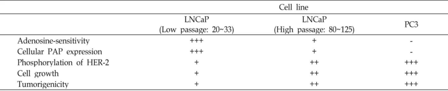
Table 2. Characteristics of different prostate cancer cells
Cell line LNCaP
(Low passage: 20~33) LNCaP
(High passage: 80~125) PC3 Adenosine-sensitivity
Cellular PAP expression Phosphorylation of HER-2 Cell growth
Tumorigenicity
+++
++++ ++
+ +++ ++++
- +++- ++++++
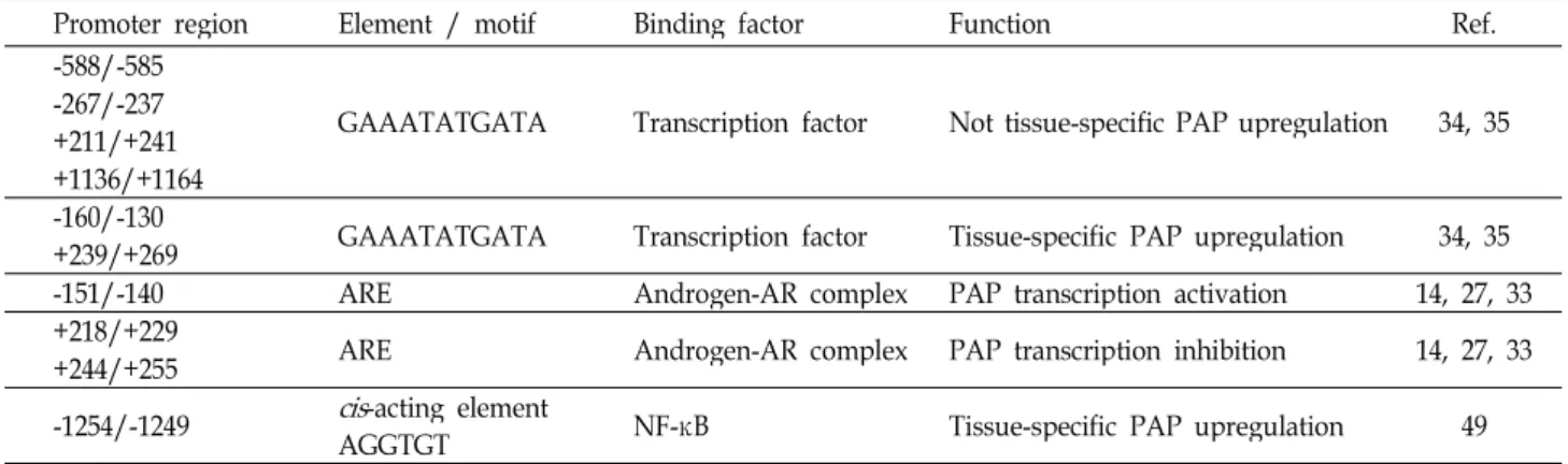
androgen receptor (AR)에 직접적으로 결합하여 전립선의 발 달과 기능에 중요한 영향을 미치게 되는데, PAP의 조절부위 에서 androgen-AR complex가 작용하는 androgen response element (ARE)가 총 세 번 발견된다(-151/-140, +218/+229, +244/+255). ARE에 androgen-AP complex가 homodimer를 이루어 ARE에 직접 결합하는데, 이 세부분 중 -151/-140 부분 은 androgen level이 정상일 때 hPAP의 transcription을 활성 화시키지만, +218/+229, +244/+255 부분은 androgen level이 낮을 때 hPAP의 transcription을 억제하는 기능을 한다[14, 27, 33]. 이러한 사실들은 hPAP의 발현 시 androgen이 중요하게 관여함을 보여준다. 한편 TNF-α와 IL-1 등이 NF-κB를 통해 PAP promoter의 활성을 증가시킴이 보고되었는데, deletion analysis를 통하여 -1356/-779 577 bp가 cis-active enhancer region임을 확인하였고, 향후 연구결과로 NF-κB가 PAP pro- moter 영역 -1254/-1249의 AGGTGT motif에 결합하여 작용 함으로써 전립선 암 세포주 특이적인 PAP 발현을 증가시킴이 확인되었다[49]. 이 외에도 PAP의 promoter region에 조직 특 이적 발현을 조절하는 element들이 상당수 존재할 것으로 예 상되는 바, PAP의 발현 조절에 관한 연구는 PAP의 기능을 연구하는데 많은 도움을 줄 것으로 기대된다.
PAP의 효소 활성
PAP의 탈 인산화 과정은 기존에 알려진 fructose-2,6-bi- sphosphatase의 탈 인산화 과정과 비슷하다[24]. Zhang 등의 연구를 통하여 PAP의 active site는 His12와 Asp258임이 밝혀 졌는데, H12D 또는 D258A mutant에서는 ErbB-2의 phospho- tyrosine level이 감소하지 않음이 확인되었다[51]. PAP의 탈 인산화 과정은 His12 잔기가 nucleophile로 작용하여 기질의 phosphate를 받아들여 phosphohistidine intermediate를 형 성하여 시작한다. 이후 Asp258가 general acid로 작용하여 기 질을 가수분해시키고 기질의 탈 인산화를 유발한다[26]. 또한 His12 뿐만이 아니라 His257, Asp258, Arg11, Arg15, Arg54, Arg79 등이 중요한 잔기임을 site-directed mutagenesis를 통 하여 확인하였으며 이들은 서로 다른 종들간에 잘 보존되어 있다[26](Fig. 1). 이 잔기들 중 His257은 에너지 장벽을 낮추어 주는 역할을 하고, Asp258이 proton donor로서의 역할을 한다 는 모델이 제시되었다[36]. 이와 같이 PAP의 catalytic active
site와 탈 인산화의 모델에 대해 알려졌는데, 현재까지 PAP의 기질로는 AMP, ErbB-2 phosphotyrosine, phosphocholine, phosphocreatine [8,18,31] 정도가 밝혀져 있다. PAP의 조직 특이적 발현 특성과 protein tyrosine phosphatase의 가능성을 볼 때, 아직 밝혀지지 않은 PAP의 기질들이 많이 존재할 것으 로 추정된다. 이러한 PAP의 기질들을 밝혀내면, 아직 이해되 지 않는 신호전달과정들이 정립되거나 새로운 신호전달 경로 들이 발견될 수 있을 것이다. 이런 노력들을 통해 PAP가 전립 선 암의 발생과 진행에 어떤 영향을 주는지를 구체적으로 이 해할 수 있을 것이며, 더 나아가 전립선 암의 진단 및 치료와 예방에 큰 도움을 줄 것으로 기대된다.
전립선 암에서의 PAP의 기능
PAP는 전립선 세포의 성장을 조절할 수 있는 것으로 알려
졌는데, 그 정확한 기작은 아직 밝혀지지 않았다[21]. 전립선
암의 초기 발생과정에서 PAP 발현의 억제는 human ErbB-2
receptor (HER-2)의 tyrosine의 hyperphosphorylation을 유발
하여 하부의 ERK와 MAPK signaling이 활성화되고 세포 성장
이 촉진되는 결과를 가져온다[22](Fig. 3). 이와는 다른 기작으
로, HER-2가 활성화됨에 따라 PI3K가 활성화 되는데, 이는
PI를 인산화시키고 Akt pathway의 활성화로 이어진다. 결국
ERK1/2와 Akt 모두 androgen이 존재하지 않는 상황에서도
androgen receptor의 인산화를 유발하여 세포 성장을 촉진시
킬 수 있게 된다[45]. 이 과정에서 PAP는 PI의 인산화를 막아
세포 성장을 억제하는 역할을 담당할 수 있다. 이들을 종합해
볼 때, PAP의 발현이 전립선 암 진행과 역의 상관관계를 가짐
을 예측할 수 있다. 즉 전립선 암의 진행 정도가 심할수록 더
낮은 수준으로 PAP가 발현되게 되고[38], 낮은 PAP의 발현은
악성종양 생성의 위험이 높다는 것을 암시한다[23]. 이러한 이
유 때문에 PAP도 tumor suppressor의 한 종류로 간주되기도
한다[42](Table 2, Fig. 3). 한편 immunohistochemical staining
시 높은 Gleason score의 전립선 암에서 PAP의 강력한 발현이
관찰되는데, 이 모순되는 결과는 서로 다른 형태의 PAP로 설
명될 수 있다[13]. 즉 암 조직에서는 cellular PAP의 발현이
감소하는 대신 secretory form의 PAP가 증가하여 분열하는
세포들의 세포막으로 결합하게 되고 이를 통해 PAP가 phos-
phatase 기능을 수행하는데 이것이 proliferative prostatic dis-
Table 3. Human PAP promoter regions and their associated factors
Promoter region Element / motif Binding factor Function Ref.
-588/-585 -267/-237 +211/+241 +1136/+1164
GAAATATGATA Transcription factor Not tissue-specific PAP upregulation 34, 35
-160/-130
+239/+269 GAAATATGATA Transcription factor Tissue-specific PAP upregulation 34, 35
-151/-140 ARE Androgen-AR complex PAP transcription activation 14, 27, 33
+218/+229
+244/+255 ARE Androgen-AR complex PAP transcription inhibition 14, 27, 33
-1254/-1249
cis
-acting elementAGGTGT NF-κB Tissue-specific PAP upregulation 49
Fig. 3. The role of cellular PAP in human ErbB-2 receptor signal- ing pathway. When HER-2 is activated by phosphor- ylation on its tyrosine residue, it undergoes homodimerization. This activated HER-2 can transduce signal to downstream molecules such as ERK1/2 and MAPK. Hyperphosphorylated HER-2 signaling can also enter PI3P-Akt pathway. When PI3K is activated by HER-2 signal, it leads to PI3P accumulation, which in turn activates the downstream Akt. Akt leads to phos- phorylation and activation of Androgen receptor (AR), which increases cell proliferation. As shown in this car- toon, PAP can act as a negative regulator of ErbB-2 path- way by dephosphorylating key signaling molecules.
Dephosphorylation of HER-2 and PI3P helps to inhibit prostate cell proliferation in an androgen-independent manner.
ease에서 중요한 역할을 담당하는 것이다[5]. 또한, PAP는 ac- tive site가 세포 밖으로 노출되어 있는 막관통단백질 형태로도 존재하는 바[30], tumorigenesis동안에 아직 알려지지 않은 기 작으로 인하여 막관통단백질 형태의 PAP 발현이 늘어났을 가능성도 있다.
전립선 암 표지자로서의 PAP
전립선 암이 진행될수록 sercetory PAP의 발현이 증가하므 로 PAP는 전립선 암의 표지자로 널리 사용될 수 있다[2,13,46].
그러나, 최근 prostate-specific antigen (PSA)이 PAP보다 전립 선 암 detection에 sensitive하다는 근거를 바탕으로 PAP를 대 신하여 전립선 암의 표지자로 많이 연구되고 있다[32]. 하지만, PAP의 농도가 PSA보다 전립선 암의 형태학적 특징과 상관관 계가 더 크고[1], 193명의 환자를 대상으로 조사한 cause-spe- cific survival (CSS)은 PAP의 농도가 1.5 U/l 이하, 1.5~2.4 U/l, 2.5 U/l 이상에서 각각 93%, 87%, 75%를 보인 반면 (p=0.013), PSA의 농도가 10 ng/ml, 10~20 ng/ml, 20 ng/ml 이상일 때 92%, 76%, 83%를 보였다(p=0.393). 이를 볼 때, PAP 가 PSA보다 높은 상관관계를 지니고 있어 CSS의 지표로 활용 될 수 있음이 확인되었다[9]. 따라서, PAP가 전립선 암의 지표 로서 갖는 유용성과 중요성에 대한 새로운 시각과 후속 연구 가 필요해졌고, 이런 노력들의 일환으로 2011년도에 면역치료 (immunotherapy) 방법인 PROVENGE (Sipuleucel-T)가 임상 시험을 마치고 FDA제품화 승인을 받아 시판 중이다. 이는 전 립선 암 세포의 95% 이상이 PAP를 발현하고 있는 것에 착안 하여, 환자로부터 antigen presenting cell (APC)을 추출해내 고, 이를 PAP와 GM-CSF에 노출시켜 활성화 시키는 protocol 이다. 이렇게 활성화된 APC를 다시 환자에게 주입하여 생체 내 T-cell의 활성화를 유도하면, 활성화된 T-cell이 전립선 암 세포를 공격하여 전립선 암을 치료하게 되는 전략이다[6,7].
한편, PAP를 발현하는 DNA vaccine도 전립선 암의 예방 및
치료를 목표로 임상시험 중에 있다[25]. 이는 임상시험 결과
22명의 환자 중 10명의 환자에게서 antigen-specific T-cell 증
식 (proliferation)과 CD8
+INFγ의 증가를 확인하여, 이 DNA
vaccine이 전립선 암의 예방 및 치료 효과를 보일 것으로 기대
하고 있다. 이상과 같이 전립선 암과 PAP의 상관관계와 PAP
기능에 관한 기초연구가 활발히 진행되어 PAP에 대한 심층적
이해가 가능해지면, 전립선 암 세포 특이적으로 발현되는
PAP의 특성을 이용하여 보다 나은 전립선 암 치료제나 백신
이 가능할 것으로 기대된다.
Prostatic acid phosphatase의 다른 기능들
정상적인 전립선 세포에서는 PAP가 human EGF re- ceptor-2 (HER-2)의 phosphotyrosine 잔기를 dephosphor- ylation시켜 downstream인 ERK와 MAPK의 신호전달을 억제 하고 이는 세포 성장과 tumorigenicity의 억제로 이어지게 된 다[20,42]. 이런 PAP의 잘 알려진 기능 외에도 PAP가 정액에 풍부하게 존재하여 수정 과정에서 중요한 역할을 할 것으로 예측되고 있다. 한 연구에서는 365명의 정액 시료를 분석한 결과, PAP의 농도가 높을수록 정자의 농도가 낮아짐을 발견 하였고[8,19,37], 이 PAP 농도와 정자부족증의 높은 상관관계 를 바탕으로 PAP가 정자부족증의 표지자로 사용되어지기도 한다. 하지만, PAP가 정자부족증을 유발하는 분자적 기작에 대해서는 자세히 밝혀지지 않았다. 또한 prostate cDNA li- brary를 대상으로 한 yeast two-hybrid screening을 통해 sex hormone-binding globulin과 PAP가 직접적으로 결합한다는 사실을 밝혔지만[28], 역시 그 의미에 관해서는 구체적으로 알 려진 바가 없다. 또, PAP의 세가지 형태 중 secretory PAP와 transmembrane PAP는 ectonucleotidase로 작용하여 AMP를 분해하여 extracellular adenosine을 만들고, 이 adenosine은 nociceptive neuron의 A1R을 활성화시켜 만성 통증의 감소를 가져오기도 한다[52,53]. 이렇듯 PAP는 전립선 암뿐만 아니라 여러 다른 기능들을 담당할 수 있는 바, PAP를 제대로 이해하 기 위해서는 그 기능과 생체 내 역할에 대한 보다 체계적인 접근이 필요할 것이다.
결 론
1935년 전립선에서 처음으로 동정된 이후, PAP는 전립선 암의 표지자로서 꾸준히 각광받아왔다. PAP는 PSA와 같은 다른 표지자들보다 더 높은 상관관계로 전립선 암의 예후를 더 잘 예측할 수 있고, 전립선 암의 형태학적 특징과도 잘 부합 하기 때문에 전립선 암의 표지자로서의 잠재력이 크다고 할 수 있다. 또한, PAP는 phosphotyrosine phosphatase로서 작 용하여 ErbB-2의 탈 인산화를 유도하고 ERK1/2와 MAPK의 비활성화를 유도하여 세포 성장 신호를 억제할 수 있는데, 아 직 cellular PAP 발현 감소의 원인이 밝혀지지 않은 바, 이의 원인 규명이 전립선 암의 발생과정에 대한 이해를 도울 수 있을 것이다. 한편, PAP가 전립선 암에서 과 발현되는 특성에 착안한 DNA vaccine이 현재 개발 중이고, 면역치료방법이 상 용화되는 등 PAP의 중요성에 부각한 연구들이 진행되고 있는 데, 무엇보다 전립선 암이 발생 후 어떤 형태의 PAP 발현이 조절되어 전체 PAP가 증가하는지에 대한 원인이 밝혀져야 한다. 즉, 여러 형태의 PAP 발현 조절의 원인 규명이 절실하고, 이런 암과 관련된 기능 외에 PAP가 전립선세포 성장과 관련
된 다른 단백질들과 어떤 상호작용을 하는가에 대한 연구들을 통해 전립선 암의 예방과 치료, 그리고, 기능이 밝혀지지 않은 다른 phosphatase의 연구에도 많은 도움을 줄 수 있을 것이다.
감사의 글
본 연구는 2008년 정부(교육과학기술부)의 재원으로 한국 학술진흥재단의 지원을 받아 수행된 연구임(KRF-2008-314- E00164).
References
1. Afzal, S., M. Ahmad, S. Mushtaq, A. Mubarik, A. H. Qureshi, and S. A. Khan. 2003. Morphological features correlation with serum tumour markers in prostatic carcinoma.
J. Coll.
Physicians Surg. Pak.
13, 511-514.2. Azumi, N., S. T. Traweek, and H. Battifora. 1991. Prostatic acid phosphatase in carcinoid tumors. Immunohistochemical and immunoblot studies.
Am. J. Surg. Pathol.
15, 785-790.3. Bais, R., A. Huxtable, and J. B. Edwards. 1983. Human pro- static acid phosphatase: properties of the native enzyme, and the enzyme-antibody complex.
Annu. Clin. Biochem.
20, 374-3804. Banas, B., D. Blaschke, F. Fittler, and W. Hörz. 1994. Analysis of the promoter of the human prostatic acid phosphatase gene.
Biochim. Biophys. Acta
1217, 188-194.5. Boissonneault, M., A. Chapdelaine, and S. Chevalier. 1995.
The enhancement by pervanadate of tyrosine phosphor- ylation on prostatic proteins occurs through the inhibition of membrane-associated tyrosine phosphatases.
Mol. Cell Biochem.
153, 139-144.6. Carballido, E. and M. Fishman. 2011. Sipuleucel-T: Prototype for development of anti-tumor vaccines.
Curr. Oncol. Rep.
13, 112-119.
7. Cheever, M. A. and C. Higano. 2011. PROVENGE (Sipuleucel-T) in Prostate Cancer: The First FDA Approved Therapeutic Cancer Vaccine.
Clin. Cancer Res.
[Epub ahead of print]8. Dave, B. N. and T. T. Rindani. 1988. Acid phosphatase activ- ity in human semen.
Int. J. Fertil.
33, 45-47.9. Fang, L. C., M. Dattoli, A. Taira, L. True, R. Sorace, and K. Wallner. Prostatic acid phosphatase adversely affects cause-specific survival in patients with intermediate to high-risk prostate cancer treated with brachytherapy.
Urology
71, 146-150.10. Gieselmann, V., P. Lemansky, A. Hasilik, K. von Figura, A.
Waheed, and R. L. van Etten. 1986. Human tartrate-in- hibitable lysosomal acid phosphatase. Purification, charac- terization, biosynthesis and intracellular transport.
Acta Biochim. Pol.
33, 119-126.11. Goldfarb, D. A., B. S. Stein, M. Shamszadeh, and R. O.
Petersen. 1986. Age-related changes in tissue levels of pro- static acid phosphatase and prostate specific antigen.
J. Urol.
136, 1266-1269.
12. Graddis, T. J., C. J. McMahan, J. Tamman, K. J. Page, and J. B. Trager. 2011. Prostatic acid phosphatase expression in human tissues.
Int. J. Clin. Exp. Pathol.
4, 295-306.13. Gunia, S., S. Koch, M. May, M. Dietel, and A. Erbersdobler.
2009. Expression of prostatic acid phosphatase (PSAP) in transurethral resection specimens of the prostate is pre- dictive of histopathologic tumor stage in subsequent radical prostatectomies.
Virchows. Arch.
454, 573-579.14. Hakalahti, L., P. Vihko, P. Henttu, H. Autio-Harmainen, Y.
Soini, and R. Vihko. 1993. Evaluation of PAP and PSA gene expression in prostatic hyperplasia and prostatic carcinoma using northern-blot analyses, in situ hybridization and im- munohistochemical stainings with monoclonal and bispe- cific antibodies.
Int. J. Cancer
55, 590-597.15. Jakob, C. G., K. Lewinski, R. Kuciel, W. Ostrowski, and L.
Lebioda. 2000. Crystal structure of human prostatic acid phosphatase.
Prostate
42, 211-218.16. Kuciel, R., A. Bakalova, A. Mazurkiewicz, A. Bilska, and W. Ostrowski. 1990. Is the subunit of prostatic phosphatase active? Reversible denaturation of prostatic acid phosphatase.
Biochem. Int.
22, 329-334.17. Kutcher, W., and H. Wolbergs. 1935. Prostataphosphatase.
Z. Physiol. Chem.
236, 237-240.18. Lee, H. C., R. E. Gaensslen, E. M. Pagliaro, and B. Novitch.
1988. Two-dimensional absorption-inhibition.
J. Forensic Sci.
33, 1127-1138.
19. Lin, M. F. and G. M. Clinton. 1986. Human prostatic acid phosphatase has phosphotyrosyl protein phosphatase activity.
Biochem. J.
235, 351-357.20. Lin, M. F., M. S. Lee, X. W. Zhou, J. C. Andressen, T. C.
Meng, S. L. Johansson, W. W. West, R. J. Taylor, J. R.
Anderson, and F. F. Lin. 2001. Decreased expression of cel- lular prostatic acid phosphatase increases tumorigenicity of human prostate cancer cells.
J. Urol.
166, 1943-1950.21. Lin, M. F., R. Garcia-Arenas, X. Z. Xia, B. Biela, and F. F.
Lin. 1994. The cellular level of prostatic acid phosphatase and the growth of human prostate carcinoma cells.
Differentiation
57, 143-149.22. Meng, T. C., M. S. Lee, and M. F. Lin. 2000. Interaction be- tween protein tyrosine phosphatase and protein tyrosine kinase is involved in androgen-promoted growth of human prostate cancer cells.
Oncogene
19, 2664-2677.23. Merrick, G. S., W. M. Butler, K. E. Wallner, A. Allen, J. L.
DeFilippo, and E. Adamovich. 2005. Enzymatic prostatic acid phosphatase in the clinical staging of patients diag- nosed with prostate cancer.
W. V. Med. J.
101, 116-119.24. Okar, D. A., D. H. Live, M. H. Devany, and A. J. Lange.
2000. Mechanism of the bisphosphatase reaction of 6-phos- phofructo-2-kinase/fructose-2,6-bisphosphatase probed by (1)H-(15)N NMR spectroscopy.
Biochemistry
39, 9754-9762.25. Olson, B. M., T. P. Frye, L. E. Johnson, L. Fong, K. L.
Knutson, M. L. Disis, and D. G. McNeel. 2010.
HLA-A2-restricted T-cell epitopes specific for prostatic acid phosphatase.
Cancer Immunol. Immunother.
59, 943-53.26. Ostanin, K., A. Saeed, and R. L. Van Etten. 1994.
Heterologous expression of human prostatic acid phospha- tase and site-directed mutagenesis of the enzyme active site.
J. Biol. Chem.
269, 8971-8978.27. Patrikainen, L., J. Shan, K. Porvari, and P. Vihko. 1999.
Identification of the deoxyribonucleic acid-binding site of a regulatory protein involved in prostate-specific and an- drogen receptor-dependent gene expression.
Endocrinology
140, 2063-2070.28. Pope, S. N. and I. R. Lee. 2005. Yeast two-hybrid identi- fication of prostatic proteins interacting with human sex hormone-binding globulin.
J. Steroid Biochem. Mol. Biol.
94, 203-208.29. Porvari, K., R. Kurkela, A. Kivinen, and P. Vihko. 1995.
Differential androgen regulation of rat prostatic acid phos- phatase transcripts.
Biochem. Biophys. Res. Commun.
213, 861-868.30. Quintero, I. B., C. L. Araujo, A. E. Pulkka, R. S. Wirkkala, A. M. Herrala, E. L. Eskelinen, E. Jokitalo, P. A. Hellström, H. J. Tuominen, P. P. Hirvikoski, and P. T. Vihko. 2007.
Prostatic acid phosphatase is not a prostate specific target.
Cancer Res.
67, 6549-6554.31. Saini, M. S. and R. L. Van Etten. 1981. A clinical assay for prostatic acid phosphatase using choline phosphate as a substrate: comparison with thymolphthalein phosphate.
Prostate
2, 359-368.32. Saito, T., N. Hara, Y. Kitamura, and S. Komatsubara. 2007.
Prostate-specific antigen/prostatic acid phosphatase ratio is significant prognostic factor in patients with stage IV pros- tate cancer.
Urology
70, 702-705.33. Shan, J. D., K Porvari, M. Ruokonen, A. Karhu, V.
Launonen, P. Hedberg, J. Oikarinen, and P. Vihko. 1997.
Steroid-involved transcriptional regulation of human genes encoding prostatic acid phosphatase, prostate-specific anti- gen, and prostate-specific glandular kallikrein.
Endocrinology
138, 3764-3770.34. Shan, J., K. Porvari, A. Kivinen, L. Patrikainen, M.
Halmekytö, J. Jänne, and P. Vihko. 2003. Tissue-specific ex- pression of the prostatic acid phosphatase promoter constructs.
Biochem. Biophys. Res. Commun.
311, 864-869.35. Shan, J., K. Porvari, and P. Vihko. 2005.
GAAAATATGATA-like elements in androgen-associated regulation of the prostatic acid phosphatase gene.
J. Steroid Biochem. Mol. Biol.
96, 245-249.36. Sharma, S., A. Rauk, and A. H. Juffer. 2008. A DFT study on the formation of a phosphohistidine intermediate in pro- static acid phosphatase.
J. Am. Chem. Soc.
130, 9708-9716.37. Singh, G., P. G. Adaikan, and Y. K. Ng. 1996. Is senimal prostatic acid phosphatase a reliable marker for male in- fertility?
Singapore Med. J.
37, 598-599.38. Sinha, A. A., D. E. Gleason, M. J. Wilson, M. R. Wick, P.
K. Reddy, and C. E. Blackard. 1998. Relationship of prostatic acid phosphatase localization in human prostate by a mono- clonal antibody with the Gleason grading system.
Prostate
13, 1-1539. Skinningsrud, A. 1983. Acid phosphatases of the human pla- centa, characterization and immunological comparison with
초록:Prostatic acid phosphatase의 전립선 암에서의 역할 공훈영
1․이학종
2․변종회
1*
(
1단국대학교 분자생물학과, 나노센서바이오텍연구소
2서울대학교 의과대학, 분당서울대학교병원 영상의학 과)
Prostatic acid phosphatase (PAP)는 전립선 암의 진단에 널리 사용되는 표지자로서 1935년 처음으로 동정되었 고 인체 전립선에 가장 많이 존재하는 탈 인산화효소이다. PAP는 prostate epithelial cells에서 합성되는 전립선 특이적인 효소로서, 산성 환경에서 효소활성을 띠는 acid phosphatase 그룹에 속한다. PAP는 전립선액에 풍부히 존재하여 수정, 정자부족증, 만성통증의 감소에 관여한다. 그러나 가장 눈에 띄는 기능은 ERK1/2와 MAPK 경로 에 관계된 HER-2와 PI3P의 탈 인산화를 유도하여 세포 성장 신호를 억제하고 전립선 암의 억제자로 작용하는 것이다. 최근 PAP DNA 백신을 이용하는 임상시험이 현재 진행 중이고, PAP를 이용한 immunotherapy를 통해 전립선 암을 치료하는 방법이 FDA의 승인을 받아 시행되고 있다. 이러한 PAP의 임상적 중요성에도 불구하고 현재까지 PAP의 분자적 조절기작에 대한 이해는 제한적이라 PAP에 대한 많은 연구가 필요한 실정이다. PAP는 NF-κB, TNF-α, IL-1 및 androgen과 androgen receptor에 의하여 promoter region이 조절된다고 알려졌다. 본 총 설에서는 현재까지 밝혀진 PAP 유전자 및 단백질의 특징들과 더불어 전립선 암에서의 PAP의 기능, 발현 조절, 역할들을 종합하였다.
prostatic acid phosphatase.
Enzyme
29, 250-259.40. Solin, T., M. Kontturi, R. Pohlmann, and P. Vihko. 1990.
Gene expression and prostate specificity of human prostatic acid phosphatase (PAP): evaluation by RNA blot analyses.
Biochim. Biophys. Acta
1048, 72-77.41. Van Etten, R. L., R. Davidson, P. E. Stevis, H. MacArthur, and D. L. Moore. 1991. Covalent structure, disulfide bond- ing, and identification of reactive surface and active site resi- dues of human prostatic acid phosphatase.
J. Biol. Chem.
266, 2313-2319.42. Veeramani, S., T. C. Yuan, S. J. Chen, F. F. Lin, J. E. Petersen, S. Shaheduzzaman, S. Srivastava, R. G. MacDonald, and M.
F. Lin. 2005. Cellular prostatic acid phosphatase: a protein tyrosine phosphatase involved in androgen-independent proliferation of prostate cancer.
Endocr. Relat. Cancer
12, 805-822.43. Vihko, P. 1978. Characterization of the principal human pro- static acid phosphatase isoenzyme, purified by affinity chro- matography and isoelectric focusing. Part II.
Clin. Chem.
244, 1783-1787.44. Vihko, P., E. Sajanti, O. Jänne, L. Peltonen, and R. Vihko.
1978 Serum prostate-specific acid phosphatase: develop- ment and validation of a specific radioimmunoassay.
Clin.
Chem.
24, 1915-1919.45. Vihko, P., I. Quintero, A. E. Ronka, A. Herrala, P. Jantti, K. Porvari, Y. Lindqvist, H. Kaija, A. Pulkka, and J.
Vuoristo. 2005. Acid phosphatase (PAcP) is PI(3)P-phospha- tase and its inactivation leads to change of the cell polarity and invasive prostate cancer. Proceedings of the AACR, 96th Annual Meeting. Anaheim, CA, USA. Abstract 5239.
46. Wang, Y., M. Harada, H. Yano, S. Ogasawara, H. Takedatsu,
Y. Arima, S. Matsueda, A. Yamada, and K. Itoh. 2005.
Prostatic acid phosphatase as a target molecule in specific immunotherapy for patients with nonprostate adenocarcinoma.
J. Immunother.
28, 535-541.47. Winqvist, R., P. Virkkunen, K. H. Grzeschik, and P. Vihko.
1989. Chromosomal localization to 3q21---qter and two TaqI RFLPs of the human prostate-specific acid phosphatase gene (ACPP).
Cytogenet. Cell Genet.
52, 68-71.48. Zelivianski, S., D. Comeau, and M. F. Lin. 1998. Cloning and analysis of the promoter activity of the human prostatic acid phosphatase gene.
Biochem. Biophys. Res. Commun.
245, 108-112.49. Zelivianski, S., R. Glowacki, and M. F. Lin. 2004.
Transcriptional activation of the human prostatic acid phos- phatase gene by NF-kappaB via a novel hexanucleo- tide-binding site.
Nucleic Acids. Res.
32, 3566-3580.50. Zelivianski, S., T. Igawa, S. Lim, R. Taylor, and M. F. Lin.
2002. Identification and characterization of regulatory ele- ments of the human prostatic acid phosphatase promoter.
Oncogene
21, 3696-3705.51. Zhang, X. Q., M. S. Lee., S. Zelivianski, and M. F. Lin. 2001.
Characterization of a prostate-specific tyrosine phosphatase by mutagenesis and expression in human prostate cancer cells.
J. Biol. Chem.
276, 2544-2550.52. Zimmermann, H. 2009. Prostatic acid phosphatase, a ne- glected ectonucleotidase.
Purinergic. Signal
5, 273-275.53. Zylka, M. J., N. A. Sowa, B. Taylor-Blake, M. A. Twomey, A. Herrala, V. Voikar, and P. Vihko. 2008. Prostatic acid phosphatase is an ectonucleotidase and suppresses pain by generating adenosine.