[논 문] 한국재료학회지 http://dx.doi.org/10.3740/MRSK.2013.23.1.031 Kor. J. Mater. Res.
Vol. 23, No. 1 (2013)
31
Photoluminescence of Y 3 (Al, Ga) 5 O 12 :Ce 3+ Nanoparticles by a Reverse Micelle Process
Min Yeong Kim and Dong-Sik Bae †
School of Nano & Advanced Materials Engineering, Changwon National Univ., Gyeongnam 641-773, Korea (Received July 30, 2012 : Received in revised form October 29, 2012 : Accepted November 20, 2012)
Abstract
Trivalent cerium-ion-doped Y3(Al, Ga)5O12 nanoparticle phosphor nanoparticles were synthesized using the reverse micelle process. The Ce doped Y3(Al, Ga)5O12 particles were obtained from nitrate solutions dispersed in the nanosized aqueous domains of a micro emulsion consisting of cyclohexane as the oil phase and poly(oxyethylene) nonylphenyl ether (Igepal CO- 520) as the non-ionic surfactant. The crystallinity, morphology, and thermal properties of the synthesized Y3(Al, Ga)5O12:Ce3+powders were characterized by thermogravimetry-differential thermal analysis (TGA-DTA), X-ray diffraction analysis (XRD), scanning electron microscopy (SEM), and transmission electron microscopy. The crystallinity, morphology, and chemical states of the ions were characterized; the photo-physical properties were studied by taking absorption, excitation, and emission spectra for various concentrations of cerium. The photo physical properties of the synthesized Y3(Al, Ga)5O12:Ce3+ powders were studied by taking the excitation and emission spectra for various concentrations of cerium. The average particle size of the synthesized YAG powders was below 1
µm. Excitation spectra of the Y
3Al5O12 and Y3Al3.97Ga1.03O12 samples were 485 nm and 475 nm, respectively. The emission spectra of the Y3Al5O12 and Y3Al3.97Ga1.03O12 were around 560 nm and 545 nm, respectively. Y3(Al, Ga)5O12:Ce3+ is a red-emitting phosphor; it has a high efficiency for operation under near UV excitation, and may be a promising candidate for photonic applications.Key words
Y3(Al, Ga)5O12: Ce3+ nanopowders, reverse micelle processing, phosphors powder.1. Introduction
Yttrium aluminum garnet(Y 3 Al 5-x Ga x O 12 :Ce 3+ , YAG) has been known as one of the most common phosphor host materials. The YAG phosphors doped with various rare earth elements are useful in a variety of display applica- tions including cathode ray tube, low voltage field emission display, and backlight source. 1-4) Among them, cerium doped YAG(Y 3 Al 5-x Ga x O 12 :Ce 3+ ) is a comprehensively stud- ied phosphor which is used as a yellow-emitting com- ponent for the production of a white light in the liquid crystal display(LCD) backlighting and the illumination light source. A combination of a blue light emitting diode (LED) and a yellow phosphor is a widely adopted type for a white light production because of its low fabrication cost and high luminous efficiency compared to the white LED mixed with red, green, and blue LEDs. 1,5,6) Which provides a basis to use YAG:Ce phosphor along with GaN LEDs to produce white light emitting diodes(WLEDs).
Compared with traditional lighting, WLEDs have more advantage of high-energy efficiency, high reliability, long life, fast response and non-polluting. It is suggested that there would be a prosperous future in lamp market. The shift of Ce emission in a YAG has been manipulated by substituting dodecahedrally coordinated Y 3+ site or octa- hedrally coordinated Al 3+ site with different sizes of ions, wherein the crystal field splitting of the 5d levels of Ce 3+
ion could be controlled by its coordination surrounding. 7-9) In this approach, there is an empirical rule that increasing the diameter of the ion on the dodecahedral site increases the crystal field splitting, while an diameter on the octahedral site has the reverse effect, 9) thereby the red and blue shift of Ce emission can be realized. The formation process, spectral properties and the effects of Ga 3+ concentration (x) on the luminescent properties of the activator ions (Ce 3+ ) of the phosphor have been inve- stigated. 7-9) Recently, much effort has been made to im- prove the brightness and resolution of projection. Y 3 Al 5-x
†
Corresponding author
E-Mail : [email protected] (D. -S. Bae, Changwon Nat'l Univ.)
© Materials Research Society of Korea, All rights reserved.
This is an Open-Access article distributed under the terms of the Creative Commons Attribution Non-Commercial License (http://creative-
commons.org/licenses/by-nc/3.0) which permits unrestricted non-commercial use, distribution, and reproduction in any medium, provided
theoriginal work is properly cited.
32 Min Yeong Kim and Dong-Sik Bae
-Ga x O 12 phosphor has been prepared using solid-state reaction technique, formed by partial substitution of Al in Y 3 Al 5-x Ga x O 12 with Ga, which exhibits considerably better saturation characteristics at higher density electron beam excitation than other Ce 3+ activated garnet. Mono-phase cubic Y 3 Al 5-x Ga x O 12 :Ce 3+ ( x = 0-5) phosphors with different Ga concentration were synthesized using nitrate-citrate sol-gel combustion process. The crystalline evolution and the particle size of the product were investigated and the influence of Ga concentration on the photoluminescence properties of the phosphors was reported. 7-9) Traditionally, YAG phosphors doped with activators are mainly syn- thesized by solid-state reaction techniques. 10,11) Phosphor powder can be fabricated through several methods, such as co-precipitation method, 12,13) sol-gel, 14,15) and combus- tion, 16,17) must of them requiring expensive equipment.
Although less expensive equipment are needed for con- ventional reverse micelle process. The reverse micelle and micro-emulsion techniques have a further(potential) advantage which is yet to be investigated. That is, depending on the location within the phase diagram, the micro-emulsion may assume a variety of structures:
spherical, cylindrical, lamellar, etc. Thus besides the size, one can even control the morphology of the particles. 18) The reverse micelle method has the unique advantage that the numerous nanoscale water pools existing in the micelle suspension are ideal microreactors for synthesizing nanoparticles. 18) In other words, this method allows the synthesis of particles with uniform small size and narrow size distribution because the reaction volume is limited to the size of the water droplets(typically a few nm). The object of this study was to investigate photoluminescence properties of the synthesized Y 3 Al 5-x Ga x O 12 :Ce 3++ ( x = 0- 5) nanoparticles by a reverse micelle process.
2. Experimental Procedure
Reverse micelle technique was used in this work to prepare Ce 3+ activated YAG phosphor powders. The starting materials were: yttrium nitrate, Y(NO 3 ) 3 ·6H 2 O (99.99 %, Daejung chemicals & metals Co., LTD); aluminum nitrate, Al(NO 3 ) 3 ·9H 2 O (98 %, Sigma-Aldrich Chemical Co., LTD.);
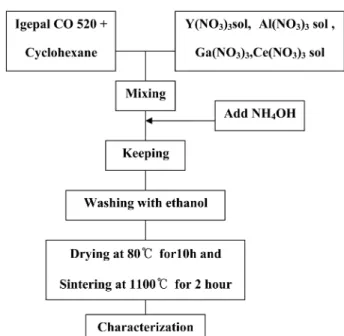
Gallium nitrate, Ga (99.9 %, Sigma-Aldrich Chemical Co., LTD.); cerium nitrate, Ce(NO 3 ) 3 ·6H 2 O (98 %, Yakuri pure chemicals Co., LTD.); Igepal[poly (oxyethylene) nonylphenly (Igepal CO-520, Aldrich Chemical Co.); Cyclohexane (Sigma-Aldrich, HPLC grades); NH 4 OH (28 % Daejung chemicals & metals Co., LTD), respectively. Fig. 1. illus- trates the experimental procedure of the synthesized YAG powders by a reverse micelle process. The amount of 20.05 g of Igepal CO-520 was mixed with about 50 ml of cyclohexane by stirring for 3 h at room temperature.
Yttrium nitrate(3 M) was added with continuous stirring.
Aluminum nitrate(1 M) sol and gallium nitrate(1 M) sol was dissolved in 10 ml[rate Al 5-x Ga x ( x = 0-5) mol] of distilled water by stirring for 2 h at room temperature.
For preparing doped samples, the corresponding quantity of cerium nitrate(0.06 mol%) was added to the solution.
Both solutions were mixed with 3.25-6.5 ml aqueous solution. NH 4 OH 3.5 ml was injected into the micro- emulsion to accelerate the hydroxylation reaction of metal ions. Which were subsequently washed by ethanol to remove any residual surfactant and water and dried at 80 o C. The dried powder were then calcined at 1100 o C for 2 h. with a heating rate of 5 o C/min. The crystal structures of powders were characterized by using X-ray diffraction(XRD, X’pert, MPD, Philips, Nedelland) in the 2 θ range of 10-80 o , with a counting time of 1 s for each step size of 0.04 o . Scanning electron microscopy(SEM) images were taken on a JEOL JSM-5610 field emission scanning electron microscope. The luminescence spectra of Ce 3+ substituted for Y 3+ in YAG have been measured on samples made-up at different Y 3 Al 5-x Ga x O 12 :Ce 3+ ( x = 0-5) rate. The synthesized powder was pressed into a pellet for the absorption and the photoluminescence(PL) mea- surements. PL spectra were taken with a steady-state flu- orescence system with a 450-W Xe lamp. The excitation light from the Xe lamp(SHIMADZU, RF-5301PC).
3. Results and Discussion
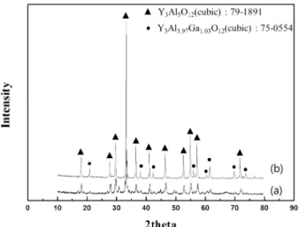
Fig. 2 shows the XRD patterns of Y 3 Al 5 O 12 :Ce 3+ and
Y 3 Al 3.97 Ga 1.03 O 12 :Ce 3+ calcined at 1100 o C for 2 h. The
diffraction pattern of the sample showed that all the
peaks of the Y 3 Al 5 O 12 and Y 3 Al 3.97 Ga 1.03 O 12 phase, which
Fig. 1. Experimental procedure for the synthesis YAG powder by
a reverse micelle process.
Photoluminescence of Y3(Al, Ga)5O12:Ce3+ Nanoparticles by a Reverse Micelle Process 33
is in good agreement with JCPDS Card (no. 79-1891) and (no. 75-0554), respectively. The luminescence properties of phosphor particles depend on the morphology of the particles such as size, size distribution, shape and defects.
Fig. 3 shows scanning electron micrographs of the syn- thesized Y 3 Al 5 O 12 :Ce 3+ and Y 3 Al 3.97 Ga 1.03 O 12 :Ce 3+ calcined at 1100 o C for 2 h. The average particles size of the syn- thesized powders by measuring linear intercept method
was below 1 µm. Fig. 4 shows the excitation spectra for the Ce 3+ emission 475 nm in the YAG phosphors. Each excitation spectrum of Y 3 Al 5 O 12 :Ce 3+ and Y 3 Al 3.97 Ga 1.03 O 12 : Ce 3+ were around 475 nm and 485 nm, respectively, which corresponds to the electronic transitions from the ground state of Ce 3+ to the crystal field splitting bands of excited state of Ce 3+ ion.
The emission of Ce 3+ is ascribed to the electron tran- sitions from the lowest crystal-splitting component of 5D 1 level to the ground state of Ce 3+ (2F 5/2 , 2F 7/2 ). Modifi- cation of the YAG chemical composition with foreign ions is a common strategy for tailoring the Ce 3+ 5d-4f emission. 9,20) The excitation of the luminescence of Ce 3+
is done by laser, and trivalent cerium is excited to a 5d state. 19) In the first case, the excited (Ce 3+ )* ion emits a 5d-4f scintillation photon hv
via(Ce 3+ )* → Ce 3+ + hv s (1)
Ce 3+ may also lose its 4f electron to form Ce 4+ either by direct ionization or by capturing a hole created in the valence states of the anions. Usually, the ionization holes are sequentially trapped on the luminescent ions, as in- dicated below, because Ce 3+ ions act as hole traps in the present materials.
Ce 3+ + h → Ce 4+ (2)
The process may be prompt if the hole is nearby or delayed if the hole diffuses to the vicinity of cerium site.
The resulting Ce 4+ may then capture an electron,
Ce 4+ + e → (Ce 3+ )* (3)
and subsequently decay by the process in eq. 3.
Ce 3+ emission in a YAG host can be tuned by partially Fig. 4. Photoluminescence excitation spectra of the synthesized powders; (a) Y
3Al
5O
12:Ce
3+and (b) Y
3Al
3.97Ga
1.03O
12:Ce
3+. Fig. 2. X-ray diffraction of synthesized YAG; (a) Y
3Al
5O
12:Ce
3+and
(b) Y
3Al
3.97Ga
1.03O
12:Ce
3+.
Fig. 3. SEM micrographs of YAG:Ce
3+particles calcined at 1100
oC
for 2 h; (a) Y
3Al
5O
12:Ce
3+and (b) Y
3Al
3.97Ga
1.03O
12:Ce
3+.
34 Min Yeong Kim and Dong-Sik Bae
or entirely substituting Al 3+ site with different types of cation respectively, substitution of Al 3+ with larger Ga 3+
ion results in blue-shifted Ce emission at the expense of emission efficiency. 20) Fig. 5 shows the emission spectra of nano-scale powders of Y 3 Al 5-x Ga x O 12 :Ce 3+ prepared at 1100 o C for 2 h in air with different (x = 0, x = 1.03) con- centrations. The emission spectra of the Y 3 Al 5 O 12 and Y 3 Al 3.97 Ga 1.03 O 12 were around 560 nm and 545 nm, re- spectively. It is clear from the figure that the emission intensity decreased from without Ga to with 1.03 Ga.
4. Conclusions
We studied the optical properties of Ce 3+ doped Y 3 (Al, Ga) 5 O 12 to understand the principle of phosphors active under NUV excitation. We synthesized Y 3 (Al, Ga) 5 O 12 : Ce 3+ phosphors by reverse micelle process. The structure and the basic properties of the phosphors were charac- terized. The diffraction pattern of the sample showed that all the peaks of the Y 3 Al 5 O 12 and Y 3 Al 3.97 Ga 1.03 O 12 phase, which is in good agreement with JCPDS Card (no. 79- 1891) and (no. 75-0554), respectively. The average size of the synthesized Y 3 (Al, Ga) 5 O 12 :Ce 3+ powders was below 1 µm. The optical properties were studied by taking excitation and emission spectra at room temperature. Ex- citation spectrum of the Y 3 Al 5 O 12 and Y 3 Al 3.97 Ga 1.03 O 12 were around 475 nm and 485 nm, respectively. The emis- sion spectra of the Y 3 Al 5 O 12 and Y 3 Al 3.97 Ga 1.03 O 12 were around 560 nm and 545 nm, respectively.
Acknowledgements
This study was financially supported by a Research Grant(2012-0071-0000) from the National Research Foun- dation, Korea.
References
1. H. S. Jang, W. B. Im, D. C. Lee, D. Y. Jeon and S. S.
Kim, J. Lumin., 126, 371 (2007).
2. K. Y. Jung and H. W. Lee, J. Lumin., 126, 469 (2007).
3. S. Zhou, Z. Fu, J. Zhang and S. Zhang, J. Lumin., 118, 179 (2006).
4. Y. Zhou, J. Lin, M. Yu, S. Wang and H. Zhang, Mater.
Lett., 56, 628 (2002).
5. U. Kaufmann, M. Kunzer, K. Kohler, H. Obloh, W.
Pletschen, P. Schlotter, J. Wagner, A. Ellens, W. Rossner and M. Kobusch, Phys. Status Solidi A, 192, 246 (2002).
6. Y. H. Won, H. S. Jang, W. B. Im, D. Y. Jeon and J. S. Lee, Appl. Phys.Lett., 89, 231909 (2006).
7. P. Dorenbos, J. Lumin., 99, 283 (2002).
8. F. Euler and J. A. Bruce, Acta Crystallogr., 19, 971 (1965).
9. J. L. Wu, G. Gundiah and A. K. Cheetham, Chem. Phys.
Lett., 441, 250 (2007).
10. K. Ohno and T. Abe, J. Electrochem. Soc., 133, 638 (1986).
11. K. Ohno and T. Abe, J. Electrochem. Soc., 134, 2072 (1987).
12. J. G. Li, T. Ikegami, J. H. Lee, T. Mori and Y. Yajima, J.
Eur. Ceram. Soc., 20, 2395 (2000).
13. M. Yada, M. Ohya, M. Machida and T. Kima, Chem.
Commun., 18, 1941 (1998).
14. M. Veith, S. Mathur, A. Kareiva, M. Jilavi, M. Zimmer and V. Huch, J. Mater. Chem., 9, 3069 (1999).
15. P. Vaqueiro and M. A. Lopez-Quintela, J. Mater. Chem., 8, 161 (1998).
16. K. T. Pillai, R. V. Kamat, V. N. Vaidya and D. D. Sood, Mater. Chem. Phys., 44, 255 (1996).
17. N. J. Hess, G. D. Maupin, L. A. Chick, D. S. Sunberg, D.
E. McCreedy and T. R. Armstrong, J. Mater. Sci., 29, 1873 (1994).
18. M. Yoshida, M. Lal, N. D. Kumar and P. N. Prasad, J.
Mater. Sci., 32, 4047 (1997).
19. A. A. Bol and A. Meijerink, J. Phys. Chem. B, 105, 10203 (2001).
20. O. Milosevic, L. Mancic, M. E. Rabanal, J. M. Torralba, B. Yang and P. Townsend, J. Electrochem. Soc., 152, G707 (2005).