∙ Received: February 29, 2012. Accepted: April 6, 2012.
∙ Corresponding author : Sung-hwan Kim
Department of Nuclear Medicine. Asan Medical Center, 388-1 Pungnap 2-dong, Songpa-gu, Seoul 138-736, Korea Tel: +82-2-3010-4609, Fax: +82-2-3010-5429 E-mail: [email protected]
Original Article
Tl-201을 이용한 심근관류 SPECT에서 Bodycontour와 Circular mode의 영상 획득 차이에 따른 영상의 질 비교
서울 아산병원 핵의학과
김성환 ․ 남기표 ․ 류재광 ․ 윤순상
The Comparison of Image Quality Using Body Contour and Circular Method with L-mode in Myocardial Perfusion SPECT
Sung Hwan Kim, Ki Pyo Nam, Jae Kwang Ryu and Soon Sang Yoon Departmetn of Nuclear Medicine, Asan Medical Center, Seoul, Korea
Purpose : In myocardial perfusion SPECT, the type of orbit (circular vs. body contouring) that affect the image quality is still on the debate. Presently in the nuclear medicine field, the body contouring orbit acquisition is widely used to improve the image quality on the myocardial perfusion SPECT. But in case of body contouring acquisition using the vertical method with dual detect machine, there is a tendency of increasing the radius. In this research, we compared body contouring orbit acquisition with circular orbit acquisition, so we suggest ideal method that reduces the radius for improving image quality. Materials and Methods : Phantom and clinical studies were performed. The anthropomorphic torso phantom was made on equally with counts from patient’s body. The study was performed under six different conditions. To compare image quality according to the radius, we increased radius sequentially per step during circular orbit acquisition. On the other hand, sensors that protect a collision and reduce the radius automatically were used to acquire image during body contouring orbit acquisition. So we compared FWHM value of apex. In clinical studies, we analyzed the 40 patients who were examined by Tl-201 gated myocardial perfusion SPECT in department of nuclear medicine at Asan Medical Center in August 2011. To acknowledge the differences according to the radius, we acquired the results two times using circular orbit acquisition and body contouring orbit acquisition. Results : In phantom study, we analyzed that increase of radius resulted in changes of FWHM value. It was 5.41, 6.24, 6.33, 6.42, 6.93 mm. On the other hand, using the body contouring orbit acquisition, FWHM value was 6.23 mm. In clinical study, difference of average radius between two methods was 2.5 cm (circular orbit acquisition was more close to patients).
Conclusion : Through the experiments using Anthropomorphic torso phantom and patients data, we found that FWHM value of circular orbit acquisition was lower than body contouring orbit acquisition. As a result, if the difference of average radius exists approximately 3 cm, circular orbit type acquisition is better than body contouring type acquisition. But clinical investigation is only aimed to average radius, so it needs more investigation in comparison of patient’s image. (Korean J Nucl Med Technol 2012;16(1):3-7)
Key Words : Circular orbit acquisition, Body contouring orbit acquisition, Myocardial perfusion SPECT.
INTRODUCTION
Cardiovascular disease is the main reason of death in South Korea. It also resents an enormous societal burden with re- spect to morbidity, health care expense, and personal hardship. With its unique ability to evaluate perfusion at the
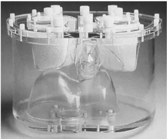
Fig. 1. Anthropomorphic torso phantom.
Table 1. Anthropomorphic torso phantom 1) Lateral outside dimension : 38 cm 2) Lateral inside dimension : 36 cm
3) Anterior-posterior outside dimension : 26 cm 4) Anterior-posterior inside dimension : 24 cm 5) Wall thickness : 9.5 mm
6) Liver volume : 1.2 liters 7) Background : 10.3 liters cellular level, myocardial perfusion SPECT (MPS) plays an
important role in diagnosing cardiovascular disease, estab- lishing prognosis, assessing the effectiveness of therapy, and evaluating viability.1) While myocardial perfusion imaging is a well established clinical procedure, one of the technical as- pects of this procedure, such as the type of orbit (circular vs.
body contouring) is still on the debate.2)
According to an academic research, B-mode, which is complimentary to the C-mode, increases the spatial resolution.
However, a lot of nuclear medicine technicians already knew that Tl-201 has more artifact than TC-99m, besides B-mode injected with Tl-201 resulted in lower quality image than Tc-99m.3,5)
In Asan Medical center, MPS injected with Tl-201 using the Infinia (General Electric Healthcare, Waukeshau, WI) has proceed B-mode with 90 degree angled double detectors, but many patients who had frozen shoulder or severe pain due to preoperative condition were difficult to make a ad- equate position. For this reason, the radius between patient and detectors increased unintentionally. Increased radius af- fects the spatial resolution on the image so that images can be misinterpreted improperly.
The purpose of this study was to examine the impact of changes in resolution according to the type of orbit (C-mode vs. B-mode) more closely, and then we suggest suitable meth- od for using in clinical practice.
MATERIALS AND METHODS
1. Phantom study
We compared image quality by using anthropomorphic torso phantom (Fig. 1, Table 1). We made a phantom on an equally with counts from patient’s body. The Tl-201 concen- trations in the compartments were 74 kBq (2 μCi)/cc in my- ocardium, 11.1 kBq (0.3 μCi)/cc in soft tissue, and 2.59 kBq (0.07 μCi)/cc in lung. The non-gated Tl-201 MPS data were acquired with the phantom. The phantom was positioned such that the collimator to phantom separation was at a mini- mum at 0 degrees.
C-mode was performed with both detectors heads set at radius of 18.8, 21.8, 23.8, 25.8, 28.8 cm respectively.
B-mode was performed with sensors to recognize the min- imum radius between patient and detectors per each step.
The study was scanned for 50 seconds per frame in 64 × 64 matrix. A total of 30 views were acquired over 180 degree (90 degree rotation/head) using a step and shoot acquisition.
Using the Xeleris ver. 2.0551, full width at half maximum (FWHM) value was compared (Fig. 3, 4). To get the FWHM value, first, set frame. Second, profile ROI. All the re- constructed conditions set equally. And lastly, we obtained the FWHM.
2. Clinical study
In clinical studies, 40 patients who had coronary artery diseases or preoperative patients were examined by Tl-201 MPS in department of nuclear medicine at Asan Medical Center from 9 to 23, August, 2011. Patients were in the 32-90 age range (mean = 61.9±11.0). All patients were examined two times at rest, using C-mode and B-mode. The patients were imaged at full time (50 second per frame) during
Fig. 2. Infinia (General Electric Healthcare, Waukeshau, WI).
Fig. 3. Profiling ROI at apex. Fig. 4. Graph of counts per pixels at apex.
C-mode. When we acquired images using B-mode, we had to reduce the scan time from 50 sec to 10 sec per each step. We only checked distance. Not image quality. And we averaged the radius each step.
3. Study protocol and image processing
For this study, Infinia (General Electric Healthcare, Waukeshau, WI) 90 degree angled double head detectors equipped with general-purpose collimator was used for all
SPECT imaging (Fig. 2). To acquire MPS images, we used dual energy window. A ±15% and a ±10% window were centered over the 70 keV and 167 keV Tl-201 photopeaks. An angular step of degree is 6. A matrix size was 64 × 64. And we used FBP reconstruction method with Butterworth filter.
C-mode conditions ; 180 degree arc, 15 stops (30 views), and 50 second-per stop, step-and-shoot m node. 8 frame per car- diac cycle gating, 100±50% irregular beat acceptance window. B-mode condition ; 180 degree arc, 15 stops (30 views), and 10 second-per stop, step-and-shoot mode. 8 frame per cardiac cycle gating, 100±50% irregular beat ac- ceptance window.
Results
Through the phantom result, images of increased radius gradually showed that more blurring artifacts were seen. In clinical result, we found that there were a little differences of radius from center of rotation. Approximately 2.5 cm (C-mode is more closer to patient’s body than B-mode).
1. Phantom study
The reconstructed vertical-axis images of anthro- pomorphic phantom employing C-mode with different radi- us showed visually homogeneous images.4) But the image
Table 2. Anthropomorphic torso phantom
Body contouring Circular + 0cm Circular + 3cm Circular + 5cm Circular + 7cm Circular + 10cm
FWHM 6.23 5.41 6.24 6.33 6.42 6.93
quality between two methods was clearly exists. When C-mode set at radius of 18.8, 21.8, 23.8, 25.8, 28.8 cm re- spectively, FWHM value were 5.41, 6.24, 6.33, 6.42, 6.93 mm.
On the other hand, FWHM value of using B-mode was 6.23 mm. As a result of these studies, C-mode, which was set a ra- dius (18.8 cm) had lower FWHM than B-mode (Table 2).
2. Clinical study
40 patients were compared with two types of rotation method. 40 patient’s average radius with C-mode was 19.54 cm. On the other hand, average radius with B-mode was 22.04 cm. The difference between two methods was 2.5 cm.
This figure indicated that B-mode was far away from patient than C-mode.
Discussion
According to the results of this study, using C-mode has higher image quality than B-mode. However, Infinia (General Electric Healthcare, Waukeshau, WI) has no auto- matic vertical movement of the table function. Also 90 degree angled double head detectors can easily affected by patient’s condition, so if B-mode is used, radius will significantly change.5) These technical and physical aspects are likely to make a possibility of variable results. Lastly, we couldn’t get a clinical image with B-mode, so we didn’t compare the ac- tual image quality.
Conclusions
Through phantom study results, we found that the C-mode with minimum radius had lower FWHM value than B-mode. When B-mode was performed, FWHM value is similar to circular +3 cm’s value, which set more far away 3 cm from minimum radius.
From clinical study results, we also found that the two types of method had difference of average radius. That is ap-
proximately 2.5 cm (C-mode was more close to patients).
Radius of approximately 3 cm causes changes in the FWHM value as mentioned previously.
Therefore C-mode is more suitable method if more than 3 cm difference of radius exists during MPS with B-mode.
However, we need to take an actual test for many patients with its analysis of quantitative indices.
요 약
심근관류 SPECT의 영상획득 방법에는 Body contour 와 Circular 방식이 있고, 환자와 검출기 사이의 거리를 줄이기 위해 Body contour 방식을 주로 사용한다. 그러나 이중(Dual) 검출기 장비에서 수직방식으로 검출기를 설 정하여 영상을 획득하는 경우에는 환자와 검출기 사이의 거리가 Body contour 방식이 Circular 방식에 비해 더 증 가하는 경향이 있다. 본 연구의 목적은 이중 검출기 장비 에서 수직방식으로 검출기를 설정하여 영상을 획득하는 경우, Body contour 방식과 Circular 방식에서 환자와 검 출기 사이의 거리를 비교하고, 거리에 따라 영상에 미치는 영향을 파악하여 영상의 질을 높이기 위한 방안을 제시하 고자 시행하였다. 실험에 사용된 장비는 GE INFINIA Gamma Camera와 Anthropomorphic torso 모형에 심장 모형을 삽입하여 영상을 획득하였고, 2011년 8월 본원에 서 심근관류 SPECT를 시행한 환자 40명을 대상으로 하 였다. 모형 제작은 실제 환자조건과 동일하게 하기 위하여 심근에 74 kBq (0.22 mCi)/cc, 폐에 2.59 kBq (60 μCi)/cc, 연부조직에 11.1 kBq (2.2 mCi)/cc의 201-Tl을 주입하여 제작하였고, 영상획득 조건은 회전각 6˚, 50 sec/frame으로 환자와 동일한 조건으로 하였다. 영상획득 방식에 따른 차이를 확인하기 위해 Body contour와 Circular 방식으로 영상을 획득하여 환자와 검출기 사이의 거리를 각각 측정 하여 비교하였고, 정상군과 비 정상군의 영상을 육안 분 석하였다. 거리에 따라 영상에 미치는 영향을 확인하기 위해, 모형 실험에서 Circular 방식으로 회전반경을 18.8, 21.8, 23.8, 25.8, 28.8 cm 로 다르게 설정하여 영상을 획득 하였고, 각 영상에서 심첨(Apex)에 관심영역을 설정하여 반치폭을 비교하였다.
모형 실험 결과, Body contour를 이용한 실험에서 산출된 반치폭은 6.23 mm이고, Circular mode에서 18.8, 21.8, 23.8, 25.8, 28.8 cm의 회전반경으로 산출된 반치폭은 각각 5.41, 6.24, 6.33, 6.42, 6.93 mm이다. 환자 데이터에서 Body con- tour의 회전반경이 -3.5 cm ~ 9.5 cm으로 차이를 보였고, Circular mode에 비해 평균 2.5 cm 더 큰 차이를 보였다. 본 연구를 통해, INFINIA 장비에서 Circular mode가 Body contour를 사용한 영상 획득 방법보다 낮은 반치폭을 보였 다. 비록 INFINIA장비의 두 가지 영상 획득 방식에 대한 비 교였지만, INFINIA장비를 이용한 심근관류 검사에서는 Circular mode 의 영상 획득 방식이 더 적합하다고 사료된다.
REFERENCES
1. Steven Burrell, MD, and Anita MacDonald, BHSc. Artifacts
and pitfalls in myocardial perfusion imaging. J Nucl Med Technol 2006;34:193-211
2. Michael K. O’Connor and Carrie B. Hruska. Effect of tomo- graphic orbit and type of rotation on apparent myocardial activity. Nucl Med Commun 2005;26:25-30.
3. Michael P. White, April Russell, Victor A. Mascitelli, Robert S.
Morris, Adel R. Shehata and Gary V. Heller. Clinical compar- ison of circular versus noncircular acquisition using techne- tium-99m myocardial perfusion SPET imaging. J Nucl Med Technol 1997;25:37-40.
4. Piorre J. Maniawski, Hugh T. Morgan, and Frans J. Th.
Wackers. Orbit-related variation in spatial resolution as source of artifactual defects in thallium-201 SPECT J Nucl Med 1991;32:871-875
5. Oktay Yapici MD, Tarik Basoglu MD, Sibel Ucak MD, Meltem Aydin MD. Auto-contouring at 90° dual head fitting angle:A potential cause of a myocardial perfusion SPECT artifact in slim patients. Hell J Nucl Med 2009;12(3):289-290.