• 검색 결과가 없습니다.

Study on the Correlation Between Physical Function and Forward Head Posture in Spastic Diplegia

N/A
N/A
Protected

Academic year: 2021

Share "Study on the Correlation Between Physical Function and Forward Head Posture in Spastic Diplegia"

Copied!
10
0
0

로드 중.... (전체 텍스트 보기)

전체 글

(1)

| Abstract |

Purpose: This study investigated the correlation between physical function and forward head posture in spastic diplegia.

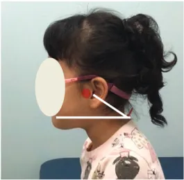
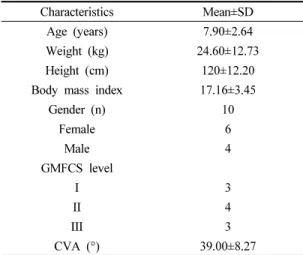
Methods: The subjects of this study were 10 spastic diplegia patients. We took pictures of the subjects’ craniovertebral angle with a digital camera to determine the degree of forward head posture and then analyzed them using the NIH image J program.

The physical function test used the TCMS, the BBT, and a spirometer. The data in this study were measured using SPSS version 23.0, and the statistical significance level α was 0.05. A Pearson correlation coefficient analysis was performed to identify the correlation between the degree of the subject’s head forward position and physical function.

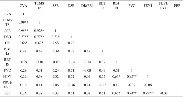
Results: When we performed the BBT and spirometer tests, the subjects’ forward head postures were not correlated (p < 0.05).

However, with the TCMS, there was a strong correlation between the forward position of the head and balance, with balance decreasing as the head position increased (p < 0.05).

Conclusion: Spastic diplegia patients with severe forward head posture showed problems with static balance, dynamic balance, and equilibrium reaction when sitting. Intervention on the right posture and preventive activities will be needed to improve the health of spastic diplegia patients and prevent future problems with physical function.

Key Words: Forward head posture, Sitting balance, Physical function, Spastic diplegia

†Corresponding Author : Eun-Ju Lee ([email protected])

Original Article Open Access

경직형 양하지 뇌성마비 아동의 전방머리자세와 신체기능간의 상관관계

조영은⋅이은주

2)

경성대학교 물리치료학과 재활과학 연구소

Study on the Correlation Between Physical Function and Forward Head Posture in Spastic Diplegia

Yong-Eun Jo, P.T., M.S.⋅Eun-Ju Lee, P.T., Ph.D.

Department of Physical Therapy and Institute for Rehabilitation Science, Kyungsung University

Received: April 13, 2021 / Revised: May 29, 2021 / Accepted: July 5, 2021

ⓒ 2021 Journal of Korea Proprioceptive Neuromuscular Facilitation Association

This is an Open Access article distributed under the terms of the Creative Commons Attribution Non-Commercial License

(http://creativecommons.org/licenses/by-nc/3.0) which permits unrestricted non-commercial use, distribution, and reproduction

in any medium, provided the original work is properly cited.

(2)

Ⅰ. 서 론

정상 만삭 아동은 태내 발달과정 중 한정된 자궁 안에서 머리를 여러 방향으로 돌리며 팔을 움직이고 발로 자궁벽을 차는 등 다양한 움직임의 경험을 통하 여 머리와 체간의 연결성과 안정성을 발달시킨다. 그 러나 양하지 뇌성마비의 주요 원인이 되기도 하는 미 숙아는 이런 과정을 충분히 경험하지 못하고 태어나 머리와 체간의 연결성이 부족하며 지연된 목 정위반 응과 제한된 움직임으로 인하여 머리 안정성이 더욱 떨어지게 된다(Hong, 2011; Ryu, 2015). 머리의 안정성 이 부족한 뇌성마비 아동은 이후 항중력 자세조절시 머리의 과도한 뒤로 젖힘(capital hyper extension)이 나 타나게 되는데 이는 추후 골격 정렬의 불균형을 야기 시키고 전방머리자세를 유발하여 일상생활에서 긴목 근(longus colli muscle)과 긴머리근(longus capitis muscle) 같은 깊은목굽힘근을 잘 사용하지 않게 만들 고 목 굽힘 근육의 두께 변화를 초래하게 된다(Ishida et al., 2015).

머리와 목 근육의 잘못된 발달은 아이의 발달 방향 원칙 중 머리에서 꼬리쪽으로 발달한다는 원칙에 따 라 등뼈와 허리뼈 및 신체 다른 부위의 근육 형성에도 영향을 주게 되어 체간이 앞쪽으로 굴곡되며 체간을 곧게 세우지 못하게 되는 구부정한 앉은 자세 (crouched sitting)를 취하게 된다(Yun, 2003). 구부정하 게 앉은 자세는 배가로근의 두께가 감소되는 등 체간 근력의 약화로 인해 균형의 저하를 발생시키고 향후 뇌성마비 아동이 자라 성년이 될 수록 관절의 구축 및 근육의 단축⋅위축, 골 밀도의 감소와 같은 또 다른 근골격계의 문제를 일으킨다(Yun & Kim, 2014). 근골 격계의 변형으로 인해 발생될 수 있는 통증은 뇌성마 비 아동의 일상활동과 사회생활 참여에 제약을 일으 키며 뇌성마비 삶의 질을 저하시킨다(Harbourne et al., 2010; Schwartz et al., 1999).

뇌성마비 아동의 전방머리자세와 구부정하게 앉은 자세는 양쪽 궁둥뼈결절(ischial tuberosities)의 체중지 지가 아니라 엉치뼈(sacrum)쪽으로 체중지지를 하게

하여 체중 기저면을 좁게 만들고 안정성과 균형을 더 욱 저하시켜 아동을 불안하게 만든다. 뇌성마비 아동 은 저하된 안정성과 균형을 보상하기 위해 어깨는 더 욱 앞으로 움츠리게 되며 손은 기능적인 조작 (manipulation)에 사용하지 못하고 지지면을 단순 고정 하며 체간을 보호하는데 주로 사용하게 된다. 균형을 보상하기 위한 손의 고정과 지지는 뇌성마비 아동의 일상활동에서 탐색과 놀이의 제한으로 이어진다 (Bobath, 1985; Brogren et al., 2001; Case-Smith &

O’brien, 2004).

전방머리자세가 있는 뇌성마비 아동들은 약화된 깊은목굽힘근 대신 목부위 얕은 근육인 목빗근과 위 등세모근을 과활성 시키는 경향이 있어 호흡에 필요 한 기도공간이 감소될 수 있다(Hruska, 1997; Jull et al., 2004; Kang & Oh., 2019). 또한, 전방머리자세로 인한 뇌성마비 아동들의 머리, 목, 혀의 비정상적이고 불충분한 근육의 발달은 흡기시 가슴우리를 안정시키 는데 필요한 근육 발달까지도 억제하여 가슴우리의 움직임을 제한시키고 불규칙하고 거친 호흡조절을 일 으키며 폐질환을 야기하기도 한다(Mcdonald et al., 1987; Wang, 2012).

이와 같이 뇌성마비 아동의 전방머리자세는 아동 의 다양한 신체 부위에 영향을 미치고 기능을 저하시 킬 수 있음에도 불구하고 뇌성마비 아동의 전방머리 자세와 신체기능의 관계를 복합적으로 다룬 연구는 현재 부족한 실정이다. 따라서 본 연구에서는 뇌성마 비 아동의 전방머리자세와 다양한 신체기능간의 관련 성을 알아보아 뇌성마비 건강관리 프로그램에 활용될 수 있는 기초 자료를 제시하고자 한다.

Ⅱ. 연구 방법 1. 연구 대상

본 연구는 B시의 M병원에 내원하는 아동 중 경직성

양하지 마비 진단을 받은 5세∼13세 의 아동 10명을

(3)

대상으로 하였다. 모든 연구 대상자 및 법적 보호자는 연구의 절차 및 목적에 대해 동의하고 자발적으로 본 연구에 참여하였다. 대상자 선정기준은 대동작 기능 분류 시스템(gross motor function classification system, GMFCS)에서 1∼3단계로 분류되어 독립적 앉기가 가 능하고 수정판 애쉬워스 경직 척도(modified Ashworth scale, MAS)에서 하지 경직이 G2 이하이며 시각적 장 애 또는 시야 결손, 청각에 문제가 없고 소아 의식 척도(rancho los amigos) 8단계에 해당되어 자유롭게 의사소통이 가능한 아동으로 하였다. 그리고 최근 6개 월 이내 경기(seizure)를 하거나 정형 외과적 수술 또는 보툴리눔 독소 주사 치료를 받은 아동은 대상자 선정 에서 제외하였다. 본 연구는 B시 K대학 임상연구윤리 위원회(institutional review board, IRB)로부터 연구 승 인을 받았다(KSU-19-08-004).

2. 측정 방법 및 도구 1) 머리척추각도 측정

뇌성마비 아동의 앉은 자세의 머리 정렬을 알아보 기 위하여 이미지 J 분석프로그램(Image J ver.1.47, Sun Microsystems, Inc, USA)을 사용하여 머리척추각 (cranio vertebral angle, CVA)을 분석하였다. 이미지 J 분석프로그램은 일반적인 이미지 작업뿐만 아니라 이 미지상의 거리, 각도 면적, 픽셀 값, 공간적 측정, 농도 등을 통계적으로 분석할 수 있는 프로그램으로 측정 자 간 신뢰도는 r=0.97이며, 측정자 내 신뢰도는 r=0.88∼

0.98이다(Amaro et al., 2007; Koppenhaver et al., 2009;

Girish & Vijayalakshmi, 2004; Raine & Twomey, 1997).

머리척추각을 분석하기 위한 사진 촬영은 대상자 와 3m 떨어져 카메라를 바닥으로부터 1.2m 높이에 위치시켜 시행하였다(Junior, 2010). 촬영 시 귀의 이주 와 목뼈 7번 가시돌기에 표식자를 부착하였고 대상자 의 자세는 등받이가 있는 의자에 엉덩이를 붙여 편안 하게 착석하게 하였고 발바닥이 지면에 닿는 상태에 서 엉덩관절과 무릎관절의 각도를 90°로 만들었으며 손은 무릎 위에 자연스럽게 놓았다(Kapreli et al.,

2009). 자가 균형위치(self-balance posture, SBP)를 통해 자연스러운 머리자세(natural head posture, NHP)를 만 들었고 자세를 유지시키기 위해 대상자의 정면에 거 울을 두어 자신의 눈을 보도록 지시하였으며 측정 오 차를 최소화하기 위해 3회 반복 측정하여 평균값을 대표값으로 사용하였다(Chae, 2002; Chung 2019)(Fig.

1). 머리척추각도는 귀의 이주와 목뼈 7번 가시돌기를 연결한 선과 수평선이 이루는 각으로 정하고 계산하 였고 전방머리자세가 심할수록 머리척추각은 감소하 게 된다(Kapreli et al., 2009; Watson & Trott, 1993).

Fig. 1. Measurement of craniovertebral angle.

2) 앉은 자세 균형 측정

앉은 자세의 균형능력을 측정하기 위해서 체간조 절 평가 척도(trunk control measurement scale, TCMS)를 사용하였다. TCMS는 독립적인 앉기가 가능한 경직성 뇌성마비 아동의 앉은 자세 균형능력을 알아보는데 적합한 평가도구로, 총 15개의 항목으로 구성되어 있 다. 세부항목은 정적앉기 균형능력 5항목, 동적앉기 균형능력 7항목, 동적 팔뻗기(평형 반응) 3항목이며 최저 0점에서 최고 58점까지 점수를 산정하여 점수가 높을수록 체간의 조절 능력이 좋다는 것을 의미한다 (Verheyden et al., 2004).

측정은 양말, 보조기, 신발 등을 모두 벗은 상태에서

(4)

등받이, 팔 걸이 그리고 발 받침대가 없는 치료 테이블 에 걸터 앉은 채로 엉덩관절 90도, 무릎관절 90도를 유지하고 양손은 다리 위에 위치하도록 하여 왼쪽 측 면에서 3회 반복 측정하였으며 평균값을 대표값으로 사용하였다(Han, 2017). TCMS의 측정자 간 신뢰도는 r=0.98이며, 측정자 내 신뢰도는 r=0.97 이다(Heyrman et al., 2011).

3) 손 기능

손 기능은 나무와 나무토막 검사(Box & Block Test) 를 사용하였다. 나무와 나무토막 검사는 일상생활에 서 많이 사용되는 손의 기민성 및 조작능력을 검사하 기 위한 평가도구로 측정자 간 신뢰도는 오른손 r=1.00, 왼손 r=0.99 이다(Mathiowetz et al., 1985). 검사 방법은 한손으로 1분 동안 1인치 크기의 나무토막을 한쪽 상자 에서 다른 쪽 상자로 옮긴 후 각각의 손이 옮긴 블록의 개수를 점수로 한다. 본 연구에서는 총 3회 반복 측정하 였으며 평균값을 대표값으로 사용하였다.

4) 호흡능력

호흡능력을 알아보기 위해 폐활량 측정계 (Microdirect Microplus Spirometer, Medical Device Depot, England)로 단순 폐기능 검사를 시행하였다. 측 정된 세부 항목은 노력성 폐활량(forced vital capacity, FVC), 1초간 노력성 호기량(forced expiratory volume in one second, FEV1), 1초간 노력성 호기량의 노력성 폐활량에 대한 비율(forced expiratory ration, FEV1/FVC), 최대 호기 유속(peak expiratory flow, PEF) 이다. FVC는 제한성 환기장애의 유무를 의미하며, FEV1는 폐쇄성 환기장애의 정도, FEV1/FVC율은 폐 쇄성 환기장애의 유무를 알 수 있고, PEF는 기도폐쇄 유무와 기도폐쇄 손상 정도를 판단하는 임상지표로 활용된다(Kwon & Lee, 2013). 아동은 스스로 들이마실 수 있는 최대한 공기를 들이 마신 후 가능한 빠르고 강하게 내쉬는 공기의 양을 FVC으로, 최대한 공기를

들어 마신 다음 1초간 가능한 빠르고 강하게 내쉬는 공기의 양을 FEV1으로, PEF는 더 이상 호기량이 없을 때 까지 ‘빨리, 세게 또는 가능한 오랫동안’ 숨을 내쉬 도록 하였다. 아동이 입으로만 숨을 쉴 수 있도록 코마 개를 사용하였으며, 평상 시 호흡을 3회 정도 한 이후 3회 반복 측정하여 평균값을 대표값으로 사용하였다 (Reeve et al., 2009; Yun & Kim, 2014).

3. 자료 분석

대상자의 머리척추각도와 신체기능 간의 상호 관 련성을 알아 보기 위해 스피어만(Spearman) 상관관계 분석을 실시하였다. 통계 프로그램은 SPSS 23.0(IBM SPSS Inc., USA)을 사용하였고 유의수준(α)은 0.05로 설정하였다.

Ⅲ. 연구 결과 1. 연구 대상자의 일반적인 특성

본 연구에 참여한 대상자 10명의 일반적인 특성은 다음과 같다. 평균 연령은 7.90±(2.64)세, 평균 체중은

Characteristics Mean±SD Age (years) 7.90±2.64 Weight (kg) 24.60±12.73 Height (cm) 120±12.20 Body mass index 17.16±3.45

Gender (n) 10

Female 6

Male 4

GMFCS level

Ⅰ 3

Ⅱ 4

Ⅲ 3

CVA (°) 39.00±8.27

GMFCS: gross motor function classification system, CVA:

craniovertebral angle

Table 1. General characteristics of subjects (n=10)

(5)

24.60±(12.73), 평균키는 120±(12.20), 체질량 지수는 17.16±(3.45), 여아(6), 남아(4), 동작 기능 분류체계는 1단계 3명, 2단계 4명, 3단계 3명이었고, 평균 머리척추 각은 39±(8.27)이었다(Table.1).

2. 머리척추각도와 균형, 손 기능, 호흡능력과의 상 관관계

대상자들의 머리척추각과 앉은자세에서의 균형능 력, 손 기능, 호흡능력 간의 상관관계 분석 결과는 다음 과 같다(Table 2). 머리척추각은 정적 앉기 균형능력 (r=0.95), 동적 앉기 균형능력(r=0.77), 동적 팔뻗기 (r=0.66) 등 앉은 자세에서의 체간 조절능력(r=0.99)과 뚜렷하고 강한 양의 상관관계를 보였다(p<0.05). 그러 나 왼쪽 손 기능(r=0.48)과 오른쪽 손 기능(r=-0.09), 호흡능력을 나타내는 FVC (r=0.29), FEV1 (r=0.36), FEV1/FVE (r=0.10), PEF (r=0.36)와 머리척추각은 상관 관계를 보이지 않았다.

Ⅳ. 고 찰

본 연구는 양하지 뇌성마비 아동의 전방머리자세 가 신체 기능과 어떤 상관성이 있는지를 알아보는 연 구이다. 연구 결과 양하지 뇌성마비 아동의 전방머리 자세 정도와 손 기능, 호흡 능력은 상관성이 없었지만 균형능력에는 강한 상관관계가 나타나 뇌성마비 전방 머리자세가 증가할수록 균형능력은 감소되는 경향을 보였다.

균형은 외부의 환경이나 스스로 신체의 이동이 나 타날 때 기저면 안에서 신체의 무게 중심을 유지하고, 자세를 지속적으로 유지할 수 있는 능력이다(Tyson et al., 2006). 균형 조절을 위해 사용되는 감각으로는 전정 감각, 시각, 고유수용성 감각이 있고 이러한 요소 중 시각은 특히 머리의 안정성에 영향을 받는다(cheng et al., 2001). 머리와 목은 신체가 주변환경에 대한 기준 을 설정하고 자세를 조절할 때 시각계, 안뜰계를 위한 안정된 기저면을 제공하게 된다(Keshner et al., 1988).

CVA TCMS

TS SSB DSB DR(ER) BBT

Lt BBT

Rt FVC FEV1 FEV1/

FVC PEF

CVA 1

TCMS TS 0.99** 1

SSB 0.95** 0.92** 1

DSB 0.77** 0.77** 0.73* 1

DR 0.66* 0.67* 0.580.22 1

BBT

Lt 0.480.49 0.39 0.32 0.49 1

BBT Rt -0.09 -0.10 -0.10 -0.14 -0.14 0.37 1

FVC 0.29 0.31 0.24 0.61 -0.080.480.51 1

FEV1 0.36 0.380.32 0.52 0.01 0.53 0.63* 0.95** 1

FEV1/

FVC 0.10 0.11 0.06 -0.36 0.24 -0.12 0.12 -0.32 -0.06 1

PEF 0.36 0.380.33 0.51 0.02 0.51 0.65* 0.94** 0.99** -0.06 1

CVA: craniovertebral angle, TCMS: trunk control measurement scale, TS: total scole, SSB: static sitting balance, DSB:

dynamic sitting balance, DR(ER): dynamic reaching(equilibrium reaction), BBT : box & block test, FVC: forced vital capacity, FEV1: forced expiratory volume in one second, FEV1/FVC: forced expiratory ration, PEF : peak expiratory flow

*p<0.05, **p<0.01

Table 2. The correlation between craniovertebral angle and body function (n=10)

(6)

뇌성마비 아동도 머리 안정성의 정도에 따라 시각 정 보와 몸통 조절, 균형능력이 달라 질 수 있는데 머리와 목의 불안정성이 자세 조절에 기여하는 시각과 안뜰 감각 조절에 부정확성을 야기하여 뇌성마비 아동으로 하여금 환경 변화에 대한 적응력을 감소시켜 공간에 서의 신체 무게 중심 유지를 어렵게 만들 수 있다 (Saavedra, 2010; Shin, 2016). 본 연구에서도 머리 안정 성이 부족하여 전방머리자세 정도가 심한 뇌성마비 아동일수록 앉은 자세에서 실시하는 체간 조절 평가 에서 정적 균형능력, 동적 균형능력, 동적 팔 뻗기(평 형반응) 능력이 다 저하되는 경향이 나타나 위의 선행 연구 결과와 일치됨을 확인 하였다.

전방머리자세는 상부 등뼈의 뒤굽음 증가시키고, 어깨뼈의 벌림, 내밈, 그리고 위쪽 돌림을 유발하며, 어깨관절의 안쪽돌림을 증가시킨다. 이러한 어깨의 자세 변화로 인한 위등세모근의 활성 변화는 어깨 근 육에 과도한 긴장을 가져와 어깨관절의 가동 범위에 제한을 주며 상지 기능에 부정적인 영향을 미친다 (Kleine, 1999). Kebaetse 등(1999)은 전방머리자세와 같이 잘못된 목과 머리의 정렬은 목 후면의 근육을 짧게 하고 목 전면 근육의 과도한 긴장을 일으켜 어깨 뼈의 위치와 움직임에 부정적인 영향을 미친다 하였 다. 경직형 뇌성마비 아동의 전방머리자세와 함께 나 타나는 구부정한 척추의 형태와 과도하게 긴장된 어 깨는 아동에게 자세 변화에 대한 심리적 두려움을 크 게 느끼게 하며 아동은 부족한 균형능력을 보상하기 위해 목과 어깨를 더 움츠리고 원위부의 근긴장도를 증가시킨다. 원위부의 근긴장도가 높은 뇌성마비 아 동은 일상생활 활동에서 기능적 조작을 위한 양손 사 용과 손의 발달이 지연되게 된다(Bly, 1983; Bobath

& Bobath, 1975; Bobath 1985; Brogren et al. 2001). 본 연구에서 뇌성마비 아동의 상지 기능은 전방머리자세 가 심할수록 동적 팔뻗기 능력은 제한이 있었지만 손 의 기민성 및 조작 능력을 알아보기 위한 나무와 나무 토막 검사에서는 제한이 없었다. 이는 동적 팔뻗기 검사 항목은 신체중심선을 지나치거나 체중지지 기저 면을 벗어나며 상지를 뻗게 하는 동작을 통해 상지

고유의 기능보다는 평형 반응을 알아보는 검사이고, 나무와 나무토막 검사는 큰 자세변화 요구 없이 안정 된 지지면 안에서 한 상자에서 다른 상자로 나무토막 을 옮기는 소동작 활동을 통해 손의 기민성과 조작 능력을 알아보는 검사라 전방머리자세 정도에 따른 손의 기능을 나무와 나무토막 검사가 민감하게 반영 하지 못했던 것으로 사료된다(Backman et al., 1991;

Lee & Jung, 2002; Trombly et al., 2001).

신체 자세 정렬은 호흡능력 및 호흡 근력 발달에 매우 큰 영향을 준다(Yun & Kim, 2014; Yun, 2016).

정상적인 호흡을 위해서는 안정적인 자세와 척추 정 렬이 선행되어야 한다(Perri & Halford, 2004). 전방머 리자세는 목의 정렬 이상과 등뼈의 뒤굽음 증가를 일 으켜 호흡보조근인 목빗근과 목갈비근의 과활성과 단 축을 유발하며 가슴우리 움직임에 장애를 일으키고, 폐 확장 능력과 폐용적(lung volume), 폐활량(vistal capacity)을 감소시켜 호흡기능 장애를 초래할 수 있다 (Troyer et al., 1985). 양하지 뇌성마비 아동의 호흡능력 에 관한 선행 연구를 살펴보면 머리 안정성 저하는 뇌성마비 아동의 자세조절 및 호흡능력을 감소시킨다 하였고 경직성 뇌성마비 아동의 구부정하게 앉은 자 세가 아동이 직립하여 앉은 자세 보다 호흡에 더 부정적인 영향을 준다고 하였다(Choi, 2015; Yun &

Kim, 2014). 그러나 본 연구에서는 경직성 양하지 뇌성마비 아동의 전방머리자세와 호흡능력(FVC, FEV1, FEV1/FVC, PEF)의 상관성은 나타나지 않았다.

이와 같은 결과는 본 연구의 대상자의 연령은 학령기

전이나 초등 저학년인 7.90±2.64 이었고, 위의 선행

연구에서는 초등 고학년과 중학생인 14.90±3.12 이라

선행 연구의 대상자들에 비해 본 연구의 대상자들의

연령이 낮아 호흡능력에 직접적인 영향을 미치는 구

부정한 척추, 가슴우리 움직임 제한, 기도 공간 감소가

상대적으로 덜 진행되어 호흡능력에 제한이 발생하지

않았다고 생각된다. 아동은 발달과정에서 연령에 따

라 다양한 환경과 상황에 놓이게 되며 학년 진급에

따른 환경적 요인은 아동의 신체구조와 기능 발달을

다르게 야기할 수 있다. Bang과 Leem(2015)은 비장애

(7)

인 청소년을 대상으로 한 연구에서 책상 앞에 구부정 한 자세로 장시간 앉아 있고 스마트폰 등 과도한 인터 넷 기기를 사용하는 습관과 환경은 청소년들에게 전 방머리자세와 같은 잘못된 신체 정렬을 유발한다고 하였다. 뇌병변으로 인한 기본적인 자세조절 문제가 있는 뇌성마비 아동은 학령기가 진행되고 성장할수록 잘못된 자세를 유발하는 환경에 추가로 노출됨으로써 비대칭적인 자세 정렬, 가동성 감소, 근력 약화 문제가 더욱 심화되어 호흡 능력의 장애로까지 이어진다 (Ersoz et al., 2006). 이는 본 연구의 대상자들도 향후 연령이 더 증가하고 잘못된 자세조절을 가중시키는 환경에 장시간 놓이게 된다면 호흡능력의 제한이 나 타날 수 있음을 의미한다.

본 연구의 제한점으로는 연구 조건을 충족하는 뇌 성마비아동의 모집이 어려워 연구대상자의 수가 적고 한정된 연령대와 비슷한 기능 수준을 가진 뇌성마비 아동에게만 연구대상자가 국한되어 있어 모든 연령대 와 다양한 기능 수준의 뇌성마비 아동들에게 본 연구 결과를 일반화시키기 어렵다는 것이다. 또한, 손기능 의 검사 도구가 적은 안정성에도 수행이 가능하여 전 방머리자세로 인해 발생할 수 있는 손의 기능 제한을 민감하게 반영하지 못하였으며 뇌성마비아동의 전방 머리자세와 함께 발생할 수 있는 구부정한 척추와 어 깨의 형태 측정을 간과하였다는 것이다. 향후에는 본 연구의 제한점을 보완하여 전방머리자세로 인한 문제 를 세밀하게 반영할 수 있는 평가 도구 사용과 인접한 신체의 형태를 측정하며 다양한 연령대와 기능 수준 을 가진 많은 뇌성마비 아동을 대상으로 하는 연구가 필요할 것으로 사료된다.

Ⅴ. 결 론

본 연구에서는 양하지 뇌성마비 아동의 전방머리 자세와 앉은 자세에서의 균형능력, 손 기능, 호흡능력 과의 상관성을 알아 보았고 그 결과 뇌성마비 아동의 전방머리자세와 손 기능, 호흡 능력은 상관성이 없었

지만 앉은 자세에서의 정적 균형능력 동적 균형능력 평형반응은 뚜렷한 양의 상관관계가 있음을 알 수 있 었다. 균형능력은 다양한 환경에서 일상생활 과제 수 행시 신체의 안정성을 선제적으로 확보하는데 필수적 이고 중요한 능력이다. 뇌성마비 아동은 전방머리자 세와 같은 잘못된 신체 배열로 인해 균형능력이 저하 될 수 있으며 자세 변화에 대한 두려움으로 인해 근긴 장도가 증가될 수 있다. 이에 본 연구자는 양하지 뇌성 마비 아동의 균형능력 저하를 예방하고 올바른 자세 를 유지할 수 있도록 하기 위해 뇌성마비 아동의 전방 머리자세에 대한 중재와 교육이 이루어져야 한다고 제안하는 바이다.

References

Amaro A, Amado F, Duarte JA, et al. Gluteus medius muscle atrophy is related to contralateral and ipsilateral hip joint osteoarthritis. International Journal of Sports Medicine. 2007;28(12):1035-1039.

Backman C, Mackie H, Harris J. Arthritis hand function test:

development of a standardized assessment tool. The Occupational Therapy Journal of Research. 1991;11 (4):245-255.

Bang HK, Leem YM. A study on shirts design for postural stability of teenagers considering human sensibility ergonomics. Journal of Korea Safety Management

& Science. 2015;17(1):139-148.

Bly L. The components of normal movement during the first year of life and abnormal motor development. Chicago.

Neuro developmental Treatment Association. 1983.

Brogren E, Forssberg H, Hadders‐Algra M. Influence of two different sitting positions on postural adjustments in children with spastic diplegia. Developmental Medicine & Child Neurology. 2001;43(8):534-546.

Bobath B, Bobath K. Motor development in the different types

of cerebral palsy. Oxford. Butterworth-Heinemann.

(8)

1975.

Bobath B. Abnormal postural reflex activity caused by brain lesion, 3rd ed. Rockvill. Aspen systems corporation.

1985.

Case-Smith J, O’brien JC. Occupational therapy for children and adolescents, 7th ed. St. Louis. Mosby. 2014.

Cheng PT, Wu SH, Liaw MY, et al. Symmetrical body-weight distribution training in stroke patients and its effection fall prevention. Archives of Physical Medicine and Rehabilitation. 2001;82(12):1650-1654.

Chae YW. The measurement of forward head posture and pressure pain threshold in neck muscle. The Journal of Korean Physical Therapy. 2002;14(1):117-124.

Choi SK. Effects of head stabilizing exercises on respiratory muscle activity and phonation of children with spastic diplegic cerebral palsy. Catholic University of Daegu.

Dissertation of Master’s Degree. 2015.

Chung EJ, Han SJ, BH Lee. The effects of cranio-cervical flexion based trunk stabilization exercise on gross motor function and posture alignment change in children with spastic cerebral palsy. Journal of Korean Physical Therapy. 2019;26(2):61-73.

Ersoz M, Selcuk B, Gunduz R, et al. Decreased chest mobility in children with spastic cerebral palsy. The Turkish Journal of Pediatrics. 2006; 48:344-350.

Girish V, Vijayalakshmi A. Affordable image analysis using NIH Image/Image J. Indian Journal of Cancer.

2004;41(1):47.

Harbourne RT, Willett S, Kyvelidou A, et al. A comparison of interventions for children with cerebral palsy to improve sitting postural control. A clinical trial.

Physical Therapy. 2010; 90(12):1881-1898.

Heyrman L, Molenaers G, Desloovere K, et al. A clinical tool to measure trunk control in children with cerebral palsy: the trunk control measurement scale. Research indevelopmental disabilities. 2011;32(6):2624-2635.

Hong JS. Cerebral palsy treatment ideas. Seoul. Gunja

Publishing House. 2011.

Hruska RJ. Influence of dysfunction respiratory mechanics on orofacial pain. Dental Clinic of North America.

1997;41(2):211-27.

Ishida H, Suehiro T, Kurozumi C, et al. Correlation between necks lope angle and deep cervical flexor muscle thickness in healthy participants. Journal of body work and movement therapies. 2015;19(4):717-721.

Jull G, Kristjansson E, Dall’Alba P. Impairment in the cervical flexor: a comparison of whiplash and insidious onset neck pain patients. Manual Therapy. 2004;9(2):89-94.

Junior ANC, Gazzola JM, Gabilan YP. Head and shoulder alignment among patients with unilateral vestibular hypofunction. Revista Brasileira de Fisioterapia.

2010;14(4):330-336.

Kang DH, Oh TY. Comparison of the muscle activity in the normal and forward head postures based on the pressure level during cranio-cervical flexion exercises.

The Journal of Korean Society of Physical Therapy 2019; 31(1):1-6.

Kapreli E, Vourazanis E, Billis E, et al. Respiratory dysfunction in chronic neck pain patients. A pilot study.

Cephalalgia. 2009;29(7):701-710.

Kebaetse M, McClure P, and Pratt NA. Thoracic position effect on shoulder range of motion, strength, and three-dimensional scapular kinematics. Archives of Physical Medicine and Rehabilitationl. 1999;80(8):

945-950.

Keshner E, Woollacott M, Debu B. Neck, trunk and limb muscle responses during postural perturbations in humans. Experimental Brain Research. 1988;71(3):

455-66.

Kleine BU, Schumann NP, Bradl I, et al. Surface EMG of shoulder and back muscles and posture analysis in secretaries typing at visual display units. International Archives of Occupational and Environmental Health.

1999;72(6):387-394.

(9)

Kwon YH, Lee HY. Differences of respiratory function in children with spastic diplegic and hemiplegic cerebral palsy, compared with normally developed children.

Journal of Pediatric Rehabilitation Medicine. 2013;

6(2):113-117.

Lee HJ. Effects of cranio-cervical flexion exercise on muscle activity of sternocleidomastoid and upper trapezius in children with spastic diplegia. Daegu University.

Dissertation of master’s Degree. 2015.

Lee SH, Jung MY. Complete Minnesota dexterity test standard data for people in their 20s. Journal of the Korean Association of Occupational Therapy. 2002;10(2):

119-126.

Mathiowetz V, Volland G, Kashman N, et al. Adult norms for the box and block test of manual dexterity.

American Journal of Occupational Therapy. 1985;

39(6):386-391.

McDonald ET. Treating cerebral palsy: for clinicians by clinicians. Austin. Pro-Ed. 1987.

Perri MA, Halford E. Pain and faulty breathing: a pilot study.

Journal of Bodywork and Movement Therapies.

2004;8:297-306.

Raine S, Twomey LT. Head and shoulder posture variations in 160 asymptomatic women and men. Archives of physical medicine and rehabilitation. 1997;78(11):

1215-1223.

Ryu HJ. Effect of core and cervical stabilization exercise on proprioception and balance in children with cerebral palsy due to premature birth. Daegu University.

Dissertation of Doctorate Degree. 2015.

Reeve A, Dilley A. Effects of posture on the thickness of transversus abdominis in pain-free subjects. Manual Therapy. 2009;14(6): 679-684.

Saavedra S, Woollacott M, van Donkelaar, P. Head stability during quiet sitting in children with cerebral palsy:

effect of vision and trunk support. Experimental brain research. 2010; 201(1):13-23.

Schwartz L, Engel JM, Jensen MP. Pain in persons with cerebral palsy. Archives of Physical Medicine and Rehabilitation. 1999;80(10):1243-1246

Shurtleff TL, Engsberg JR. Changes in trunk and head stability in children with cerebral palsy after hippotherapy:

a pilot study. Physical & occupational therapy in pediatrics. 2010;30(2):150-163.

Shin JW. Effect of neck and torso stabilization exercise on the motor function and balance and vision and perception of children with cerebral palsy. Daegu University. Dissertation of Doctorate Degree. 2016.

Trombly CA, Randomski MV. Occupational therapy for physical dysfunction, 5th ed. Baltimor. Lippincott Williams Wilkins. 2001.

Tyson SF, Hanley M, Chillala J, et al. Balance disability after stroke. Physical therapy. 2006;86(1): 30-38.

Troyer AD, Kelly S, Macklem PT, et al. Mechanics of intercostal space and actions of external andinternal intercostal muscles. The Journal of Clinical Investigation.

1985;75(3):850-857.

Yun CK. The effect of neck stabilization exercise in sitting posture on deep neck flexor muscle thinkness, gait, balance of children with cerebral palsy. Daegu University. Dissertation of master’s Degree. 2016.

Yun CK, Kim WB. Effects of different sitting postures on transverse abdominis muscle thickness and sitting balance in children with cerebral palsy. Physical Therapy Korea. 2014;21(3):11-19.

Yun JY. The effects of trunk strengthening exercise using therapeutic ball on the gross function movements for the cerebral palsy children with spastic type.

Dankook University. Dissertation of Master’s Degree.

2003.

Verheyden G, Nieuwboer A, Mertin J, et al. The trunk

impairment scale: a new tool to measure motor

impairment of the trunk after stroke. Clinical

rehabilitation. 2004;18(3):326-334.

(10)

Wang HY, Chen CC, Hsiao SF. Relationship between respiratory muscle strength and daily living function in children with cerebralpalsy. Research in developmental disabilities. 2012;33(4):1176-1182.

Watson DH, Trott PH. Cervical headache: an investigation of natural head posture and upper cervical flexor muscle performance. Cephalalgia. 1993;13(4):

272-284.

수치

Fig.  1.  Measurement  of  craniovertebral  angle.
Table  1.  General  characteristics  of  subjects      (n=10)
Table  2.  The  correlation  between  craniovertebral  angle  and  body  function                                                              (n=10)

참조

관련 문서

The effect of Combined Exercise program on Body composition, Physical fitness and thickness of subcutaneous fat in. Obese

This study aims to examine the effects of fun factors on exercise commitment and schol happiness in school sports club activity.. The population was

Based on the above results, the 12-week complex type correction program had a positive effect on the shoulder angle, trunk rotation angle, and body shape

In this regard, this study analyzes the effect of the job characteristics and work environment of workers with physical disabilities on job satisfaction

Wrestler anaerobic exercise on body composition and function by analyzing the impact of physical man wrestler necessary for the efficient exercise

The purpose of this research is to confirm whether voluntary early morning physical exercise effects on the emotional and behavioral development of

The Effects of Inhaling Rosa Rugosa Aroma Essential Oil on Brain Function Quotient by EEG Analysis

This study aims for investigating the relationship between the satisfaction based on one’s participation in physical education class and a physical self-concept