강연희
1∙박세현
1∙서동준
1∙차인호
1,3,4∙이충국
1,3∙김현실
4∙김진
2,4∙김형준
1,3,41
연세대학교 치과대학 구강악안면외과학교실,
2구강병리학교실,
3
구강과학연구소,
4구강종양연구소
소아의 하악골을 침범한
랑거한스세포 조직구 증식증의 임상적 고찰
LANGERHANS CELL HISTIOCYTOSIS IN THE JUVENILE MANDIBLE
Yeon-Hee Kang1, Se-Hyun Park1, Dong-Jun Seo1, In-Ho Cha1,3,4, Choong-Kook Yi1,3, Hyun Sil Kim4, Jin Kim2,4, Hyung Jun Kim1,3,4
1
Department of Oral and Maxillofacial Surgery, College of Dentistry, Yonsei University, Seoul, Korea
2
Department of Oral Pathology,
3Oral Science Research Center,
4Oral Cancer Research Institute
Langerhans cell histiocytosis (LCH) is characterized by proliferation of pathological Langerhans cells within different organs. It mainly affects children, but adult cases also occur, with an incidence rate of one to two per million.
1,2)LCH results from the clonal proliferation of Langerhans cells. And its etiopathogenesis is still unknown.
2,3)The hypothesis that it is a neoplastic or inflammatory disease, as well as the existence or not of immunological, viral or genetic predisposing factors, has been widely discussed in the literature, but no conclusive proof has ever been provided.
4)Although lesions may appear in tissues of various origins such as skin, hypothalamus, liver, lung, or lym- phoid tissue, bone is the most common site of the disease. The head and neck are affected in almost 90% of cases. The maxillary and mandibular bones are affected in 5 to 10% of cases.
1,5)In our report, we present four cases of LCH in patients aged 3, 4, 7 and 9 years respectively, with prima- ry manifestation in maxillofacial area.
Key words: Langerhans cell histiocytosis, Histiocytosis-X, Hand-Schuller-Christian disease, Eosinophilic granuloma, Letterer-Siwe disease
Ⅰ. 서 론
랑 거 한 스 세 포 조 직 구 증 식 증 (Langerhans Cell Histiocytosis)은 과거 조직구증-X (Histiocytosis-X)로 불려졌고 이를 다시 임상적으로 호산구성 육아종(Eos- inophilic granuloma), Hand-Schuller-Christian병, 그 리고 Letterer-Siwe병으로 분류하였으나 최근에는 병태생 리학적으로 같은 질환군으로 생각하여 랑거한스세포 조직 구증식증 (LCH)이라는 명칭이 사용되고 있다.
6)단구∙대 식 세포계의 조직구가 증식을 일으켜 여러 장기에 침범하여 질환을 일으키는 증후군으로 전신을 침범하기도 하며 장기 침범의 정도에 따라 다양한 증세가 나타나는데, 하나 또는 여러 증세가 동시에 나타나기도 한다.
5,7)주로 피부, 뼈, 간, 폐, 골수 등에 침범하고 나이가 어릴수록, 침범된 기관이 많
을수록, 기관장애가 많을수록, 조직학적으로 섬유화나 괴사 가 많을수록 그 예후가 좋지 않다.
1,8)원인은 확실하게 알려 져 있지 않으나 면역학적인 측면에 의해 발생하는 것으로 추정된다.
1)LCH 는 소아 환자에서 약 0.0001~0.0002%의 유병률 을 보이는 매우 드문 질환으로 초진 시 이환 된 부위의 치아 동요도, 치은의 부종, 증식, 방사선학적으로 골 파괴 양상을 보여 육종, 골수염 등으로 오진되기 쉬워 감별이 필요하 다.
1,7,9,10)저자 등은 각각 다른 병기로 진단되어 수술을 동반한 치료 를 받은 소아의 하악골에 이환된 LCH 희유한 네 증례를 치 험하여 양호한 결과 및 다소의 지견을 얻은바 이를 보고하 고자 한다.
Abstract Abstract
#31,41,42번 치아의 심한 동요도와 43번치아의 약한 동요 월 추적관찰 결과는 양호하였다.
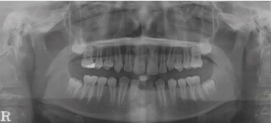
Fig. 1.Panoramic view & CT at the first medical examination; There is radiolucent lesion in anterior mandible.
Fig. 2.Photograph at the operation.
Fig. 3 .Histological examination revealed a diffuse proliferation of Langerhans histiocytic cells with occasional multinucleated cells (arrow), B. Large hitiocytes with eosinophilic cytoplasm with interspersed eosinophils (arrowhead) were also noted. (hematoxylin-eosin stain, original magnification Ax100, B:x200).
2.증례2; 이OO, 4세, 남자
2004년 4월 23일 , 4주전 개인치과에서 우측 하악 유전 치 발거 후 부터 잇몸에서 계속 피가 나고 3주전 넘어져 턱 을 부딪힌 후의 부종을 주소로 본과에 내원하였으며 하악 이부의 부종과 점상 출혈, 하악 전치부 주위의 부종, 골 팽 창, 32, 31, 41, 42번 치아의 동요도를 보이고 있었다. 특 이한 전신병력은 없었으며, CT (Fig.5), 교합방사선 사진
과 파노라마 방사선 사진, 해당 부위의 치근단 방사선 사진 을 촬영하였다. 동년 동월 24일 LCH로 인한 하악 이부의 병적 골절로 가진 하에 본원에 입원하였으며 동년 동월 30 일 전신마취 하 31,32,41 치아의 발거, 해당부위의 소파술 을 시행하였다 (Fig.6). 당시 하악골의 내고정술을 하기에 는 건전한 골이 부족하여 고정술은 시행하지 않았다. 술 후 3년까지 f/u 시행 (Fig.7)하였으며 특별한 재발소견은 관찰 되지 않았다.
Fig. 4 . 1year post-operative; It shows good healing process on symphysis area.
Fig. 5 . CT images at the first medical examination; There is fracture line on symphysis .
Fig. 6 . A. Clinical photograph at the first medical examination, B. Photograph at the operation
3.증례 3; 최OO, 7세, 남자
1999년 9월 15일 , 1달 전부터 왼쪽 뺨이 부어 보인다는 주소로 내원하였다. 입 크게 벌릴 때 통증 호소 하였으며 본 과 내원 2주전 개인 이비인후과에 상기 증상으로 내원하여 급성 이하선염 가진하에 항생요법 시행 후 증상 호전되었으 나 CT 상 좌측 하악지와 하악 과두 부위 육종 의심되어 본 과 의뢰되었다. 초진 시 국소적 열감은 없었으며 병소 부위 의 종창, 약간의 개구장애 (MMO=29mm), 동측의 악하
림프절종대를 보였으며 파노라마 사진 (Fig.8)상 좌측 하악 지와 하악 과두부위의 방사선투과성 병소를 관찰 할 수 있 었다. 치성 육종 가진하에 이학적 검사, 전신골스캔, 복부 초음파, MRI촬영 후 동년 동월 27일 좌측 하악지 부위의 종물 적출술, 소파술 시행하였으며 조직 생검 결과 랑거한 스세포 조직구증으로 진단되어 동년 10월 6일 소아과로 화 학요법 위해 전과 하였다. 소아과로 2006년까지 경과관찰 위해 내원하였으며 특별한 재발소견 관찰되지 않았다.
4.증례 4; 신OO, 3세, 남자
1993년 5월 20일, 약 1년 전부터 왼쪽 볼 부위 통증으로 개인치과에서 충치치료 하였으나 증상 호전되지 않고 지속 적인 열과 갈증 있어 한양대 소아과(1992년 4월말)에서 검 진 받았으나 특기할 이상소견 발견하지 못하다가 약 3달 전 부터 부종이 더 심해져 개인 내과 경유하여 개인치과 에서 파노라마방사선 사진 찍고 지방 대학병원 경유하여 본과로 내원하였다. 초진 시 좌측의 안면부종과 촉진 시 동통을 호 소 하였으며 국소적 열감은 없었다. 좌측 하악 제1유구치의 동요도는 없었고 타진 시 동통이 있었다. 전신병력으로는 요붕증이 있었다. 좌측 하악 유구치부의 골 팽창소견과 좌
측 악하림프절 종대가 관찰되었으며 파노라마방사선 사진 상의 좌측 하악체 부위의 불규칙한 경계를 관찰 (Fig.9)할 수 있었으며 두피의 비정상적인 발적이 있다. 좌측 하악지 와 하악체 부위의 골수염 가진 하에 항생제 요법 시행하였 고, 전신골스캔 촬영하였으며, 소아과, 이비인후과 협진 하 였다. 동년 동월 27일 전신마취 하 이환 부위의 종물 적출 술 및 소파술 시행하였으며 화학치료 위해 소아과로 전과 하였다. LCH의 다발성 장기로의 이환으로 소아과에서 경 과 관찰 중 1996년 8월 9일 술후 3년째에 다시 골 팽창 소 견으로 본과에 내원하여 LCH재발 가진하에 동년 10월 22 일 외과적 절제술 시행하였으며 동년 동월 28일 화학치료 를 시행하였다.
Fig. 7. 3years Post-operative panoramic view; There is no evidence of recurrence.
Fig. 8 . A. Panoramic view at the first medical examination; There is radiolucent lesion on the left ramus & condylar area, B. 1month post-operative panoramic view at the operation.
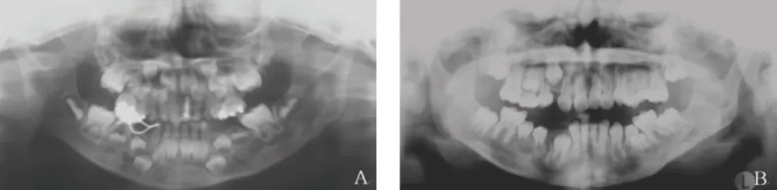
Fig. 9 . A. Panoramic view at the first medical examination, B. 5years post-operative panoramic view at the operation.
Table 1. Summary
Case 1 Case 2 Case 3 Case 4
Age/Gender 9/Male 4/ Male 7/ Male 3/ Male
Site Symphysis Symphysis Left ramus & condyle Left body & ramus Size(largest
diameter) 4cm 6cm 5cm 5cm
Clinical Gingival redness, Gingival bleeding, Left facial swelling. Left facial swelling.
Manifestations swelling & mobility redness & swelling. Lymphadenophathy. Bony expansion.
of the tooth. Mobility of the tooth. Lymphadenophathy.
Pathologic fracture of symphysis.
Image Studies Panoramic view, MRI, Panoramic view, CT Panoramic view, Panoramic view,
Previous to WBBS* WBBS, Abdominal WBBS
Surgery ultrasonography, MRI
Radiological Bone loss, Fracture line, Bone Cystic lesion Multiple cystic lesion
Findings MRI : Cystic lesion loss, WBBS : hepatomegaly
WBBS : uptake CT : Mass of soft
on symphysis tissue
Time* 12 days 4 weeks 4 weeks 1 year
CTx* X 6-Mercaptopurine Vinblastine 6-Mercaptopurine
50mg/day for 3years 3mg/2weeks 50~100mg/day from
Vinblastine for 1year for 3years
3mg/2weeks for 1year Vinblastine
3mg/2weeks for 1month
PMH* Tic disordor Nothing special Nothing special diabetes insipidus
Evolution Nothing special Pediatrics follow Nothing special 4times recurrences,
Following up until now Glaucoma,
Surgery Pediatrics follow up
until now
Clinical Group* I I I III
Stage* I II II IIIa
* PMH*: Premedical history
* Time*: Time elapsed between first symptoms and first consultation.
* CTx*: Chemotherapy
* WBBS*: Whole Body Bone Scan
* Cinical Group*: by Osband ME, 1981
* Stage*: Staging by Greenberger JS, 1981
경변, 신경장애 등의 후유증이 남을 수 있으며. 드물게 갑상 선암, 급성 골수구성 백혈병, 림프종, 간암 등 2차성 악성종 양이 발생할 수 있다.
13-16)랑거한스세포 조직구증식증은 뼈에만 병변이 나타난 경우 에는 외과적인 절제나 단순한 소파술로도 효과가 있다. 자 연적으로 치유되는 경우도 있고 방사선 조사에도 잘 반응하 며 약 5회 방사선조사에 총 10Gy정도 주며, 치료 후 약 5 개월 정도에 효과가 현저하다.
7,14,15)뇌실질, 뇌막, 요붕증, 폐, 내장을 침범한 경우 방사선 치료가 효과가 있다. 그러나 뼈에 국한된 경우에도 다른 부위의 골격에 병소가 생길 가 능성이 있고 재발하기도 한다. 미만성인 경우에는 화학요법 이 필요하다. 수혈, 항생제 투여 등 일반적인 요법을 시행하 면서 스테로이드, 빈블라스틴, 빈크리스틴, 사이클로포스파 마이드, 메토트렉세이트와 같은 약을 단독 또는 복합적으로 투여한다. 그러나 상기 방법의 치료에도 경과가 좋지 않은
당하는 증례4에서 급속도의 다발성 골 병변 재발과 장기 침 범 때문에 예후가 좋지 않음을 확인할 수 있었다. 증례 1의 경우 stage I (by Greenberger JS, 1981, Table 3.) 에 해당하는 호산구성 육아종으로 병소가 뼈에 국한되어 화학 요법 없이도 예후가 좋은 것을 확인할 수 있었다.
랑거한스세포 조직구증은 조직 병리학적인 확진이 없으면 임상적으로 골수염이나 다른 악성질환과의 감별이 어렵기 때문에 진단 시 주의가 필요하다. 또한 질환의 희귀성 때문 에 치료의 적절한 프로토콜을 수립하는 데 어려움이 있어 현재까지 명확한 치료 프로토콜이 수립되지 않은 것이 실정 이다. 랑거한스세포 조직구증 환자의 치료를 위해서는 초기 진단으로 잘못된 치료계획 수립을 예방하고 질병의 진행 단 계를 명확하게 파악하여 전체적인 프로토콜을 수립하는 것 이 필요하리라 사료된다.
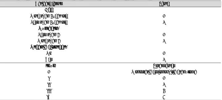
Table 2. Cinical Grouping of Langerhans cell histiocytosis by Osband ME, 19813,17)
Variable fators Score
A Aggee
More than 2year old 0
Less than 2year old 1
N
Nuummbbeerrss**
Less than 4 0
More than 4 1
O
Orrggaanniicc ddiissoorrddeerr**
No 0
Yes 1
G
Grroouupp Total scores
0 Monostatic eosionophilic granuloma
I 0
II 1
III 2
IV 3
* Numbers* : The numbers of the affected organs among the followings; Skin, Liver, Lung, Skeleton, Pituitary gland
* Organic disordoers* :
Liver : More than one of followings; Protein<5.5gm/dl, Albumin<2.5gm/dl, jaundice>1.5gm/dl, Swelling, Ascites Lung : More than one of followings ;Tachypnea, Dyspnea, cough, cyanosis, Pleural fluid, Pneumothorax Bone marrow : More than one of followings; Hemoglobin<10gm/dl, Platelet<100x109/L, Neutrophil<1.5x10⁹/L
References
1. Weitzman S, Egeler RM : Langerhans cell histiocytosis:
update for the pediatrician. Curr Opin Pediatr 20 : 23, 2008.
2. Campos MK, Viana MB, de Oliveira BM et al : Langerhans cell histiocytosis: a 16-year experience. J Pediatr (Rio J) 83 : 79, 2007.
3. Osband ME : Histiocytosis X. Langerhans’cell histiocyto- sis. Hematol Oncol Clin North Am 1 : 737, 1987.
4. Hicks J, Flaitz CM : Langerhans cell histiocytosis: current insights in a molecular age with emphasis on clinical oral and maxillofacial pathology practice. Oral Surg Oral Med Oral Pathol Oral Radiol Endod 100 : S42, 2005.
5. Margo CE, Goldman DR : Langerhans cell histiocytosis.
Surv Ophthalmol 53 : 332,2008.
6. Hoover KB, Rosenthal DI, Mankin H : Langerhans cell histiocytosis. Skeletal Radiol 36 : 95, 2007.
7. Valdivielso M, Bueno C : [Langerhans cell histiocytosis].
Actas Dermosifiliogr 96 : 275, 2005.
8. Garcia de Marcos JA, Dean Ferrer A, Alamillos Granados F et al : Langerhans cell histiocytosis in the maxillofacial area in adults. Report of three cases. Med Oral Patol Oral Cir Bucal 12 : E145, 2007.
9. Satter EK, High WA : Langerhans cell histiocytosis: a review of the current recommendations of the Histiocyte
Society. Pediatr Dermatol 25 : 291, 2008.
10. Hauser C : [The Langerhans cell histiocytoses]. J Dtsch Dermatol Ges 1 : 725, 2003. ;
11. Nquyen K, Tazi A : [Langerhans cell histiocytosis in adults]. Rev Prat 56 : 1863, 2006.
12. Greenberger JS, Crocker AC, Vawter G et al : Results of treatment of 127 patients with systemic histiocytosis.
Medicine (Baltimore) 60 : 311, 1981.
13. Bechan GI, Egeler RM, Arceci RJ : Biology of Langerhans cells and Langerhans cell histiocytosis. Int Rev Cytol 254 : 1, 2006.
14. Kim KY : Pediatric Oncology, 1 ed., Chang, Ju Young, 380,1999.
15. Greenberger JS, Cassady JR, Jaffe N et al : Radiation therapy in patients with histiocytosis: management of dia- betes insipidus and bone lesions. Int J Radiat Oncol Biol Phys 5 : 1749, 1979.
16. Chang KL, Snyder DS : Langerhans cell histiocytosis.
Cancer Treat Res 142 : 383, 2008.
17. Osband ME, Pochedly C : Histiocytosis-X: an overview.
Hematol Oncol Clin North Am 1 : 1, 1987.
18. Osband ME, Lipton JM, Lavin P et al : Histiocytosis-X. N Engl J Med 304:146, 1981.
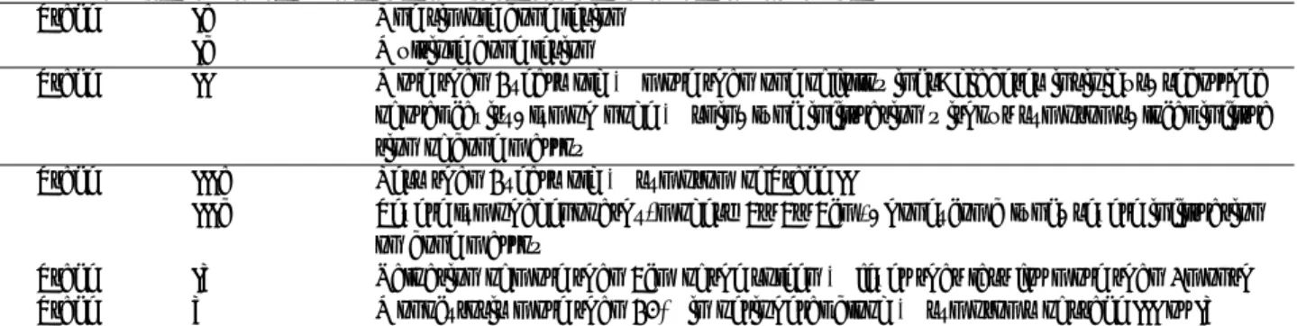
Table 3. The staging of Langerhans cell histiocytosis by Greenberger JS, 19813,15)
Stage Ia One simple bone lesion
Ib Multiple bone lesion
Stage II More than 2years old & more than one of followings; diabetes insipidus, seborrhea of oral cavity, lymph node & skin, lung infiltration without symptoms, local infiltra tion of bone marrow
Stage IIIa Less than 2years old & symptom of Stage II
IIIb Severe lymphadenopathy(modes>5×5×5cm), honeycomb lung, severe infiltration on bone marrow
Stage IV Palpation of more than 5cm of the spleen & fever that last for more than 1month Stage V Monocytosis more than 20% in peripheral blood & symptoms of stage III or IV
저자 연락처
우편번호 120-752 서울 서대문구 성산로 250
연세대학교 치과대학병원 구강악안면외과학교실 김 형 준
원고 접수일 2008년 9월 29일
Reprint Requests
Hyung Jun Kim
Dept. of OMFS, College of Dentistry, 250 Seong-sanno, Seodaemun-gu, Seoul 120-752, Korea
Tel. 82-2-2228-3132, Fax. 82-2-364-0992 E-mail : [email protected]
Paper received September 29 2008