Research in Plant Disease pISSN 1598-2262, eISSN 2233-9191 www.online-rpd.org
The Korean Society of Plant Pathology
This is an open access article distributed under the terms of the Creative Commons Attribution Non-Commercial License (http://creativecommons.org/licenses/
by-nc/4.0/), which permits unrestricted non-commercial use, distribution, and reproduction in any medium, provided the original work is properly cited.
Research Article Open Access https://doi.org/10.5423/RPD.2020.26.3.170
딸기바이러스 진단을 위한 최적의 RNA 추출 방법 및 주요 7종 딸기바이 러스의 진단법 개발
Optimal RNA Extraction Methods and Development of Synthetic Clones for Seven Strawberry Viruses
권선정1,2 · 윤주연1 · 조인숙1 · 정봉남1*
1국립원예특작과학원 원예특작환경과, 2서울대학교 그린바이오과학기술연구원
Sun-Jung Kwon1,2, Ju-Yeon Yoon1, In-Sook Cho1, and Bong-Nam Chung1*
1Horticultural and Herbal Crop Environment Division, National Institute of Horticultural and Herbal Science, Rural Development Administration,Wanju 55365, Korea
2Institute of Green Bio Science and Technology, Seoul National University, Pyeongchang 25354, Korea
Most strawberry viruses exist relatively low titers in tissues, and strawberry tissues include high levels of contamination by polysaccharides and phenolic compounds. These traits make the efficiency of strawberry diagnosis difficult. In this study, we tested different commercially available kits and reagents to secure optimal RNA extraction methods to determine virus detection from strawberry leaves. Total RNA was isolated from leaves of strawberry mottle virus (SMoV)-infected strawberry cultivar ‘Mihong’. The efficiency of total RNA for virus diagnosis was confirmed through SMoV detection by one-step or two-step reverse transcription and polymerase chain reaction (RT-PCR). Among those, the RNeasy plant RNA kit was best to isolate RNA and the isolated RNA was good enough for further applications. To ensure a reliable detection for strawberry viruses, synthetic diagnosis clones for major seven strawberry viruses such as strawberry mild yellow edge virus, SMoV, strawberry latent ring spot virus, strawberry crinkle virus, strawberry pallidosis associated virus, straw- berry vein banding virus and strawberry necrotic spot virus have been constructed. Based on the synthetic genes in each clone, primer sets for seven strawberry viruses were designed and tested an RT-PCR condition through a simultaneous application of the same annealing temperature that allowed to achieve an efficient and convenient diagnosis.
Keywords: RNA extraction, RT-PCR, Strawberry, Synthetic clone
서 론
전 세계적으로 딸기(Fragaria spp.)에 발생하는 바이러스 는 30여종이 알려져 있으며 야생딸기(F. virginiana, F. vesca, F.
chiloensis) 및 재배딸기(F. ananassa) 품종에 따라 주요 바이러 스의 피해율은 다르다. 딸기는 주로 영양체로 증식하는데 생 육기간 동안 한번 감염되면 런너를 통해 지속적으로 바이러스 를 보유하여 피해를 야기할 수 있다. 딸기바이러스 중 경제적으 로 가장 피해를 주는 바이러스에는 진딧물이 전염하는 straw- berry crinkle virus (SCV), strawberry mottle virus (SMoV), strawberry mild yellow edge virus (SMYEV) 및 strawberry
*Corresponding author Tel: +82-63-238-6320 Fax: +82-63-238-6305 E-mail: [email protected] ORCID
https://orcid.org/0000-0001-7909-2152
Received August 13, 2020 Revised September 12, 2020 Accepted September 12, 2020
vein banding virus (SVBV) 등 4종과 그 외 매개 곤충이 전반하 는 strawberry pallidosis associated virus (SPaV), strawberry
latent ringspot virus (SLRSV), strawberry necrotic shock virus (SNSV) 등이 있다(Bonneau 등, 2019; Converse, 1987; Martin과 Table 1. Information on the selection of synthetic genes for diagnostic clones of major seven strawberry viruses
Virus GenBank database Gene selection location Selected gene size (bp) Gene region Cloning vector
SMYEV AJ577359 5,074–5,902 829 CP pGEM-T Easy
KR350471 5,075–5,903
KR707814 5,073–5,901
SMoV MG418837 0,001–782 782 3' UTR pGEM-T Easy
SLRSV AY860979 1,144–1,640 497 Polyprotein
(MP–CP)
pGEM-T Easy
SCV AY250986 00 25–587 563 L protein (RdRp) All in One
KJ748457 0004–566
AY331390 00 58–620
AY331385 AY331386 AY331387 AY331388 AY331389
SPaV AY262160 0 01–747 747 R2 (CP) pTOP-Blunt-V2
AY262159 MF401547 MF420344
AY488138 4,286–5,032
SNSV AY363228 1,264–1,998 736 R3 (CP) pTOP-Blunt-V2
JF781586 1,265–2,000
KU053514 1,265–2,000
NC008706 1,264–1,998
SVBV KX787430 1,916–2,874 959 ORFIV
(CP)
pTOP-Blunt-V2 KR080547
MH894295 KX249738 KX249737 KX249736 KX249735
X97304 1,902–2,863
KX950836 1,911–2,869
HE681085
KP311681 1,919–2,877
KT250632
KX950840 105–1,063
KX950837 KX950837
Gene synthesis: Macrogene (Seoul, Korea).
SMYEV, strawberry mild yellow edge virus; SMoV, strawberry mottle virus; SLRSV, strawberry latent ringspot virus; SCV, strawberry crinkle virus (SCV); SPaV, strawberry pallidosis associated virus (SPaV); SNSV, strawberry necrotic shock virus; SVBV, strawberry vein banding vi- rus; CP, coat protein; UTR, untranscribed spacer; MP, movement protein.
Tzanetakis, 2006; Spiegel과 Martin, 1998) (Table 1). 국내의 경 우 2007년–2008년에 딸기바이러스병에 대한 발생조사에서 일 부 국내품종과 일본계통 유전자원에서 SMoV, SMYEV, SVBV 및 SPaV가 검출되었으며(Choi 등, 2009), 최근 2017–2019년 동안 국내 육성 품종에 대한 발생조사 결과에서는 SMYEV와 SMoV 2종이 주요 발생 바이러스인 것으로 조사되었다(Cho 등, 2011;
Kwon 등, 2018, 2019).
SCV는 1932년에 미국 오레곤에서 처음 보고되었다(Zeller와 Vaughan, 1932). 이 바이러스는 딸기에 가장 피해를 주는 바이 러스 가운데 하나로 SPaV와 복합감염되거나 그 외 진딧물에 의 해 전염되는 바이러스와 복합감염되는 경우 피해가 크다(Fra- zier 등, 1987). SCV는 증식성 영속전염 방식으로 Chaetosiphon 속에 속하는 진딧물에 의해 10–19일간의 잠복기를 거쳐 전염 된다(Krczal, 1982; Richardson 등, 1972; Vaughan, 1933). SMoV 는 딸기 재배지에 가장 흔하게 발생하는 바이러스로 Chaeto- siphon속에 속하는 진딧물에 의해 반영속 전염방식으로 전염 되며, 목화진딧물(Aphis gossypii)에 의해서도 전염된다(Frazier 와 Sylvester, 1960). 바이러스 계통이 다양하여 대부분 감염증 상이 없는 경우가 많지만 병원성이 강한 바이러스 계통의 경우 딸기 생육과 과일 수확량을 30%까지 감소시킨다(Cieślińska, 2019; Freeman과 Mellor, 1962). SMYEV는 1922년에 미국 캘 리포니아에서 최초로 보고한 바이러스로 딸기 재배지에서 가 장 흔한 바이러스 가운데 하나이다. 바이러스 계통이 다양하 여 감염시 수확량 손실은 0–30%로 차이가 많다(Tzanetakis 와 Martin, 2013). SMYEV는 Chaetosiphon속 진딧물에 의해 영 속전염방식으로 전염된다(Craig과 Stultz, 1964). SVBV는 딸기 재배지에서 많이 발생하지는 않는다. 단독으로 감염시에는 증 상을 나타내지 않지만 다른 바이러스와 복합감염시에는 피해 가 커진다(Mahmoudpour, 2004). SPaV는 F. virginiana에 속하 는 품종 감염 시 잎 가장자리가 황화되며 위축된다. 그러나 F.
vesca에 속하는 품종은 무증상으로 감염된다(Converse, 1987).
SLRSV는 기주 범위가 매우 넓어 27과 125종의 식물을 감염시 키며, Xiphinema속에 속하는 선충 또는 종자에 의해 전염된다 (Murant, 1974). 최근 미국에서 발생한 경우에는 증상이 없거 나 증상이 있는 경우에는 다른 바이러스와 복합감염되어 있다 (Milkus, 2001; Stace-Smith, 1970). SNSV는 1956년에 처음 알려 진 바이러스로 딸기에 단독 감염되었을 때는 병징을 보이지 않 지만 다른 바이러스와 복합감염되면 생육이 감소되는데 특히 런너 생산에 영향을 미친다. SNSV는 종자에 의해 35% 이상 전 염되며 식물조직에서 매우 낮은 농도로 존재하여 정밀 진단이 필요한 바이러스로 알려져 있다(Veetil 등, 2016).
식물 병의 진단은 계절, 온도 또는 식물의 생리적 변화와 같
은 환경 변화에 의해 영향을 받는다(Dal Zotto 등, 1999; Hele- guera 등, 2001; Posthuma 등, 2002; Tzanetakis 등, 2004). 이는 식물 병원체의 농도에 직접적인 영향을 미치든지 핵산과 함께 추출되는 식물 혼합물에 분자수준의 병원체의 검출을 위해 사 용되는 효소를 억제할 수 있는 물질의 존재에 의한 것이다.
딸기는 세계적으로 생장점 배양을 통하여 양성된 우량묘를 증식용 모주로 사용한다. 왜냐하면 딸기에 발생하는 바이러스 의 종류가 많고 특히 바이러스를 매개하는 곤충이 딸기진딧물, 담배가루이 및 선충 등으로 다양하여 바이러스에 감염될 가능 성이 있고, 감염된 모주를 증식하여 사용할 경우 딸기 수확량 에 미치는 피해를 우려해서이다. 우량묘 양성은 열처리와 식물 조직배양 기술을 이용한 생장점 배양을 하는 과정을 통하여 바 이러스와 같은 병원체를 제거한 조직배양묘를 만들어 이루어 진다. 생장점배양을 통하여 얻는 또 다른 효과는 식물체의 생 육 활성을 높이는 것이다. 특히 육묘업체에서는 증식을 위한 런 너 생산을 늘리기 위해서 우량묘를 사용한다.
우리나라에서도 딸기 품종육성 기관을 중심으로 조직배양 묘를 만들어 기본묘, 원원묘 단계를 거쳐 우량묘를 양성하는 체 계를 갖추는 중이다. 생장점 배양을 통해서 조직배양묘를 만든 다고 바이러스가 모두 제거되는 것은 아니기 때문에 병에 감염 되지 않은 우량묘를 만들기 위해서는 생장점 배양 후 반드시 바 이러스 검정을 해서 바이러스가 존재하지 않는 것을 확인해야 한다. 그러나 딸기 우량묘를 만드는 것이 초기단계로, 바이러스 를 검정하는 체계가 갖추어져 있지 않다. 특히 딸기 바이러스는 단독으로 감염된 경우 계절에 따라 증상이 나타나지 않으며, 감 염되었 어도 바이러스 검출이 잘 되지 않는 경우가 많다. 생장 점 배양을 한 딸기묘는 바이러스가 존재하더라도 농도가 낮아 서 진단 방법의 민감도가 높지 않으면 검출되지 않을 가능성이 높다. 본 연구에서는 딸기 우량묘 생산에 필요한 바이러스 검정 을 위하여 국내 육성 품종인 ‘미홍’의 SMoV 감염주를 대상으 로 잎으로부터 RNA를 추출하는 방법 및 reverse transcription and polymerase chain reaction (RT-PCR) 방법에 관한 연구를 하여 딸기에서 바이러스 검출을 위한 최적의 방법을 비교 분석 하고 주요 7종 딸기 바이러스 대한 유전정보를 바탕으로 진단 용 합성 바이러스 클론을 제작하여 정확한 진단법 적용에 따른 딸기 바이러스 진단법을 제시하기 위해 연구를 수행하였다.
재료 및 방법
딸기식물체의 다양한 핵산 추출법 및 유전자 진단법. 핵산 추출법 및 유전자 진단법은 SMoV에 감염된 딸기 식물체를 대 상으로 수행하였다. RNA 추출 여부를 확인하기 위하여 딸기 유
전체에서 항상 발현되는 ACTIN1 유전자를 대조로 사용하였다.
ACTIN1 유전자의 증폭을 위한 프라이머는 기 보고에서 사용 한 프라이머를 사용하였다(Zhang 등, 2018).
RNA를 추출하는 방법은 식물 전체 RNA를 추출하도록 만들 어진 5개 시판 제품과 Trizol을 이용하였다. Trizol (Invitrogen, Carlsbad, CA, USA)을 이용한 RNA를 추출하는 방법은 제조사 에서 지시하는 사용법대로 하였다. 추출된 식물체 전체 RNA 로부터 바이러스를 검출하기 위해서 두 가지 방법으로 RT-PCR 을 수행하였다. One-step RT-PCR은 한 튜브에 RT-PCR에 필요 한 모든 시약이 들어있어서 cDNA 합성과 PCR을 한번에 이루어 질 수 있도록 상업화된 제품으로, 이 가운데 하나인 Genetbio (Nonsan, Korea)의 제품을 제조사의 사용 설명서대로 이용하 였다. RT-PCR 반응을 위한 온도 조건은 다음과 같다. 60oC에서 45분간 RT 반응을 수행 후 94oC에서 2분 동안 변성 후, 94oC 30 초 변성, 52oC 40초 annealing, 72oC 1분 extension을 35회 수 행 후 마지막으로 72oC에서 extension 시간을 10분 수행 후 완 료하였다. Two-step PCR은 cDNA을 만드는 과정을 완료한 후 2 차 PCR 반응을 수행하였는데 방법은 기보고 내용과 동일하게 수행하였다(Chung 등, 2009). RT-PCR 반응에 사용한 프라이머는 Table 2에 표시된 SMoV 프라이머를 사용하였다.
딸기로부터 바이러스를 검출하기 위해서 잎을 채집할 때 바 이러스 검출에 가장 적합한 잎을 사용하기 위해서 채집하는 잎 이 부착된 부위를 다르게 하였다. 이 실험을 위하여 식물체로
부터 전체 RNA를 추출하는 방법은 앞 실험에서 가장 안정적으 로 바이러스 검출이 되는 Qiagen plant mini kit (Hilden, Ger- many)를 사용하였다. 그리고 RT-PCR 반응은 one-step 방법으 로 하였으며, 앞의 실험에서 사용한 Genetbio의 제품을 사용하 였다. RNA 추출용으로 판매되는 상업용 제품의 적성 검정에 관 한 실험에 사용한 7개 샘플은 SMoV에 감염된 7개의 별도의 식 물체로부터 병징이 나타나는 성엽을 이용하여 RNA를 추출한 것이다.
SMoV에 감염된 딸기 식물체로부터 잎을 채취하는 시기는 5 월 중순, 6월 중순, 7월중순에 RNA 추출용으로 판매되는 상업 용 제품의 적성 검정에 관한 실험에 사용한 7개 식물체 가운데 4개의 식물체를 대상으로 샘플 채취시기를 다르게 하여 동일한 식물체를 사용하였다. 모주와 해당 모주로부터 증식한 런너로 구분하여 완전히 펼쳐진 성엽과 아직 펼쳐지지 않은 새롭게 나 오는 어린 잎을 RNA 추출에 사용하였다.
주요 7종 딸기바이러스의 유전자 진단 클론 합성 및 프라이 머 제작. 진단법 구축을 위해 가장 먼저 바이러스 감염시료를 확보하고자 했다. 기존연구를 통해 딸기 재배지에서 SMYEV와 SMoV 감염주를 확보하였고 SLRSV의 경우 감염건조시료를 확 보하여 각각 RNA를 추출하였다. 각 바이러스에 대한 진단 프라 이머는 GenBank에 등록된 유전정보를 바탕으로 제작하였다.
3종 바이러스에 대한 각 바이러스 유전자 증폭산물은 유전자
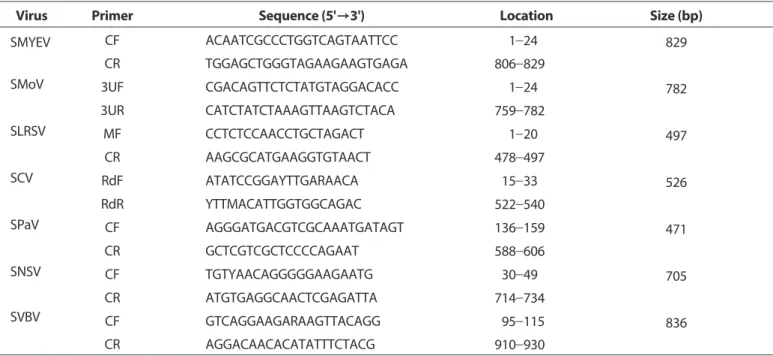
Table 2. List of primers used to detect major seven strawberry viruses based on the selected gene sequences within each positive clone
Virus Primer Sequence (5'→3') Location Size (bp)
SMYEV CF ACAATCGCCCTGGTCAGTAATTCC 001-24 829
CR TGGAGCTGGGTAGAAGAAGTGAGA 806-829
SMoV 3UF CGACAGTTCTCTATGTAGGACACC 001-24 782
3UR CATCTATCTAAAGTTAAGTCTACA 759-782
SLRSV MF CCTCTCCAACCTGCTAGACT 001-20 497
CR AAGCGCATGAAGGTGTAACT 478-497
SCV RdF ATATCCGGAYTTGARAACA 015-33 526
RdR YTTMACATTGGTGGCAGAC 522-540
SPaV CF AGGGATGACGTCGCAAATGATAGT 136-159 471
CR GCTCGTCGCTCCCCAGAAT 588-606
SNSV CF TGTYAACAGGGGGAAGAATG 030-49 705
CR ATGTGAGGCAACTCGAGATTA 714-734
SVBV CF GTCAGGAAGARAAGTTACAGG 095-115 836
CR AGGACAACACATATTTCTACG 910-930
Diagnosis: 60°C/45 min–94°C/2 min (94°C/30 sec–52°C/40 sec–72°C/1 min) 35 cycles–72°C/10 min.
SMYEV, strawberry mild yellow edge virus; SMoV, strawberry mottle virus; SLRSV, strawberry latent ringspot virus; SCV, strawberry crinkle virus; SPaV, strawberry pallidosis associated virus; SNSV, strawberry necrotic shock virus; SVBV, strawberry vein banding virus.
분석 후 클로닝 벡터(pGEM-T easy vector, Promega, Madison, WI, USA)에 삽입하여 클론으로 확보하였다. 그 외 국내에서 감 염시료 확보가 되지 않는 SPaV, SCV, SVBV, SNSV에 대해서는 GenBank database에 등록된 다양한 분리주들의 염기서열 정 보를 수집하여 clustalX2에 의해 정렬하여 유전자 부위를 선 발하고 그에 해당하는 염기서열을 합성 의뢰하였다(Bioneer, Daejeon, Korea; Macrogen, Seoul, Korea). 합성된 유전자는 진 단용 클론으로 활용하기 위하여 벡터에 삽입하고 클론으로 제 작하였다. 각 바이러스별 합성 선발 유전자 부위와 클론 정보는 Table 1에 기재하였다. 7종 딸기바이러스의 진단 프라이머는 클 론의 삽입유전자 내의 염기서열을 대상으로 각각 선별하였다 (Table 2).
결과 및 고찰
딸기식물체의 다양한 핵산 추출법 및 유전자 진단법. 딸기는 다당류 및 페놀화합물 함유량이 높아 핵산 분리가 까다로운 작 물 중 하나이다. 딸기 바이러스의 정확한 진단을 위하여 가장 중요한 것은 고품질의 RNA를 추출하는 것으로 본 연구에서는 기존 연구에서 확보한 SMoV 감염주인 ‘미홍’ 딸기품종을 대상 으로 다양한 방법을 활용하여 바이러스 진단을 위해 가장 효 율이 높은 핵산추출법을 비교하였고 일반적으로 널리 사용되 는 RNA 추출방법과 키트들 중심으로 실험을 수행하였다. 먼저 SMoV 감염주인 딸기 품종 ‘Mihong’ 은 SMoV에 감염되면 Fig.
1A에 나타난 바와 같이 잎 끝이 황화되고 엽맥을 중심으로 잎
이 뒤로 말리면서 위축되는 증상이 나타난다. 과일도 그림에서 와 같이 과탁에 가까운 과일 부위의 과피 형성이 불량한 것을 볼 수 있다(Fig. 1A). SMoV에 감염된 모주로부터 분리한 직후의 런너 식물체는 잎 가장자리에 약한 황화 증상과 함께 잎이 컵 모양으로 위로 말리는 것을 볼 수 있다(Fig. 1B). SMoV에 감염된 식물체는 건전과 비교하여 보면 잎 크기가 다소 작다(Fig. 1C).
딸기에서 SMoV 증상은 5월 이후에 잘 나타났으며, 특히 런너가 많이 생성되는 시기에 증상이 매우 잘 나타났다.
핵산추출법의 방법 별 정확한 비교를 위해 잎 시료을 한번에 분쇄하고 6개로 나눈 후 각 키트별로 핵산을 추출하였고, 분리 된 RNA의 추출효율을 판단하기 위해 바이러스 진단에 앞서 내 재유전자인 actin 유전자에 대한 진단을 통한 RNA 효율을 분 석하였다. Trizol을 이용한 RNA시료의 경우 actin 유전자가 증 폭되지 않았으며 Transzol 또한 증폭산물 감도가 낮게 나와 키 트별 핵산 추출 효율에 차이가 있음을 알 수 있었다(Fig. 2C). 딸 기 내재 유전자 증폭결과에 따라 좀 더 정밀한 바이러스 진단 효율을 비교하기 위해 one-step RT-PCR와 two-step RT-PCR로 나누어 검정 결과를 비교 분석하였다. One-step RT-PCR 방법 을 사용시 6종류의 RNA추출 방법 중 Qiagen 제품의 경우 모든 샘플에서 바이러스 검출이 잘 되었으나 그 외의 제품에서는 바 이러스 검출이 되지 않았다(Fig. 2A). 반면 two-step PCR의 경 우 Easy MAG, Ambion, Qiagen 제품의 경우 대체로 모든 샘플 에서 바이러스 검출이 잘 되었다(Fig. 2B). 5월부터 7월에 채취 한 샘플에서는 성엽과 유엽, 모주와 런너에 상관없이 모든 부위 에서 바이러스 검출이 잘 되었다(Fig. 3).
Fig. 1. Symptoms of strawberry cv. ’Mihong’ infected with strawberry mottle virus (SMoV). (A) SMoV-infected plants (mother plants) show- ing that leaves are stunted, crinkled and curved upward, and malformation of fruits. (B) Runner plants detached from SMoV-infected moth- er plants showing symptom of leaf curving upward in the young leaves. Arrows indicate adult or young leaves used for RNA extraction in this study. (C) SMoV-infected plants are stunted with a yellow edge in the young leaves (arrow) compared to healthy plant.
Constable 등(2010)의 보고에 의하면, SPaV를 포함한 대부분 의 딸기 바이러스가 5월에서 10월까지는 잘 검출되었으나 11월 부터 1월에는 상대적 바이러스 검출이 잘 되지 않았으며 이는 바이러스가 식물체에 고르게 분포되지 않고 농도가 균일하지 않기 때문이라고 하였다. 본 연구에서 실험한 기간인 5월에서 7 월까지는 딸기 바이러스 검출이 잘 되었다. 포도의 경우 바이러 스 종류에 따라서 동일 식물체의 여러 가지별로 바이러스가 불 균일하게 분포되어 있으며, 따라서 대부분의 경우 영양생장을 하기 전의 휴면 줄기가 포도 바이러스 검정을 위해서는 가장 좋 은 부위라고 하였다(Komínek 등, 2009). 딸기의 경우에도 계절 에 따라 바이러스 검출이 잘 되지 않을 수 있으므로 조직배양 묘의 경우 온실에서 키우면서 생육이 활발한 여름철을 지나면 서 몇 차례 검정하는 것이 필요할 것으로 판단된다.
주요 딸기바이러스 7종의 진단법 개발 및 조건 설정. 딸기 조직배양묘와 증식묘에서의 정확한 바이러스 검정을 위하여 주요 7종 바이러스에 대한 양성 대조용 진단 클론을 제작하였 다. 진단 클론 제작은 바이러스 종류별로 다양한 분리주들 간 에 가장 보존이 잘 되는 유전자를 선별하여 합성하고 벡터에 삽입하였으며 7종 바이러스의 삽입 유전자 크기 및 클로닝 벡 터에 대한 정보는 Table 1에 요약하였다. 바이러스 진단용 유전 자 합성은 Macrogen에 의뢰하여 제작하였다. 양성 클론을 이 용한 유전자 진단을 위해 진단 프라이머는 각 클론의 삽입된 유전자의 염기서열 내에서 각각 선발하였고 각 프라이머의 위 치 및 PCR 산물 크기는 Table 2에 정리하였다. 최적 진단조건 을 설정하기 위해 플라스미드 DNA 사용량의 적정농도와 바이
러스 프라이머에 대한 PCR 반응 조건을 검사하였다. DNA 농도 는 1 ng/μl, 5 ng/μl, 10 ng/μl으로 구분하고 각 바이러스별 프 라이머 세트의 결합온도(annealing temperature)는 각 프라 이머 Tm값에 근거하여 52, 56, 60oC로 나누어 PCR 반응을 비 교하였다. 그 결과, 플라스미드의 농도 차이에 따른 유의성은 없는 것으로 나타났고(data not shown) PCR 결합온도 실험은 SMYEV, SVBV의 경우 온도 차이에 따른 감도 차이가 거의 없었 으나 SMoV, SLRSV, SCV, SPaV, SNSV의 경우 60oC보다 52oC 및 56oC에서 감도가 더 좋게 나왔다(Fig. 4A). 모든 바이러스의 진 단 클론에 대해 설정한 각 프라이머 세트의 특이도는 모두 높게 잘 나타났다. 이들 결과를 바탕으로 확보된 SMYEV, SMoV 및 SLRSV의 RNA 시료에 대한 RT-PCR을 실시하였다. 대조구인 양 성 클론과 함께 바이러스 감염 RNA 시료를 대상으로 3가지 결 합온도에 대해 RT-PCR을 수행한 실험에서 52°C에서 가장 높은 감도의 PCR 산물이 증폭되는 것을 확인할 수 있었다(Fig. 4B).
종합적으로, RT-PCR 진단 시 플라스미드 DNA 1 ng/μl의 사 용과 52°C의 결합온도 PCR 조건을 최적조건으로 설정하였다 (Table 2).
유전자 진단은 검정시료의 특성, 증폭 유전자에 대한 프라이 머의 설계 및 PCR 조건 설정에 따라 검출감도와 결과 정확도의 성패가 좌우된다. 따라서 유전자 진단을 수행할 때 검정 결과 의 신뢰도를 높이기 위해서는 반드시 양성대조구와 음성대조 구를 사용하는 것이 필요하다. 특히 바이러스는 특성상 변이가 많고 다양한 병원성의 strain이 다수 존재하므로 정확한 진단 을 위해서는 바이러스의 양성시료 사용과 다양한 분리주들의 유전자 분석을 통한 최적 프라이머 설계가 매우 중요하다. 특히 Fig. 2. Comparison of detection sensitivity of strawberry mottle virus viral RNA prepared with various commercial products from straw- berry leaves. Total RNA was extracted from a total of seven leaves, one from each of the seven plants using different commercial products.
1, Easy MAG; 2, Ambion; 3, Qiagen; 4, Bio Cube; 5, Transzol; 6, Trizol; P, positive control; N, negative control. (A) One-step reverse transcription and polymerase chain reaction (RT-PCR). (B) Two-step RT-PCR. (C) RT-PCR with a primer pair Actin I as an internal control using one-step method.
주요 딸기 바이러스의 대부분은 RNA 바이러스들로 유전자 진 단 시 RT-PCR을 수행하여야 하므로 양성시료로 감염주의RNA 를 사용하는 것이 좋다. 감염주 확보를 위해 2017–2019년 동 안 발생조사를 수행하였고 SMYEV와 SMoV의 국내 분리주 및 SLRSV의 감염건조시료를 확보하였다. 감염주 확보가 되지 않은 그 외 딸기바이러스는 유전자 합성을 통한 진단 클론 제작으로 양성대조구를 확보하였다. 또한 한 시료에서 다종의 바이러스
를 효율적으로 검정하기 위해 동일한 결합온도에서 PCR 반응을 할 수 있도록 최적의 반응 조건을 설정하였다. 본 연구에서 확보 된 7종의 딸기 바이러스 양성대조구와 진단조건 확립으로 딸기 우량묘 바이러스 검정법에 유용하게 활용될 것으로 기대한다.
요 약
바이러스의 정확한 진단법 확립은 바이러스의 피해 및 확산 Fig. 3. Comparison of detection sensitivity of strawberry mottle virus viral RNA from different leaves in strawberry. Total RNA was extracted from fully unfolded or folded young leaves from mother plants and runner plants. (A) 1–4, four independent strawberry plants; P, positive control; N, negative control. (B) Reverse transcription and polymerase chain reaction with a primer pair actin I as an internal control.
Fig. 4. Reverse transcription and polymerase chain reaction (RT-PCR) detection for seven major strawberry viruses using diagnostic syn- thetic clones (A) and plant RNA samples (B). The corresponding lengths of the expected products using primer sets for each virus are de- scribed in Table 2. SMYEV, strawberry mild yellow edge virus; SMoV, strawberry mottle virus; SLRSV, strawberry latent ringspot virus; SCV, strawberry crinkle virus; SPaV, strawberry pallidosis associated virus; SVBV, strawberry vein banding virus; SNSV, strawberry necrotic shock virus; NC, negative control (RNA from healthy plant); PC, positive control (1 ng/μl of diagnostic synthetic clone).
을 예방하는데 매우 중요하게 작용한다. 대부분의 딸기 바이러 스는 조직내에 낮은 역가로 분포하여 진단이 어렵고, 특히 딸 기 조직은 다당류 및 페놀화합물의 함유가 많아 RNA 추출이 어려운 것으로 알려져 있다. 딸기 우량묘 생산에 필요한 바이러 스 검정기술을 확립하기 위해 본 연구에서는 딸기 잎에서 바이 러스 진단을 위해 가장 최적의 RNA 추출방법 정립을 위해 다 양한 상용 키트와 시약을 이용하여 RNA 추출효율 비교하였다.
바이러스 진단을 통한 RNA 추출효율을 분석하기 위해 SMoV 감염주인 미홍 딸기 품종을 이용하여 다양한 단계에서 잎조 직으로부터 RNA를 추출하고 바이러스 진단을 수행하였다. 식 물 RNA 추출 방법 가운데 상업용으로 판매되는 RNeasy plant mini kit (Qiagen)를 이용하는 경우 본 연구에서 살펴본 one- step 또는 two-step RT-PCR 방법과 무관하게 SMoV의 검출이 잘 되었다. 또한, 딸기 우량묘의 바이러스 검정에 대한 신뢰있는 진단방법을 구축하기 위해 주요 딸기 바이러스인 strawberry mild yellow edge virus (SMYEV), strawberry mottle virus (SMoV), strawberry latent ringspot virus (SLRSV), strawberry crinkle virus (SCV), strawberry pallidosis associated virus (SPaV), strawberry vein banding virus (SVBV) 및 strawberry necrotic shock virus (SNSV) 7종에 대한 유전자 합성을 통해 진 단클론을 제작하였다. 각 클론의 합성유전자를 기반으로 7종 의 딸기바이러스 프라이머 세트를 설계하고 편리한 진단법 수 행을 위해 동일한 PCR 조건을 설정하였다.
Conflicts of Interest
No potential conflict of interest relevant to this article was re- ported.
Acknowledgments
This work was carried out with the support of the “Cooperative Research Program for Agriculture Science & Technology Devel- opment (Project No. PJ PJ01186302)” of the Rural Development Administration, Republic of Korea.
References
Bonneau, P., Hogue, R., Tellier, S. and Fournier, V. 2019. Evaluation of various sources of viral infection in strawberry fields of Qubec, Canada. J. Econ. Entomol. 112: 2577-2583.
Cieślińska, M. 2019. Genetic diversity of seven Strawberry mottle virus isolates in Poland. Plant Pathol. J. 35: 389-392.
Cho, J.-D., Choi, G.-S., Chung, B.-N., Kim, J.-S. and Choi, H.-S. 2011.
Strawberry mild yellow edge potexvirus from strawberry in Ko- rea. Plant Pathol. J. 27: 187-190.
Choi, G.-S., Lee, J.-A., Cho, J.-D. Chung, B.-N., Cho, I.-S. and Kim, J.-S.
2009. Strawberry virus diseases occurring in Korea, 2007-2008.
Res. Plant Dis. 15: 8-12.
Chung, B. N., Cho, I. S. and Cho, J. D. 2009. Effective application of CF11 cellulose for detection of Apple scar skin viroid in Apple.
Plant Pathol. J. 25: 291-293.
Constable, F. E., Bottcher, C., Kelly, G., Nancarrow, N., Milinkovic, M., Persely, D. M. et al. 2010. The seasonal detection of strawberry viruses in Victoria, Australia. Julius-Kuhn-Archiv 427: 27-34.
Converse, R. H. 1987. Virus and viruslike diseases of Fragaria (straw- berry). In: Virus Diseases of Small Fruits, Agriculture Handbook No. 631, ed. by R. H. Converse, pp. 1-100. U.S. Department of Agriculture, Agricultural Research Service, Washington, DC, USA.
Craig, D. L. and Stultz, H. T. 1964. Aphid dissemination of strawberry viruses in Nova Scotia. Can. J. Plant Sci. 44: 235-239.
Dal Zotto, A., Nome, S. F., Di Rienzo, J. A. and Docampo, D. M. 1999.
Fluctuations of Prunus necrotic ringspot virus (PNRSV) at vari- ous phenological stages in peach cultivars. Plant Dis. 83: 1055- 1057.
Frazier, N. W. and Sylvester, E. S. 1960. Half-lives of transmissibility of two aphid-borne viruses. Virology 12: 233-244.
Frazier, N. W., Sylvester, E. S. and Richardson, J. 1987. Strawberry crinkle. In: Virus Diseases of Small Fruits, Agriculture Handbook No. 631, ed. by R. H. Converse, pp. 20-55. U.S. Department of Agriculture, Washington, DC, USA.
Freeman, J. A. and Mellor, F. C. 1962. Influence of latent viruses on vigor, yield and quality of British Sovereign strawberries. Can. J.
Plant Sci. 42: 602-610.
Heleguera, P. R., Taborda, R., Docampo, D. M. and Ducasse, D. A.
2001. Immunocapture reverse transcription-polymerase chain reaction combined with nested PCR greatly increases the de- tection of Prunus necrotic ring spot virus in the peach. J. Virol.
Methods 95: 93-100.
Krczal, H. 1982. Investigation on the biology of the strawberry aphid (Chaetosiphon fragaefolii), the most important vector of strawberry viruses in West Germany. Acta Hortic. 129: 63-68.
Komínek, P., Glasa, M. and Komínková, M. 2009. Analysis of multiple virus-infected grapevine plant reveals persistence but uneven virus distribution. Acta Virol. 53: 281-285.
Kwon, S.-J., Cho, I.-S., Yoon, J.-Y. and Choi, G.-S. 2018. Genetic diver- sity of two isolates in Strawberry mild yellow edge virus from Korea. Res. Plant Dis. 24: 285-291.
Kwon, S.-J., Yoon, J.-B., Cho, I.-S., Yoon, J.-Y. and Kwon, T.-R. 2019.
Incidence of aphid-transmitted strawberry viruses in Korea and phylogenetic analysis of Korean isolates of Strawberry mottle virus. Res. Plant Dis. 25: 226-232.
Mahmoudpour, A. 2004. Diagnosis and quantification of Strawber- ry vein banding virus using molecular approaches. Acta Hortic.
656: 69-74.
Martin, R. R. and Tzanetakis, I. E. 2006. Characterization and recent advances in detection of strawberry viruses. Plant Dis. 90: 384- 396.
Milkus, B. N. 2001. Incidence of four NEPO viruses in Missouri vine- yards. Am. J. Enol. Vitic. 52: 56-57.
Murant, A. F. 1974. CMI/AAB Description of Plant Viruses. No. 126.
Strawberry Latent Ringspot. Commonwealth Mycological Insti- tute, Kew, Surrey, UK. 4 pp.
Posthuma, K. I., Adams, A. N., Hong, Y. and Kirby, M. J. 2002. Detec- tion of Strawberry crinkle virus in plants and aphids by RT-PCR using conserved L gene sequences. Plant Pathol. 51: 266-274.
Richardson, J, Frazier, N. W. and Sylvester, E. S. 1972. Rhabdovirus- like particles associated with strawberry crinkle virus. Phytopa- thology 62: 491-492.
Spiegel, S. and Martin, R. R. 1998. Virus and viruslike disease. In:
Compendium of Strawberry Diseases, ed. by J. L. Maas, pp. 62- 72. American Phytopathological Society, St. Paul, MN, USA.
Stace-Smith, R. 1970. CMI/AAB Description of Plant Viruses. No. 18.
Tomato Ringspot Virus. Commonwealth Mycological Institute, Kew, Surrey, UK. 4 pp.
Tzanetakis, I. E., Mackey, I. C. and Martin, R. R. 2004. Strawberry ne- crotic shock virus is a distinct virus and not a strain of Tobacco streak virus. Arch. Virol. 149: 2001-2011.
Tzanetakis, I. E. and Martin,R. R. 2013. Expanding field of strawberry viruses which are important in North America. Int. J. Fruit Sci. 13:
184-195.
Vaughan, E. K. 1933. Transmission of the crinkle disease of straw- berry. Phytopathology 23: 738-740.
Veetil, T. T., Ho, T., Moyer, C., Whitaker, V. M. and Tzanetakis, I. E. 2016.
Detection of Strawberry necrotic shock virus using convention- al and Taqman quantitative RT-PCR. J. Virol. Methods 235: 176- 181.
Zeller, S. M. and Vaughan, E. K. 1932. Crinkle disease of strawberry.
Phytopathology 22: 709-713.
Zhang, Y., Peng, X., Liu, Y., Li, Y., Luo, Y., Wang, X. et al. 2018. Evalua- tion of suitable reference genes for qRT-PCR normalization in strawberry (Fragaria×ananassa) under different experimental conditions. BMC Mol. Biol. 19: 8.