© 2015 The Korean Ophthalmological Society
This is an Open Access article distributed under the terms of the Creative Commons Attribution Non-Commercial License (http://creativecommons.org/licenses /by-nc/3.0/) which permits unrestricted non-commercial use, distribution, and reproduction in any medium, provided the original work is properly cited.
Original Article
Normative Data of Videonystagmography in Young Healthy Adults under 40 Years Old
Sunah Kang
1, Ungsoo Samuel Kim
1,21
Department of Ophthalmology, Kim’s Eye Hospital, Seoul, Korea
2
Department of Ophthalmology, Konyang University College of Medicine, Daejeon, Korea
Purpose: The purpose of this study was to establish a set of normative data values for saccade movements us- ing videonystagmography and to evaluate the effects of manual correction on this data.
Methods: We examined 25 healthy subjects (9 men and 16 women). All tests were carried out by one well-in- structed physician. Errors such as the wrong detection of the inflection point, missing movement, and prediction occurred during some tests. Thus, the same physician manually corrected the data by deleting error data from row results.
Results: We established a set of normative data for horizontal saccade movements (amplitude size 15 and 30 degrees) for mean peak velocity, latency, and accuracy. Manual correction only impacted latency and accura- cy at 30 degrees horizontal, which is likely related to possible errors during the test.
Conclusions: The present study provides clinically useful videonystagmography-based normative data for clinicians regarding saccade movements in Korean individuals.
Key Words: Accuracy, Latency, Saccades, Velocity
The main objectives of voluntary eye movements are ei- ther to position or maintain images of interest on the fovea, the small central retinal area of highest visual acuity. Sac- cades and smooth pursuit are controlled by different neural structures. These differing anatomical pathways include several cortical areas that mediate cognitive control of eye movements, in contrast to brainstem structures that are mainly concerned with the motor control of eye move- ments [1]. The analysis of eye movements has been shown to provide key contributions to the diagnosis of some neu- rodegenerative, hereditary, and metabolic disorders.
Videonystagmography (VNG) was designed to acquire video images of eye movements, especially in nystagmus, and is potentially useful to clinicians in otorhinolaryngolo- gy, neurology, and ophthalmology [2]. Traditionally, elect- ronystagmography (ENG), which relies on the corneo-reti- nal potential to record eye movements, is considered the gold standard for evaluating dizzy patients [3]. In contrast to ENG, VNG records eye movements using digital video image technology employing infrared illumination to de- termine eye position. The use of VNG enables simultane- ous subjective observation of eye movements together with objective data collection and analysis of eye movement waveforms via computer algorithms. VNG has the benefit of not requiring skin preparation or electrode application and wiring. Moreover, adjustments are seldom required as VNG does not depend on changes in corneo-retinal poten- tial over time in contrast to ENG. However, VNG is unable
Received: May 21, 2014 Accepted: September 1, 2014
Corresponding Author: Ungsoo Samuel Kim, MD, PhD. Department of
Ophthalmology, Kim’s Eye Hospital, #136 Yeongsin-ro, Yeongdeung-
po-gu, Seoul 150-034, Korea. Tel: 82-2-1577-2639, Fax: 82-2-2677-9214,
E-mail: [email protected]
to record eye movements when the eyes are closed [4].
To date, there has been no study reporting normative data of saccade movements obtained from an Asian patient population, including Koreans. The aim of this study was to establish a set of normative data of saccade movements obtained from 25 healthy Korean adults using VNG (SLVNG; SLMED, Seoul, Korea) and to evaluate the ef- fects of manual correction on this data.
Materials and Methods
We examined 25 healthy volunteers (9 men and 16 wom- en). The following subjects were included: those with no history of vertigo, balance problems, otologic problems, neurologic diseases, or ocular diseases, who were not on any medications. The inclusion criteria were assessed by history taking and basic examination for visual acuity and eye movement. All subjects were asked for their informed consent prior to entering this study. This study was re- viewed and approved by the institutional review board of Kim’s Eye Hospital, and all procedures conformed to the guidelines of the Declaration of Helsinki.
Eye movements were recorded with an infrared camera (resolution 640 × 480 pixels, frame rate 60 Hz) and dis- played on a computer monitor (SLMED). In the saccade test, the patient was seated in the VNG chair, which was at a fixed, pre-calibrated distance from the VNG monitor, and asked to follow the movements of the object on the screen. A stimulus randomly appeared on the screen at the edge of the visual field in horizontal directions (amplitude
size 15 and 30 degrees), and the patient was asked to follow the stimulus only with his eyes, keeping his head stable;
eye movements were displayed and recorded and a com- puter analyzed the results.
Latency was defined as the delay between the onset of target movement and the initiation of eye movement. Ac- curacy was defined as the amplitude of the eye movement relative to the target. Velocity was defined as the time tak- en to complete the saccade once it was initiated.
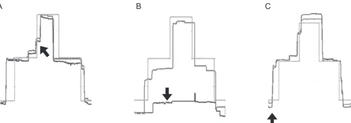
All tests were carried out by one well-instructed physi- cian. Errors such as wrong detection of the inflection point, missing movement, and prediction occurred during some tests (Fig 1). Thus, correction was required, and the same physician manually corrected the data by deleting error data from the row results.
The effect of manual correction was studied by compar- ing parameters before and after correction using the paired t-test (SPSS ver. 12.0, SPSS Inc, Chicago, IL, USA; p <
0.05).
Results
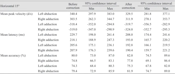
Our normative database contains over 25 saccadic eye movements from 25 normal subjects ranging from 21 to 38 years of age. Table 1 shows the mean peak velocity, laten- cy, accuracy, and 95% confidence interval at 15 degree horizontal saccade movements. The results of 30 degree horizontal saccade movements are shown in Table 2. The effect of movement direction on saccade parameters was not significant, and there was no significant difference in
Fig. 1. Possible errors during the test. (A) Wrong detection of inflection point, (B) missing movement, and (C) prediction.
A B C
the parameters between 15 and 30 degrees.
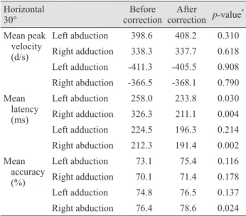
Analysis of the effects of manual correction in which one physician deleted extremely high or low data from row results showed significant differences only with respect to latency and accuracy at 30 degrees horizontal (Table 3).
Discussion
The present study provides VNG-based normative data of saccade movements in Korean individuals. From the
time the first accurate measurements of saccadic eye movements were obtained [5,6], several studies have re- ported peak velocity values in normal subjects, but these have usually been obtained from small numbers of young volunteers [6-11]. With respect to the effects of age on sac- cade eye movements, Abel et al. [12] found that there was no correlation, while Spooner et al. [13] reported that elder- ly subjects had lower peak velocity values.
Furthermore, Warabi et al. [14] reported that elderly peo- ple had slower large (40°) saccades than younger subjects, but that there were no differences at smaller saccade an- Table 1. Parameters of saccade testing at 15 degrees horizontal
Horizontal 15° Before
correction 95% confidence interval After
correction 95% confidence interval
Min Max Min Max
Mean peak velocity (d/s) Left abduction 340.4 297.9 383.0 329.5 281.8 377.1
Right adduction 303.5 262.3 344.7 311.9 270.1 353.7
Left adduction -318.4 -352.0 -284.8 -319.7 -356.5 -282.9
Right abduction -319.0 -347.0 -290.9 -324.0 -352.7 -295.3
Mean latency (ms) Left abduction 229.7 198.0 261.4 208.0 174.4 241.6
Right adduction 218.1 188.9 247.3 193.0 165.7 220.2
Left adduction 205.6 175.1 236.1 192.8 166.1 219.5
Right abduction 207.9 176.3 239.6 190.4 159.7 221.2
Mean accuracy (%) Left abduction 80.0 73.0 87.0 82.0 74.5 89.6
Right adduction 74.8 66.5 83.1 77.8 69.1 86.4
Left adduction 74.3 68.4 80.3 75.3 67.8 82.8
Right abduction 79.4 72.9 85.9 81.9 74.7 89.0
Table 2. Parameters of saccade testing at 30 degrees horizontal
Horizontal 30° Before
correction 95% confidence interval After
correction 95% confidence interval
Min Max Min Max
Mean peak velocity (d/s) Left abduction 398.6 339.0 458.1 408.2 336.4 480.1
Right adduction 338.3 298.8 377.7 337.7 297.9 377.4
Left adduction -411.3 -488.7 -333.9 -405.5 -485.2 -325.9
Right abduction -366.5 -405.6 -327.4 -368.1 -406.8 -329.4
Mean latency (ms) Left abduction 258.0 214.8 301.2 233.8 188.6 279.0
Right adduction 326.3 137.3 515.3 211.1 163.0 259.2
Left adduction 224.5 192.8 256.2 196.3 163.6 229.1
Right abduction 212.3 178.8 245.8 191.4 153.1 229.7
Mean accuracy (%) Left abduction 73.1 65.7 80.5 75.4 66.8 84.0
Right adduction 70.1 62.8 77.3 71.4 63.7 79.0
Left adduction 74.8 66.8 82.9 76.5 67.3 85.7
Right abduction 76.4 69.2 83.7 78.6 70.9 86.4
gles. In a study by Wilson et al. [15], time of day and age had weak but significant effects on saccade movements, but the effects of age only became apparent for large sac- cades (≥35°). Diurnal changes in performance have also been observed, with a tendency for slightly slower values in the early afternoon. Another study revealed that gender and education level did not influence eye movement met- rics, and with age, the latency of leftward and vertical pro- saccades and antisaccades increased, the velocity of up- ward prosaccades decreased, the gain of rightward and upward prosaccades diminished, and the error rate of anti- saccades increased [1].
In the current study, we established a set of normative data values for saccade movements using VNG and identi- fied the effects of manual correction on said data. To ob- tain reliable results from VNG, we acquired data in a clin- ical setting, and to reduce variability between inspectors,
tests and manual correction were carried out by one well-instructed physician. None of our healthy subjects complained of any form of dizziness or other side effects during the test. Values obtained with our equipment are in agreement with those of previous studies (Table 4). There has been no study reporting normative data values for sac- cade movements obtained from an Asian population. In comparison with previous studies (Table 4), there was no significant difference in the velocity of eye movements based on ethnicity. Manual correction only impacted laten- cy and accuracy at 30 degrees horizontal, which is likely related to possible errors during the test. For instance, the wrong detection of inflection point can cause delayed la- tency or decreased accuracy (Fig. 1A). Secondly, the infra- red camera can miss the eye movement for various reasons such as thick eye make-up using eyeliner and false eye- lashes or eyelid problems like ptosis, entropion, or small fissures (Fig. 1B). Thirdly, the participant’s eye can move more quickly than the target when the patient predicts tar- get movement (Fig. 1C). These errors can affect the latency and accuracy of VNG results.
There are some limitations to this study. First, the nor- mative data were obtained from a small number of young healthy volunteers. Second, we did not attempt to investi- gate the effect of age or systemic diseases like diabetic mellitus on VNG results. Therefore, the normative data from this study may not be applicable to the elderly and patients with systemic diseases. Furthermore, nysta- gmography is more frequently used for the evaluation of eye movements in pediatric examinations, but there are no data or measurements from young children in this study.
Future studies regarding the effects of specific conditions on VNG results are needed. Third, VNG might be very variable based upon the examiner and his or her level of expertise. Consequentially, its reliability needs to be con- firmed for interpretation when using this normative data
Table 4. Comparison with other studies
15° 30° Mims et al. [10]
(20°) Raab [11]
(20°) Mean peak velocity (d/s) Left abduction 329 ± 115 408 ± 174 404 ± 51 368 ± 42
Right adduction 312 ± 101 338 ± 96 388 ± 58 428 ± 45
Left adduction -320 ± 89 -406 ± 193 425 ± 56 379 ± 39
Right abduction -324 ± 69 -368 ± 94 365 ± 46 398 ± 53 Values are presented as mean ± SD.
Table 3. Comparison of parameters before and after correction Horizontal
30° Before
correction After
correction p-value
*Mean peak
velocity (d/s)
Left abduction 398.6 408.2 0.310 Right adduction 338.3 337.7 0.618 Left adduction -411.3 -405.5 0.908 Right abduction -366.5 -368.1 0.790 Mean
latency (ms)
Left abduction 258.0 233.8 0.030 Right adduction 326.3 211.1 0.004 Left adduction 224.5 196.3 0.214 Right abduction 212.3 191.4 0.002 Mean
accuracy (%)
Left abduction 73.1 75.4 0.116 Right adduction 70.1 71.4 0.178 Left adduction 74.8 76.5 0.137 Right abduction 76.4 78.6 0.024
*