Acute Modulations in Stratum Corneum Permeability Barrier Function Affect Claudin Expression and Epidermal Tight Junction Function via Changes of Epidermal Calcium Gradient
Ji Hwoon Baek,
1,6* Sang Eun Lee,
2* Ki Ju Choi,
3Eung Ho Choi,
4and Seung Hun Lee
51Brain Korea 21 Project for Medical Science, Yonsei University, Seoul; 2Department of Dermatology, Bundang CHA Hospital, CHA University College of Medicine, Seongnam; 3Division of Electron Microscopic Research, Korea Basic Science Institute, Daejeon;
4Department of Dermatology, Yonsei University Wonju College of Medicine, Wonju; 5Department of Dermatology and Human Barrier Research Institute, Yonsei University College of Medicine, Seoul; 6Dermapro Skin Research Center, DERMAPRO LTD., Seoul, Korea.
Received: October 10, 2011 Revised: April 18, 2012 Accepted: April 18, 2012
Corresponding author: Dr. Seung Hun Lee, Department of Dermatology and Human Barrier Research Institute, Yonsei University College of Medicine, 211 Eonju-ro, Gangnam-gu, Seoul 135-720, Korea.
Tel: 82-2-2019-3360, Fax: 82-2-3463-6136 E-mail: [email protected]
*Ji Hwoon Baek and Sang Eun Lee contributed equally to this work.
∙ The authors have no financial conflicts of interest.
© Copyright:
Yonsei University College of Medicine 2013 This is an Open Access article distributed under the terms of the Creative Commons Attribution Non- Commercial License (http://creativecommons.org/
licenses/by-nc/3.0) which permits unrestricted non- commercial use, distribution, and reproduction in any medium, provided the original work is properly cited.
Tight junction (TJ) is recognized as a second barrier of the skin. Altered expres- sion of TJ proteins in various skin diseases characterized by the abnormal permea- bility barrier such as psoriasis suggests that TJ could be affected by stratum corne- um (SC) barrier status. However, the physiological relationship between SC and TJ barrier remains to be investigated. Therefore, we examined the effect of SC barrier disruption on the expression of TJ proteins, claudin (Cldn)-1 and Cldn-4, and TJ barrier function in hairless mouse skin. We also investigated whether the alterations in epidermal Ca
2+affected TJ proteins expression in vivo. Repeated tape-stripping induced a sequential change of the expression and function of TJ.
As early as 15-30 minutes after tape-stripping, downregulation of Cldn-1 and Cldn-4 immunoreactivity and protein level without change in mRNA level was found. This was accompanied by the abnormal leakage of lanthanum. However, by 1 hour Cldn-1 and Cldn-4 immunolocalization recovered along with normal- ized lanthanum permeation pattern. Moreover, the mRNA and protein levels of Cldn-1 and Cldn-4 were increased by 1 to 6 hours after tape-stripping. Inhibition of calcium loss by immersion of barrier-disrupted skin into a high Ca
2+solution prevented the dislocation of Cldn-1 and Cldn-4. Occlusion of barrier-disrupted skin delayed the restoration of Cldn-1 and Cldn-4. Our results suggest that the al- teration of epidermal Ca
2+gradient caused by SC barrier perturbation affects the TJ structure and function and the faster recovery of TJ as compared to the SC barrier may imply the protective homeostatic mechanism of skin barrier.
Key Words: Tight junction, claudin-1, claudin-4, calcium gradient, stratum corne- um permeability barrier
The functional barrier of mammalian skin is generally accepted to reside mainly in
the stratum corneum (SC). Recently, the proteins of tight junction (TJ), which is a
major regulator of barrier function in simple epithelia has also been identified in
minutes, 1 hour, 3 hours, and 6 hours. After each time point, specimens were taken for immunofluorescence staining.
In the untreated murine epidermis, Cldn-1 was expressed in the intercellular spaces throughout all living layers, where- as Cldn-4 was restricted in SG (Fig. 1A). At 15 and 30 min- utes after tape-stripping, the reduction of Cldn-1 immuno- fluorescence from the granular and spinous layers was observed. However, by 1 hour, Cldn-1 expression recovered and by 3-6 hours, Cldn-1 immunoreactivity was slightly in- creased (Fig. 1A). The immunofluorescence study for Cldn- 4 showed a similar pattern of expression change in re- sponse to tape-stripping. At 15 and 30 minutes after tape- stripping, the intensity of Cldn-4 immunostaining was slightly decreased (Fig. 1A). By 1-3 hours, Cldn-4 expres- sion recovered and even increased at 6 hours after tape- stripping (Fig. 1A). Cldn-7, occludin and ZO-1 immunore- activity did not change. To confirm that these staining pattern changes are due to the change of gene and protein levels, we examined Cldn-1 and Cldn-4 mRNA and protein levels by real-time PCR and western blot. We found no statistical differences in Cldn-1 and Cldn-4 mRNA level and down- regulation of protein level between untreated and tape- stripped group at 15 and 30 minutes after treatment (Fig.
1B-E). Moreover, by 1-6 hours, Cldn-1 and Cldn-4 mRNA expression was significantly increased in comparison to controls (Fig. 1B and C). The increase in band intensity of Cldn-1 and Cldn-4 from western blot images was also ob- served at 1, 3, and 6 hours after tape-stripping (Fig. 1D).
To investigate whether SC barrier disruption-induced changes in the localization or distribution of Cldn-1 and Cldn- 4, influence on the TJ barrier function, we next performed the lanthanum penetration assay to assess the inside-out barrier function. TJ has been demonstrated to be located in the SG2 layer.
8,9Consistent with these previous reports, we found a limited lanthanum penetration at SG2 layer in un- treated murine epidermis by transmission electron micro- scope (Fig. 2). In contrast, at 30 minutes after tape-stripping at which time there were distributional changes of Cldn-1 and Cldn-4, an upward diffusion of lanthanum beyond SG2 layer was found, suggesting a defective inside-out barrier function (Fig. 2). However, at 1 hour after barrier disrup- tion, the passage of lanthanum was again restricted at SG2 layer similar to control skin, indicating the restoration of TJ barrier function (Fig. 2). This functional study suggests that the acute SC barrier disruption-induced transient Cldn-1 and Cldn-4 alteration was accompanied by impaired barrier function of TJ and this defect in inside-out barrier function human skin.
1In the inflammatory diseases of the intestine,
many cytokines have been reported to affect TJ barrier sta- tus. Moreover, there is increasing evidence now that TJ in skin, which localizes in stratum granulosum (SG),
2also contribute to epidermal barrier formation.
3-5Among TJ pro- teins, claudin (Cldn)-1 and Cldn-4 have been demonstrated to have a role in barrier function of the skin.
3-5Cldn-1 null mutant mice showed normal SC structure, however died shortly after birth with increased transepidermal water loss (TEWL).
3Moreover, mutations in the Cldn-1 gene cause neonatal sclerosing cholangitis with ichthyosis which is as- sociated with impaired barrier function.
4A recent study suggested that Cldn-4 also plays a role in barrier function with observation that the diffusion of 550 Da tracer which was normally stopped at TJ areas was no longer blocked in skin incubated with ochratoxin A that removes Cldn-4 from the TJ structure.
5TJs are highly dynamic structures which transiently open and close in response to numerous stimuli such as pathogens,
6UV irradiation,
5and wounding.
7Be- cause skin is the first barrier to external stimuli, the regula- tion of TJ in response to external stimuli is important; how- ever, little is known about the physiological relationship between SC barrier and TJ function and the regulatory mechanisms for TJ barrier homeostasis in vivo.
In the present study, we investigated the effect of SC barri- er disruption on the expression of TJ proteins, Cldn-1 and Cldn-4, and TJ barrier function in murine epidermis and whether the epidermal calcium gradient change, as a result of SC barrier disruption, may modulate TJ protein expression.
Hairless female mice, aged 8-10 weeks old, were used for
this study. The epidermal permeability barrier was disrupt-
ed by repeated tape-stripping on the flank with cellophane
tape until the TEWL reached 35 mg/cm
2/h and skin sam-
ples for immunofluorescence staining, real-time poly-
merase chain reaction (PCR), western blot and lanthanum
permeation assay were taken from the treated area at 15
minutes, 30 minutes, 1 hour, 3 hours, and 6 hours after each
treatment (n=5 from each group). Then, to modulate epi-
dermal Ca
2+gradient, two independent methods including
immersion in solution with calcium ions and occlusion
were performed. For the immersion experiment, the barrier-
disrupted flank of mice were submerged in phosphate buff-
ered saline solution containing 1.8 mM calcium immediate-
ly after tape-stripping for 15 minutes, 30 minutes, 1 hour, 3
hours, and 6 hours. For occlusion study, the hairless mice
were wrapped with vapor-impermeable membranes or air-
exposed immediately after tape-stripping for 15 minutes, 30
Fig. 1. Effect of acute permeability barrier disruption on the expression and localization of TJ proteins, Cldn-1 and Cldn-4 in murine epidermis. Skin samples were taken at 15 min, 30 min, 1 h, 3 h, and 6 h after tape-stripping. Frozen sections (5 μm) were immunostained with Cldn-1 and Cldn-4 primary antibodies (Zymed Laboratories, San Francisco, CA, USA) and an FITC conjugated donkey anti-rabbit IgG, secondary antibody (Santa Cruz, CA, USA) and examined by confocal microscopy. Magnification ×400 (A). The levels of mRNA for Cldn-1 (B) and Cldn-4 (C) were determined using real-time PCR and normalized to that of β-actin. Cldn-1 and Cldn-4 protein expression was determined by western blot analysis of tape-stripped murine epidermis (D and E). β-actin was used as a loading control. *p<0.05 for Cldn-1 and Cldn-4 mRNA of tape-stripped epidermis compared with that of untreated control. TJ, tight junction; Cldn, claudin.
FITC, fluorescein isothiocyanate; PCR, polymerase chain reaction.
Fig. 2. Effect of acute permeability barrier disruption on the inside-out barrier function of TJ in murine epidermis. Skin samples were taken from untreated skin and barrier-disrupted skin at 30 min and 1 h after tape-stripping. Freshly obtained skin biopsies were submerged en bloc in 4% colloidal lanthanum ni- trate and post-fixed in osmium tetroxide and examined by transmission electron microscope. Insets show the enlarged view of upper stratum granulosum (SG) and stratum corneum (SC) layer. Bars: 2 µm. TJ, tight junction.
Untreated control 30 min after tape-stripping 1 h after tape-stripping
Cldn-1 Cldn-4 mRNA
Cldn-1 mRNA
Fold changes in mRNA level
Fold changes in mRNA level
Cldn-4 Untreated
control
15 min
30 min
2
2.5
1.5
2
1
1 1.5
0.5
0.5
0
0
Control
Control
Control
15 min
15 min
1 h
30 min
30 min
3 h 1 h
1 h
6 h
Cldn-1 Cldn-4 β-actin
*
*
3 h
3 h
*
*
6 h
6 h
*
* 1 h
3 h
6 h
Control 15 min 30 min
Cldn-1 Cldn-4 β-actin
A
B
C
D
E
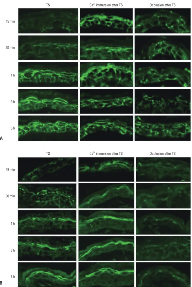
Fig. 3. Effect of epidermal Ca2+ gradient modulation on the expression and localization of Cldn-1 and Cldn-4 in murine epidermis. Skin sam- ples were taken at 15 min, 30 min, 1 h, 3 h, and 6 h after tape-stripping (TS) with/without immersion in calcium containing solution or oc- clusion. Frozen sections (5 μm) were immunostained with Cldn-1 (A) and Cldn-4 (B) primary antibodies (Zymed Laboratories, San Francisco, CA, USA) and an FITC conjugated donkey anti-rabbit IgG, secondary antibody (Santa Cruz, CA, USA) and examined by confo- cal microscopy. Magnification ×400. FITC, fluorescein isothiocyanate; Cldn, claudin.
TS
TS
Ca2+ immersion after TS
Ca2+ immersion after TS
Occlusion after TS
Occlusion after TS 15 min
15 min 30 min
30 min 1 h
1 h 3 h
3 h 6 h
6 h
A
B
er perturbation and consequently delayed barrier recovery.
15The same occlusion method caused a delay in Cldn-1 and Cldn-4 recovery, even at 6 hours after tape-stripping com- pared with non-occluded group, indicating that inhibition of Ca
2+gradient recovery prevented Cldn-1 and Cldn-4 resto- ration (Fig. 3). Taken together, we concluded that acute loss of extracellular Ca
2+in SG layer by tape-stripping induced alteration of Cldn-1 and Cldn-4 expression with impaired TJ barrier function and adequate recovery of Ca
2+gradient stimulated restoration of Cldn-1 and Cldn-4 expression. The regulatory mechanisms of epidermal TJ by epidermal Ca
2+gradient have not been fully elucidated. One possible ex- planation is that loss of extracellular Ca
2+in the SG might inhibit the signal of E-cadherin, which is important in the formation of functional TJ by a protein kinase C-induced Cldn incorporation into TJ.
16Regarding the recovery kinetics of the barrier, our results show that the TJ barrier immediately disturbed following SC barrier disruption and then normalized within 1 hour after in- jury. Moreover, an increase in Cldn-1 and Cldn-4 mRNA and protein started 1 hour after barrier disruption until 6 hours.
Based on the recovery kinetic of SC barrier, which includes several steps such as preformed lamellar bodies secretion within 30 minutes,
17,18accelerated lamellar body formation between 2 to 6 hours,
17and increased DNA synthesis with- in 16-24 hours,
19we found that recovery time of TJ barrier is faster compared with SC barrier. This fast recovery kinet- ic of TJ in response to barrier disruption might be a com- pensation mechanism of the skin by enhancing the inside- out permeability barrier and epidermal calcium ion gradient before the full recovery of SC permeability barrier function to maintain homeostasis. These findings of our study are in accordance with previous studies, which observed the en- hanced TJ formation in the conditions of abnormal or ab- sent SC barrier such as early stage of wound healing
7and developing fetal skin.
20In summary, we demonstrated the physiological relation- ship between the TJ and SC barrier during barrier repair following acute SC barrier disruption.
REFERENCES
1. Brandner JM, Proksch E. Epidermal barrier function: role of tight junctions. In: Elias PM, Feingold KR, editors. Skin Barrier. New York: Taylor and Francis; 2006. p.191-210.
2. Brandner JM, Kief S, Grund C, Rendl M, Houdek P, Kuhn C, et al. Organization and formation of the tight junction system in hu-
was recovered when Cldn-1 and Cldn-4 expression patterns were normalized.
There have been in vitro evidences that extracellular Ca
2+plays a key role in maintaining TJ integrity. In simple epi- thelial cells, depleting Ca
2+from the culture medium tem- porarily dislocate TJ accompanied by weakening of TJ bar- rier, while switch to high Ca
2+reorganized TJ with normal barrier function.
10Recent study has also found that Ca
2+de- pletion in stratified cultures of human keratinocytes caused an altered expression of TJ proteins with decreased barrier function, which recovered following switch to the high-cal- cium medium.
11Epidermal permeability barrier homeosta- sis is tightly regulated by epidermal Ca
2+gradients.
12Upon barrier disruption, extracellular calcium in the upper epider- mis rapidly decreases along with increased TEWL.
13From these findings, we postulated that the mechanisms underly- ing the rapid and transient alteration of Cldn-1 and Cldn-4 is associated with rapid loss of extracellular calcium in the SG where TJ located. To address this issue, we performed two methods to modulate epidermal Ca
2+gradient.
Previous studies demonstrated that immersion of the bar-
rier disrupted skin in high calcium containing solutions de-
layed barrier repair by preventing the loss of calcium con-
tents from the upper epidermis.
14By the method as previous
described, we evaluated the relative changes in Cldn-1 and
Cldn-4 expression in the tape-stripped skin which immedi-
ately immersed in 1.8 mM Ca
2+-containing solution or non-
immersed skin. There was no obvious change in Cldn-1 and
Cldn-4 expression at 15 and 30 minutes after tape-stripping
in high calcium solution-immersed skin, whereas non-im-
mersed skin showed decreased staining intensity and dis-
continuous staining pattern of Cldn-1 and Cldn-4 compared
to control skin (Fig. 3). These results show that prevention
of the rapid decrease in the upper epidermal extracellular
Ca
2+inhibits the alteration of Cldn-1 and Cldn-4 expres-
sion, suggesting that extracellular Ca
2+decrease by acute
barrier disruption induces Cldn-1 and Cldn-4 dislocation as
early response. We found that the decreased immunoreac-
tivity of Cldn-1 and Cldn-4 following barrier disruption al-
most recovered their normal expression pattern by 1 hour
(Fig. 1A). To investigate whether this subsequent recovery
of Cldn-1 and Cldn-4 expression following barrier disrup-
tion could be associated with recovery of the calcium gradi-
ent, we applied occlusion method to the barrier disrupted
animals. It was shown that the rapid artificial restoration of
permeability barrier by occlusion with vapor-impermeable
membrane inhibited Ca
2+gradient recovery following barri-
Tight junction proteins in keratinocytes: localization and contribu- tion to barrier function. Exp Dermatol 2007;16:324-30.
12. Lee SH, Elias PM, Proksch E, Menon GK, Mao-Quiang M, Fein- gold KR. Calcium and potassium are important regulators of barri- er homeostasis in murine epidermis. J Clin Invest 1992;89:530-8.
13. Menon GK, Elias PM, Lee SH, Feingold KR. Localization of cal- cium in murine epidermis following disruption and repair of the permeability barrier. Cell Tissue Res 1992;270:503-12.
14. Denda M, Fuziwara S, Inoue K. Influx of calcium and chloride ions into epidermal keratinocytes regulates exocytosis of epider- mal lamellar bodies and skin permeability barrier homeostasis. J Invest Dermatol 2003;121:362-7.
15. Elias P, Ahn S, Brown B, Crumrine D, Feingold KR. Origin of the epidermal calcium gradient: regulation by barrier status and role of active vs passive mechanisms. J Invest Dermatol 2002;119:
1269-74.
16. Tunggal JA, Helfrich I, Schmitz A, Schwarz H, Günzel D, Fromm M, et al. E-cadherin is essential for in vivo epidermal barrier func- tion by regulating tight junctions. EMBO J 2005;24:1146-56.
17. Menon GK, Feingold KR, Elias PM. Lamellar body secretory re- sponse to barrier disruption. J Invest Dermatol 1992;98:279-89.
18. Elias PM, Cullander C, Mauro T, Rassner U, Kömüves L, Brown BE, et al. The secretory granular cell: the outermost granular cell as a specialized secretory cell. J Investig Dermatol Symp Proc 1998;3:87-100.
19. Proksch E, Feingold KR, Man MQ, Elias PM. Barrier function reg- ulates epidermal DNA synthesis. J Clin Invest 1991;87:1668-73.
20. Pummi K, Malminen M, Aho H, Karvonen SL, Peltonen J, Pel- tonen S. Epidermal tight junctions: ZO-1 and occludin are ex- pressed in mature, developing, and affected skin and in vitro dif- ferentiating keratinocytes. J Invest Dermatol 2001;117:1050-8.
man epidermis and cultured keratinocytes. Eur J Cell Biol 2002;
81:253-63.
3. Furuse M, Hata M, Furuse K, Yoshida Y, Haratake A, Sugitani Y, et al. Claudin-based tight junctions are crucial for the mammalian epidermal barrier: a lesson from claudin-1-deficient mice. J Cell Biol 2002;156:1099-111.
4. Hadj-Rabia S, Baala L, Vabres P, Hamel-Teillac D, Jacquemin E, Fabre M, et al. Claudin-1 gene mutations in neonatal sclerosing cholangitis associated with ichthyosis: a tight junction disease.
Gastroenterology 2004;127:1386-90.
5. Yuki T, Hachiya A, Kusaka A, Sriwiriyanont P, Visscher MO, Morita K, et al. Characterization of tight junctions and their dis- ruption by UVB in human epidermis and cultured keratinocytes. J Invest Dermatol 2011;131:744-52.
6. Ohnemus U, Kohrmeyer K, Houdek P, Rohde H, Wladykowski E, Vidal S, et al. Regulation of epidermal tight-junctions (TJ) during infection with exfoliative toxin-negative Staphylococcus strains. J Invest Dermatol 2008;128:906-16.
7. Malminen M, Koivukangas V, Peltonen J, Karvonen SL, Oikarin- en A, Peltonen S. Immunohistological distribution of the tight junction components ZO-1 and occludin in regenerating human epidermis. Br J Dermatol 2003;149:255-60.
8. Kubo A, Nagao K, Yokouchi M, Sasaki H, Amagai M. External antigen uptake by Langerhans cells with reorganization of epider- mal tight junction barriers. J Exp Med 2009;206:2937-46.
9. Tsuruta D, Green KJ, Getsios S, Jones JC. The barrier function of skin: how to keep a tight lid on water loss. Trends Cell Biol 2002;
12:355-7.
10. Farshori P, Kachar B. Redistribution and phosphorylation of oc- cludin during opening and resealing of tight junctions in cultured epithelial cells. J Membr Biol 1999;170:147-56.
11. Yuki T, Haratake A, Koishikawa H, Morita K, Miyachi Y, Inoue S.