• 검색 결과가 없습니다.

Decursin induces apoptosis in glioblastoma cells, but not in glial cells a mitochondria-related caspase pathway

N/A
N/A
Protected

Academic year: 2022

Share "Decursin induces apoptosis in glioblastoma cells, but not in glial cells a mitochondria-related caspase pathway"

Copied!
7
0
0

로드 중.... (전체 텍스트 보기)

전체 글

(1)

Author contributions: S.T.O. and S.M.L. carried out cell experiments and western blotting and C.H. carried out primary glial cell culture. B.S.K., S.C.P.

and D.I.K. wrote the draft manuscript. S.H.J. and B.A.S. organized the study.

This is an Open Access article distributed under the terms of the Creative Commons Attribution Non-Commercial License, which permits unrestricted non-commercial use, distribution, and reproduction in any medium, provided the original work is properly cited.

Copyright © Korean J Physiol Pharmacol, pISSN 1226-4512, eISSN 2093-3827

INTRODUCTION

Angelica gigas (Dang Gui) is a monocarpic biennial plant from China, Korea and Japan, and is used in traditional herbal medicine. It inhabits forests, grasslands and banks of streams.

The dried roots are used in the treatment of female reproductive health issues such as dysmenorrhea, amenorrhea and menopausal syndromes by enhancing blood circulation and new blood syn- thesis [1].

Decursin, a coumarin derivative, is one of major constituents of this plant [2] and has been reported to inhibit the growth and

survival of some metastatic prostatic cancer cells [3,4]. However, there have been no studies conducted on the use of decursin for the prevention or treatment of brain tumors. The most common primary central nervous system tumor, glioblastoma, represents about 30% of all brain tumors and 80% of all malignant tumors.

It can be categorized into four grades (I to IV), where grades I and II reflect low-grade gliomas with grades III and IV (glioblastoma) being high-grade gliomas. Nearly 60% of high-grade gliomas are glioblastoma, and the incidence rate for these tumors is approxi- mately 3 per 100,000 [5]. The current clinically used treatment strategies for glioblastoma include surgery followed by concurrent

Original Article

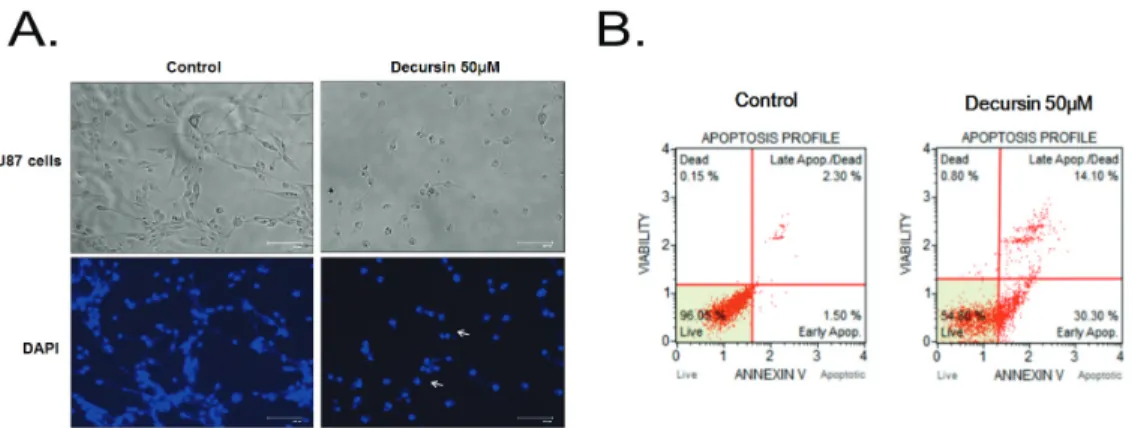
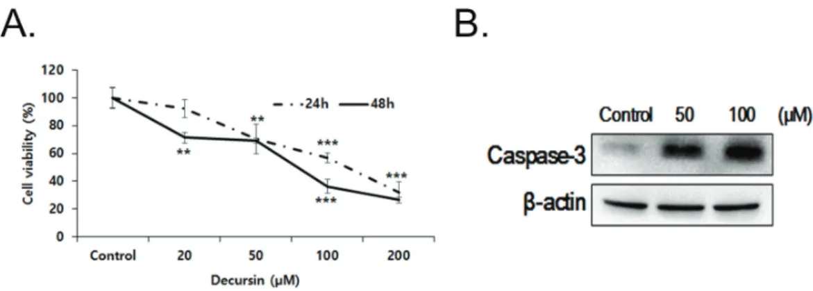
Decursin induces apoptosis in glioblastoma cells, but not in glial cells via a mitochondria-related caspase pathway

Seung Tack Oh 1 , Seongmi Lee 2 , Cai Hua 3 , Byung-Soo Koo 4 , Sok Cheon Pak 5 , Dong-Il-Kim 6, *, Songhee Jeon 3, *, and Boo Ahn Shin 7, *

1

Research Institute, Dongkwang Pharmaceutical Company, Ltd., Seoul 04535, Korea,

2

Department of Child and Adolescent Psychiatry, National Center for Mental Health, Seoul 04933, Korea,

3

Department of Biomedical Sciences, Center for Creative Biomedical Scientists at Chonnam National University, Gwangju 61469, Korea,

4

Department of Neuropsychiatry, College of Korean Medicine, Dongguk University, Goyang 10326, Korea,

5

School of Biomedical Sciences, Charles Sturt University, Bathurst, NSW 2795 Australia,

6

Department of Obstetrics & Gynecology, College of Korean Medicine, Dongguk University, Goyang 10326, Korea,

7

Department of Microbiology and Immunology, Chonnam National University Medical School, Gwangju 61469, Korea

ARTICLE INFO Received January 9, 2017 Revised October 10, 2018 Accepted November 6, 2018

*Correspondence Dong-Il Kim

E-mail: obgykdi@hanmail.net Songhee Jeon

E-mail: jsong0304@jnu.ac.kr Boo Ahn Shin

E-mail: bashin@jnu.ac.kr

Key Words Anti-cancer activity Apoptosis Cell cycle arrest Decursin Glioblastoma

ABSTRACT Decursin is a major biological active component of Angelica gigas Nakai and is known to induce apoptosis of metastatic prostatic cancer cells. Recently, other reports have been commissioned to examine the anticancer activities of this plant.

In this study, we evaluated the inhibitory activity and related mechanism of action of decursin against glioblastoma cell line. Decursin demonstrated cytotoxic effects on U87 and C6 glioma cells in a dose-dependent manner but not in primary glial cells.

Additionally, decursin increased apoptotic bodies and phosphorylated JNK and p38

in U87 cells. Decursin also down-regulated Bcl-2 as well as cell cycle dependent pro-

teins, CDK-4 and cyclin D1. Furthermore, decursin-induced apoptosis was dependent

on the caspase activation in U87 cells. Taken together, our data provide the evidence

that decursin induces apoptosis in glioblastoma cells, making it a potential candidate

as a chemotherapeutic drug against brain tumor.

(2)

ionizing radiation and chemotherapy. Glioblastoma patients have poor prognosis with a median survival time of only approximate- ly one year due to the rapid proliferation and accelerated activities of tumor cells. Unfortunately, the 5-year survival rate is only 9.8%

[6]. Decursin-mediated glioblastoma treatment has rarely been studied. Thus, the purpose of this study was to evaluate the ef- fects of decursin on glioblastoma using a human glioblastoma cell line, U87 and rat glioma cell line, C6.

METHODS

Cell culture

A human glioblastoma cell line, U87 and rat glioblastoma cell line, C6 were purchased from the American Type Culture Collec- tion (Rockville, MD, USA). Cells were maintained in Dulbecco’s Modified Eagle’s Medium (DMEM) supplemented with 10% fetal bovine serum (FBS) (Welgene, Seoul, Korea), 100 U/ml penicillin and 0.1 mg/ml streptomycin (Welgene, Seoul, Korea) in condition of 95% air and 5% CO

2

at 37°C.

Mixed glial cell culture

Primary mixed glial cells were prepared from postnatal day 2 pups of mouse. Briefly, cerebral cortices were subjected to Trypsin-EDTA mix digestion (Invitrogen, CA, USA) for 30 min at 37°C. The digested tissue was carefully triturated into single cells using increasingly smaller pipette tips. Cells were then cen- trifuged at 250 × g for 5 min and re-suspended in plating medium supplemented with DFF10 (DMEM:F12 1:1 + 10% FBS) including 100 U/ml streptomycin and 100 Pg/ml penicillin. Dissociated cells were seeded into a poly-L-lysine coated 6-well plate and in- cubated at 37°C.

Cell viability

Cells were seeded into a 24-well plate at a density of 1×10

5

/ml.

The next day, cells were treated with various concentration of decursin (10, 20, 50, 100 and 200 PM) for 24 h or 48 h incuba- tion. MTT solution was treated for 1 h at 37°C. Then, dimethyl sulfoxide was added to dissolve formazan crystals and incubated for 30 min. The optical density of solubilized formazan crystals was measured by using a spectrophotometer (Molecular Device, Sunnyvale, CA, USA) at 580 nm. The half maximal inhibitory concentration (IC

50

) was calculated using Calcusyn software (Bio- soft, Cambridge, UK). Caspase apoptosis dependency was ana- lyzed using 50 PM of pan caspase inhibitor (Cas I), p38 inhibitor (SB203580) and JNK inhibitor (SP600125) which were acquired from Calbiochem (EMD Millipore, Darmstadt, Germany). Cells were pre-treated for 30 min prior to treatment of 25 or 50 PM of decursin. At 24 h, cell viability was examined by MTT assy.

Immunoblotting

The cells were lysed with 1x lysis buffer (2% SDS, 1% 2-mercap- toethanol, 10% glycerol and 0.1 mg/ml bromophenol blue in Tris- HCl, pH 6.8) and heated at 100°C for 10 min. Thirty Pg of cell lysates were electrophoresed in 10% or 15% SDS-PAGE and gels were transferred on nitrocellulose membrane. Then, the mem- brane was incubated with anti-Bcl-2, anti-Bax, anti-phospho- p38, anti-p38 anti-phospho-JNK, anti-JNK, anti-caspase-3, anti- cleaved caspase-3, anti-caspase-7, anti-cleaved caspase-7, anti- caspase-9, anti-cleaved caspase-9, anti-cleaved PARP-1, anti- CDK-4, anti-CyclinD1 and anti- E actin antibody (Cell Signaling Technology, Beverly, MA, USA) for overnight at 4°C. To obtain band image, horseradish peroxidase-conjugated anti-mouse or anti-rabbit IgG was incubated on the blot for 1 h at room tem- perature. After washing the blot, bands were visualized using the ECL system (Thermo Fisher Scientific, Waltham, MA, USA).

Band images were obtained by using Molecular Imager Chemi- Doc XRS+ (Bio-Rad, Hercules, CA, USA) and band intensity was measured by Image Lab

TM

software version 2.0.1 (Bio-Rad).

Annexin V

U87 cells (1×10

5

/ml) were treated with decursin (50 or 100 PM) for 24 h. Then, cells were trypsinized and re-suspended with 500 Pl of 1x binding buffer. Apoptotic cells were calculated by double staining with Muse

TM

Annexin V & Dead Cell reagent (Merck KGaA, Darmstadt, Germany) and examined by using the Muse

TM

Cell Analyzer (Merck KGaA, Darmstadt, Germany).

Cell cycle analysis

U87 cells were harvested, fixed in ethanol, and stored at –20°C.

Muse

TM

Cell Cycle Reagent (Merck KGaA, Darmstadt, Germany) was added in the ethanol-fixed cells and cell cycle population was acquired by using the Muse

TM

Cell Cycle software module (Merck KGaA, Darmstadt, Germany).

Data analysis

All statistical analyses were conducted with SPSS (ver. 21,

Somers, NY, USA). The data expressed as mean±SD were ana-

lyzed by one-way ANOVA, and the dissimilarity among means

were analyzed using the Dunnett’s test or Tukey–Kramer’s mul-

tiple comparison test. Differences were considered significant at

p<0.05.

(3)

RESULTS

Decursin at high doses has a cytotoxic effect in U87 cells

To examine cytotoxic effect of decursin in U87 cells, human glioblastoma cell lines were treated with various concentrations (0-200 PM) of decursin for 24 h and then analyzed by MTT assay.

As shown in Fig. 1, cell viability was gradually decreased by 100 PM of decursin treatment after 24 h, but primary glia cells were not changed by 100 PM. U87 glioblastoma cells almost died when treated with 200 PM of decursin, but primary glial cells showed a survival rate of more than 50% (Fig. 1B), implying that high concentration of decursin showed cytotoxic effect on the normal cells. The half maximal inhibitory concentration (IC

50

) value for decursin was calculated and it was 49.01 PM in U87 cells. Thus, in the present study, we used 50 PM to examine the mechanisms of action of decursin in the U87 cells.

Decursin induces apoptosis in U87 cells

The morphological characteristics of apoptosis were captured

at 24 h after decursin treatment in U87 cells. As shown in Fig. 2A, decursin induced chromatin condensation and apoptotic bodies.

To evaluate the potential apoptotic ability of decursin in U87 cells, we stained the cells with Annexin V and PI. At a dose of 50 PM, decursin induced apoptosis by 44.4% (Fig. 2B). To examine which signaling pathway may be involved in apoptosis, Western blotting was utilized in the decursin-treated U87 cells. Activations of c- Jun NH2-terminal kinase (JNK) and p38 were examined by using phospho-specific antibodies. Increased phosphorylation of P38 and JNK was observed from 0.5 h until 1 h of decursin treatment (Fig. 3A). We also examined the expression ratio of Bcl-2/Bax in U87 cells after decursin treatment. Bcl-2 inhibits apoptosis by pre- venting mitochondrial membrane depolarization, whereas Bax promotes apoptosis by enhancing mitochondrial membrane de- polarization and cytochrome c release. As shown in Fig. 3B, Bcl-2 expression was decreased by treatment of 50 or 100 PM decursin, but Bax expression was not changed. Moreover, the ratio of Bcl-2/

Bax was significantly decreased by decursin. Next, we examined the caspase activity and PARP-1 cleavage after 50 or 100 PM of decursin treatment. Cleaved caspase-3, -7, or -9 activities were in- creased by decursin treatment in a dose-dependent manner and cleaved PARP-1 also increased by 100 PM of decursin, suggesting

Fig. 2. Decursin induces apoptosis in U87 cells. (A) The image shows that decursin induced chromatin condensation and apoptotic bodies. (B) U87 cells were treated with 50 PM of decursin for 24 h, stained with Annexin V and PI, and analyzed with FACS system.

Fig. 1. Decursin has a cytotoxic effect in U87 cells but not in primary glia cells. (A) Chemical structure of decursin. (B) U87 cells were treated with various concentrations of decursin (10, 20, 50, 100 and 200 PM) for 24 h. (C) Primary glia cells were treated with different concentrations of decursin (10, 20, 50, 100 and 200 PM) for 24 h. Cell viability was determined by the MTT assay. Values are mean±SD (n=3). **p<0.01 vs. control, and ***p<0.001 vs.

control.

(4)

that decursin induced apoptotic cell death in U87 cells (Fig. 3C).

Decursin induces apoptosis by caspase-dependent manner in U87 cells

To examine the signaling pathway responsible for the cell death, JNK or p38 kinase inhibitor was pre-treated in the decur- sin-treated cells. Either SB203580 as p38 inhibitor or SP600125 as JNK inhibitor did not suppress decursin-induced U87 cell death (Fig. 4A). In order to verify the importance of caspase activation in the decursin-induced apoptosis, U87 cells were pre-treated with a pan caspase inhibitor (Cas I) for 30 min prior to treatment with 50 PM of decursin. At 24 h, cell viability was examined by MTT assay. Pre-treatment with a pan caspase inhibitor signifi- cantly suppressed decursin-induced cell death in U87 cells, in- dicating that decursin-induced cell death is mediated by caspase activation in U87 cells (Fig. 4B).

Decursin increases G1 population apoptosis by caspase-dependent manner in U87 cells

Next, we analyzed cell cycle by using flow cytometry analysis.

Decursin increased the amount of sub-G1 DNA population dose- dependently in U87 cells compared with that of control (Fig. 5A).

In order to confirm the cell cycle arrest by decursin treatment, we examined the expression level of cell cycle-dependent proteins, cyclin dependent protein kinase-4 (CDK-4) and cyclin D1, in decursin-treated U87 cells. The expression of both proteins was decreased by 100 PM of decursin in U87 cells ( Fig. 5), which indi- cates a cell cycle arrest in G1/S stage.

Decursin induces apoptosis in C6 cells

To confirm the cytotoxic effect of decursin on other glioma cell, rat glioma C6 cells were treated with various concentrations of decursin. After 24 or 48 h, cell viability was examined by MTT method. Decursin induced cell death and caspase activation

Fig. 3. Decursin induces phosphorylation of JNK and p38 in U87 cells, alters Bcl-2 family protein levels, and activates caspases and PARP-1 in U87 cells. (A) U87 cells were treated with 50 PM of decursin for various periods of time (0, 0.5, 1, 3 and 4 h). The cell lysates were subjected to Western blot analysis for phospho-JNK, total-JNK, phospho-p38 and total p38. (B) U87 cells were treated with 50 or 100 PM of decursin for 24 h. The cell lysates were subjected to Western blot analysis for Bax and Bcl-2 (C), cleaved caspase-3, 7, 9, PARP-1 and E-actin. Values are mean±SD (n=3). *p<0.05 vs. con- trol. **p<0.01 vs. control.

Fig. 4. Decursin induces apoptosis in a caspase-dependent manner in U87 cells. (A) U87 cells were pre-treated with 5 PM of SB203580 (p38 in- hibitor) and SP600125 (JNK inhibitor) for 30 min prior to decursin treatment (50 PM). Cell viability was determined by the MTT assay. (B) U87 cells were pre-treated with 10 PM of pan caspase inhibitor (Cas Ι) for 30 min prior to decursin treatment (50 PM). Cell viability was determined by the MTT assay.

Values are mean±SD (n=4). **p<0.01 vs. control.

(5)

dose-dependently in C6 cells (Fig. 6), suggesting decursin induces apoptotic cell death in glioma C6 cells.

DISCUSSION

Previous studies have shown that decursin induced apoptosis in several cancer cell lines such as prostate cancer, myeloid leu- kemia, breast cancer and colon cancer [7-9], however there have been no reports of the effect that decursin may have on brain tumor cells. In this study, we examined the anticancer effect of decursin on glioblastoma cell lines, U87 and C6. We showed that decursin induced apoptosis of U87 and C6 cells in a dose-depen- dent manner but not in primary glial cells. The effect of decursin on primary glial cells is in accordance with the previous report that decursin has neuroprotective effect in the glutamate-injured cortical cells [10].

Apoptosis is a mechanism of regulated cell death employed during normal development [11] where mitochondrial apoptotic pathways act as important signals of apoptotic cell death in mam- malian cells [12]. The Bcl-2 family proteins are the major regula- tors of apoptosis and consist of three subfamilies such as Bcl-

2, Bax and Bik. While Bcl-2 is an anti-apoptotic member of the family, Bax is a pro-apoptotic member, and it is thought that the ratio between anti-apoptotic and pro-apoptotic members of this family may contribute to the vulnerability of a cell to apoptosis [13]. In this study, we demonstrated that decursin decreased Bcl- 2 expression without any change to Bax expression in U87 cells, which resulted in a significant decrease in the Bcl-2/Bax ratio and therefore may be involved in the apoptosis process.

In addition, decursin caused PARP cleavage. A decrease in the Bcl-2/Bax ratio may have resulted in the release of cytochrome c and the initiation of apoptosis. After its release, cytochrome c interacts with procaspase-9 and apoptotic protease activating fac- tor-1 (Apaf-1) to activate caspase-9, and caspase-9 instigates a cas- cade of further caspase activation by processing caspases-3 and -7, which leads to PARP cleavage and eventually apoptosis [14,15].

Caspase-3 is the most well-known effector caspase and its activa- tion leads to cellular death by the proteolytic cleavage of a large variety of cellular proteins and the activation of pro-apoptotic factors that destroy the cell [16,17]. Moreover, PARP cleavage dur- ing caspase activation facilitates cellular disassembly and serves as a marker of cells undergoing apoptosis [18]. This PARP cleav- age was observed in U87 cells treated with decursin at concentra-

Fig. 5. Decursin increases G1 population of cell cycle and down-regulates cell cycle proteins in U87 cells. U87 cells were treated with 50 PM of decursin for 24 h. The cell lysates were subjected to flow cytometric analysis and Western blot analysis to examine the expression of CDK-4, cyclin D1 and E-actin. Percentage of each cell population was presented in graph (A). The intensity of the each band was normalized to E-actin and presented in bar graphs (B). Values are mean±SD (n=3). **p<0.01 vs. control.

Fig. 6. Decursin induces apoptosis and caspase-3 activation in C6 cells. (A) C6 cells were treated with various concentrations of decursin (10, 20,

50, 100 and 200 PM) for 24 h and 48 h. Values are mean±SD (n=4). **p<0.01 vs. control, ***p<0.001 vs. control. (B) The cell lysates were subjected to

Western blot analysis for cleaved caspase-3 and E-actin.

(6)

tions of 50 and 100 PM, suggesting that decursin activates this mitochondria apoptotic pathway in neuroblastoma cells.

Furthermore, it has been reported that the JNK and p38 kinase pathways are related with an increased rate of apoptosis, whereas the ERK1/2 pathway activated by cell growth has been shown to be important for cell survival by inhibiting apoptosis [19-21].

However, alternate studies have demonstrated that the JNK path- way, when activated, is not involved in apoptosis of neuronal cells [22]. Other studies showed that JNK is also important for the de- velopment and survival of macrophages [23]. Furthermore, where UV and S-nitrosoglutathione were administered, p38 inhibition was unable to prevent apoptosis [24,25]. This is consistent with this study, where inhibition p38 or JNK did not block decursin- induced apoptosis. These differences suggest the existence of marked alterations in the regulation of stress reactions depending on the type of cell being examined, so that the implication of ei- ther JNK or p38 is difficult to explain and may be confounded by many factors.

Cancer is unchecked cell growth. Mutations in genes can cause cancer by accelerating cell division rates or inhibiting normal controls on the system, such as cell cycle arrest or programmed cell death. Glioma cells are distinguished from normal glial cells by abnormal proliferation and anti-apoptosis. In this study, we found that decursin markedly inhibited the viabilities of U87 and C6 cells and increased the apoptosis of glioma cells in a dose-de- pendent manner. Besides, decursin down-regulated cyclin D1 and CDK-4 expression, which are involved in the arrest of G1 in the prostate cancer [4]. This is in concert with previous reports that decursin induced G1 arrest and apoptosis in the human prostate, bladder and colon cancer cells [26]. Thus, it is suggested that the decursin-induced G1 cell cycle arrest is through a CDK4/cyclin D1-mediated pathway in the rapidly proliferating cells, resulting in cell death of glioma cells but not primary glial cells.

In this study, IC

50

of decursin was 49.01 PM in U87 cells. How- ever, the development of a natural substance as a new drug has the potential to become an IC

50

in nM units. Indeed, decursin has an anticancer effect over 50 PM in the various cancer cell line [4,26-29] and many studies have shown that decursin has syner- getic effects with doxorubicin or c-myc inhibitor [27-29]. Combi- nation therapy has been widely used as a method for overcoming the limitations of chemotherapy in the treatment of cancer [30].

Thus, decursin can be used in combination with other anticancer drugs to treat glioblastoma.

Taken together, decursin inhibited proliferation of neuroblas- toma cell line via G1/S cell cycle arrest and induction of apopto- sis through caspase-3 activation, and down-regulation of Bcl-2 protein expression without negative effect on primary glia cells.

Malignant gliomas are the most common type of primary brain tumor that even after aggressive therapy the survival rate is poor, and thus there is an urgent need to develop specific and effective therapies [31]. Moreover, intraperitoneal injection of decursin strongly inhibited selective neuronal death, astrogliosis, and oxi-

dative stress induced by KA administration [32], suggesting that decursin could penetrate brain blood barrier. Hence, decursin could be developed as an antitumor drug to treat glioblastoma patients without damage on normal brain cells.

ACKNOWLEDGEMENTS

This work was supported by a grant of the Korean Health Tech- nology R&D Project, Ministry of Health and Welfare, Republic of Korea (No. HN12C0062, HI15C0198).

CONFLICTS OF INTEREST

The authors declare no conflicts of interest.

REFERENCES

1. Heo J. DONGUIBOGAM Treasured Mirror of Eastern Medicine Part I Internal Bodily Elements. Seoul: Chin Young; 2013.

2. Ahn MJ, Lee MK, Kim YC, Sung SH. The simultaneous determina- tion of coumarins in Angelica gigas root by high performance liquid chromatography-diode array detector coupled with electrospray ionization/mass spectrometry. J Pharm Biomed Anal. 2008;46:258- 266.

3. Rhim JS. In vitro human cell culture models for the study of pros- tate cancer. Prostate Cancer Prostatic Dis. 2000;3:229-235.

4. Yim D, Singh RP, Agarwal C, Lee S, Chi H, Agarwal R. A novel an- ticancer agent, decursin, induces G1 arrest and apoptosis in human prostate carcinoma cells. Cancer Res. 2005;65:1035-1044.

5. de Groot JF. High-grade gliomas. Continuum (Minneap Minn).

2015;21:332-344.

6. Stupp R, Mason WP, van den Bent MJ, Weller M, Fisher B, Ta- phoorn MJ, Belanger K, Brandes AA, Marosi C, Bogdahn U, Cur- schmann J, Janzer RC, Ludwin SK, Gorlia T, Allgeier A, Lacombe D, Cairncross JG, Eisenhauer E, Mirimanoff RO; European Organisa- tion for Research and Treatment of Cancer Brain Tumor and Radio- therapy Groups; National Cancer Institute of Canada Clinical Trials Group. Radiotherapy plus concomitant and adjuvant temozolomide for glioblastoma. N Engl J Med. 2005;352:987-996.

7. Jiang C, Guo J, Wang Z, Xiao B, Lee HJ, Lee EO, Kim SH, Lu J.

Decursin and decursinol angelate inhibit estrogen-stimulated and estrogen-independent growth and survival of breast cancer cells.

Breast Cancer Res. 2007;9:R77.

8. Ahn Q, Jeong SJ, Lee HJ, Kwon HY, Han I, Kim HS, Lee HJ, Lee EO, Ahn KS, Jung MH, Zhu S, Chen CY, Kim SH. Inhibition of cyclooxygenase-2-dependent survivin mediates decursin-induced apoptosis in human KBM-5 myeloid leukemia cells. Cancer Lett.

2010;298:212-221.

9. Son SH, Park KK, Park SK, Kim YC, Kim YS, Lee SK, Chung WY.

Decursin and decursinol from Angelica gigas inhibit the lung me- tastasis of murine colon carcinoma. Phytother Res. 2011;25:959-964.

10. Kang SY, Kim YC. Decursinol and decursin protect primary cul-

(7)

tured rat cortical cells from glutamate-induced neurotoxicity. J Pharm Pharmacol. 2007;59:863-870.

11. Waxman DJ, Schwartz PS. Harnessing apoptosis for improved anti- cancer gene therapy. Cancer Res. 2003;63:8563-8572.

12. Chen M, Li B, Zhao X, Zuo H, He X, Li Z, Liu X, Chen L. Effect of diallyl trisulfide derivatives on the induction of apoptosis in human prostate cancer PC-3 cells. Mol Cell Biochem. 2012;363:75-84.

13. Niture SK, Jaiswal AK. INrf2 (Keap1) targets Bcl-2 degradation and controls cellular apoptosis. Cell Death Differ. 2011;18:439-451.

14. Susin SA, Daugas E, Ravagnan L, Samejima K, Zamzami N, Loef- fler M, Costantini P, Ferri KF, Irinopoulou T, Prévost MC, Brothers G, Mak TW, Penninger J, Earnshaw WC, Kroemer G. Two distinct pathways leading to nuclear apoptosis. J Exp Med. 2000;192:571- 580.

15. Wang C, Youle RJ. The role of mitochondria in apoptosis. Annu Rev Genet. 2009;43:95-118.

16. Nicholson DW. Caspase structure, proteolytic substrates, and func- tion during apoptotic cell death. Cell Death Differ. 1999;6:1028- 1042.

17. Yacobi K, Wojtowicz A, Tsafriri A, Gross A. Gonadotropins en- hance caspase-3 and -7 activity and apoptosis in the theca-inter- stitial cells of rat preovulatory follicles in culture. Endocrinology.

2004;145:1943-1951.

18. Oliver FJ, de la Rubia G, Rolli V, Ruiz-Ruiz MC, de Murcia G, Mur- cia JM. Importance of poly(ADP-ribose) polymerase and its cleav- age in apoptosis. Lesson from an uncleavable mutant. J Biol Chem.

1998;273:33533-33539.

19. Johnson GL, Lapadat R. Mitogen-activated protein kinase pathways mediated by ERK, JNK, and p38 protein kinases. Science. 2002;

298:1911-1912.

20. Olson JM, Hallahan AR. p38 MAP kinase: a convergence point in cancer therapy. Trends Mol Med. 2004;10:125-129.

21. Sah JF, Balasubramanian S, Eckert RL, Rorke EA. Epigallocatechin- 3-gallate inhibits epidermal growth factor receptor signaling path- way. Evidence for direct inhibition of ERK1/2 and AKT kinases. J Biol Chem. 2004;279:12755-12762.

22. Rockwell P, Martinez J, Papa L, Gomes E. Redox regulates COX-2 upregulation and cell death in the neuronal response to cadmium.

Cell Signal. 2004;16:343-353.

23. Himes SR, Sester DP, Ravasi T, Cronau SL, Sasmono T, Hume DA.

The JNK are important for development and survival of macro- phages. J Immunol. 2006;176:2219-2228.

24. Franklin CC, Srikanth S, Kraft AS. Conditional expression of mitogen-activated protein kinase phosphatase-1, MKP-1, is cyto- protective against UV-induced apoptosis. Proc Natl Acad Sci U S A.

1998;95:3014-3019.

25. Callsen D, Brüne B. Role of mitogen-activated protein kinases in S- nitrosoglutathione-induced macrophage apoptosis. Biochemistry.

1999;38:2279-2286.

26. Kim WJ, Lee SJ, Choi YD, Moon SK. Decursin inhibits growth of human bladder and colon cancer cells via apoptosis, G1-phase cell cycle arrest and extracellular signal-regulated kinase activation. Int J Mol Med. 2010;25:635-641.

27. Jang J, Jeong SJ, Kwon HY, Jung JH, Sohn EJ, Lee HJ, Kim JH, Kim SH, Kim JH, Kim SH. Decursin and doxorubicin are in synergy for the induction of apoptosis via STAT3 and/or mTOR pathways in human multiple myeloma cells. Evid Based Complement Alternat Med. 2013;2013:506324.

28. Choi HS, Cho SG, Kim MK, Kim MS, Moon SH, Kim IH, Ko SG.

Decursin in Angelica gigas Nakai (AGN) enhances doxorubicin chemosensitivity in NCI/ADR-RES ovarian cancer cells via inhibi- tion of P-glycoprotein expression. Phytother Res. 2016;30:2020- 2026.

29. Kim E, Nam J, Chang W, Zulfugarov IS, Okhlopkova ZM, Olen- nikov D, Chirikova NK, Kim SW. Angelica gigas Nakai and decur- sin downregulate Myc expression to promote cell death in B-cell lymphoma. Sci Rep. 2018;8:10590.

30. Palmer AC, Sorger PK. Combination cancer therapy can confer benefit via patient-to-patient variability without drug additivity or synergy. Cell. 2017;171:1678-1691.e13.

31. Westphal M, Lamszus K. The neurobiology of gliomas: from cell biology to the development of therapeutic approaches. Nat Rev Neurosci. 2011;12:495-508.

32. Lee JK, Jeong JW, Jang T, Lee GW, Han H, Kang JS, Kim IH. Decur- sin attenuates kainic acid-induced seizures in mice. Neuroreport.

2014;25:1243-1249.

수치

Fig. 1. Decursin has a cytotoxic effect in U87 cells but not in primary glia cells. (A) Chemical structure of decursin
Fig. 4. Decursin induces apoptosis in a caspase-dependent manner in U87 cells. (A) U87 cells were pre-treated with 5  PM of SB203580 (p38 in- in-hibitor) and SP600125 (JNK inin-hibitor) for 30 min prior to decursin treatment (50  PM)
Fig. 6. Decursin induces apoptosis and caspase-3 activation in C6 cells. (A) C6 cells were treated with various concentrations of decursin (10, 20,  50, 100 and 200  PM) for 24 h and 48 h

참조

관련 문서

1) Effects of methanol extracts of Capsella bursa-pastoris on cyclooxygenase-2 (COX-2) and Inducible Nitric oxide synthase (iNOS) expression in human prostate cancer cell

We found that FoxO1 is constantly increased in MCF-7/ADR, adriamycin-resistant breast cancer cells, and FoxO1 has a critical role in the MDR1 gene expression (16).. There is

Results: The human breast cancer cell subline MCF-7/MX5 cells selected in the presence of 5 µg/ml mitoxantrone (MX) were more resistant to MX (15.7... Western blot and

PI3K inhibition decreased antioxidants/GD-induced apoptosis in A549 cells, and PI3K inhibitor LY294002 had inhibitory effect on antioxidants/GD-induced caspase-3

Taken together, these results suggested that latex containing the ficin inhibited the cell growth and induced apoptosis by caspase and Bcl-2 family signaling pathway in

In the present study, consistent to that report, up-regulation of VEGF mRNA was observed in the colon cancer cell with the acquired resistance to 5-FU and this

Bone is a common site of distant metastasis in prostate cancer. Association of bone and metastatic cancer cells are important in this site-specific manifestation

Results: Poorly differentiated colorectal cancer was frequently located at right colon and in advanced stage.. During follow-up, double primary cancer had occurred