1071 www.eymj.org
INTRODUCTION
Percutaneous coronary intervention (PCI) for chronic total occlusion (CTO) has a lower initial success rate than non- CTO lesions, however, it has many clinical benefits. Current meta-analysis reported that a successful PCI of CTO lesions resulted in an improvement of left ventricular ejection frac- tion (LVEF), reduced adverse remodeling, and improvement of survival rate.1 In the published ABSORB III study, CTO le- sions were one of exclusion criteria.2 Bioresorbable vascular scaffolds (BVS) for CTO Pilot study demonstrated the safety and feasibility in midterm efficacy.3 BVS is able to overcome many of the complications and limitations of metal stents.4 One of technical difficulties of BVS implantation is difficulty in identifying edge markers on the fluoroscopy for adequate stent positioning, particularly when performing ‘overlapping
stenting’ using BVSs. The present case shows clinical useful- ness of combined optical coherence tomography (OCT) and stent boost imaging guided ‘overlapping’ BVS implantation via antegrade approach for a typical right coronary artery (RCA) CTO lesion.
CASE REPORT
A 57-year old male patient with history of a failed PCI of a RCA CTO lesion since 6 months ago was readmitted to our center with a chief complaint of effort chest pain and background history of hypertension, dyslipidemia and diabetes mellitus.
The transthoracic echocardiogram showed preserved systolic function (LVEF 54%), and akinesia of the basal to mid inferior and basal septum and mid posterior wall. The bilateral angi- ography, done through both femoral artery approaches dem- onstrated total occlusion of the mid RCA and visualization of grade 2 collateral flow from left anterior descending artery (Fig. 1). First, therefore, we planned an antegrade RCA CTO in- tervention. A 7 French Amplatz Left 1 guiding catheter (Med- tronic, Launcher, CA, USA) was used to engage the RCA osti- um and 0.014" Runthrough guide wire (Terumo, Tokyo, Japan) was advanced through Corsair microcatheter (Asahi, Tokyo, Ja- pan). Then, the Runthrough guide wire was exchanged with a Fielder XT-A wire (Asahi Intecc, Aichi, Japan), which success-
Optical Coherence Tomography and Stent Boost Imaging Guided Bioresorbable Vascular Scaffold
Overlapping for Coronary Chronic Total Occlusion Lesion
Hu Li1,2, Seung-Woon Rha2, Cheol Ung Choi2, and Dong Joo Oh2
1Department of Cardiovascular, The Second Afliated Hospital of Kunming Medical University, Kunming, China;
2Cardiovascular Center, Korea University Guro Hospital, Seoul, Korea.
We report herein the optical coherence tomography (OCT) and stent boost imaging guided bioresorbable vascular scaffold (BVS) implantation for right coronary artery (RCA) chronic total occlusion (CTO) lesion. The gold standard for evaluating BVS expan- sion after percutaneous coronary intervention is OCT. However, stent boost imaging is a new technique that improves fluorosco- py-based assessments of stent overlapping, and the present case shows clinical usefulness of OCT and stent boost imaging guid- ed ‘overlapping’ BVS implantation via antegrade approach for a typical RCA CTO lesion.
Key Words: CTO, BVS, OCT, stent boost
Case Report
pISSN: 0513-5796 · eISSN: 1976-2437
Received: August 25, 2016 Revised: November 7, 2016 Accepted: November 11, 2016
Corresponding author: Dr. Seung-Woon Rha, Cardiovascular Center, Korea Uni- versity Guro Hospital, 148 Gurodong-ro, Guro-gu, Seoul 08308, Korea.
Tel: 82-2-2626-3020, Fax: 82-2-864-3062, E-mail: [email protected]
•The authors have no financial conflicts of interest.
© Copyright: Yonsei University College of Medicine 2017
This is an Open Access article distributed under the terms of the Creative Com- mons Attribution Non-Commercial License (http://creativecommons.org/licenses/
by-nc/4.0) which permits unrestricted non-commercial use, distribution, and repro- duction in any medium, provided the original work is properly cited.
Yonsei Med J 2017 Sep;58(5):1071-1074 https://doi.org/10.3349/ymj.2017.58.5.1071
1072
BVS Overlapping for CTO Lesion
https://doi.org/10.3349/ymj.2017.58.5.1071 fully crossed the CTO segment. Then, the Fielder XT-A wire
was exchanged to Runthrough wire for balloon dilation. Sequ- ential predilation using Laxa balloon 1.3×10 mm (Goodman, Nagoya, Japan) and Genoss 2.0×15 mm (Genoss, Suwon, Korea)
was done for lesion pretreatment. OCT examination of RCA revealed that reference lumen diameter was 2.89 mm and le- sion length 41 mm.
First BVS (Abbott Vascular, Santa Clara, CA, USA) 3.0×18
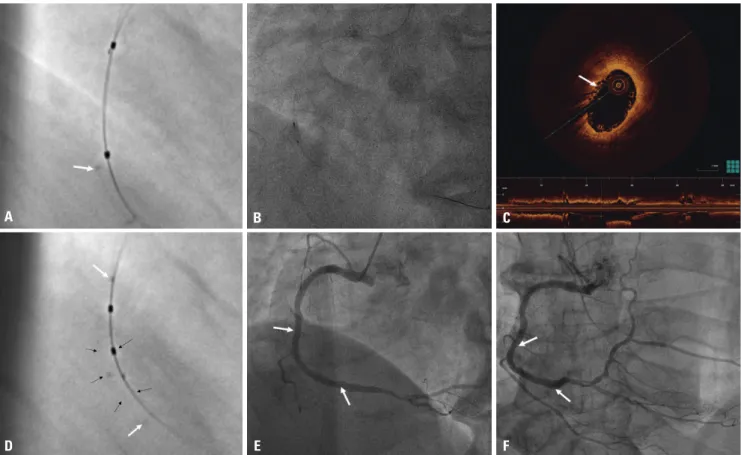
Fig. 2. Revascularization for CTO in RCA. (A) Runthrough guide wire was advanced through Corsair microcatheter. (B) Fielder XT-A wire successfully crossed the CTO segment. (C) Fielder XT-A was exchanged to Runthrough soft wire, and after balloon dilation (1.3×10 mm Laxa and 2.0×15 mm Laxa balloon), we performed an OCT study of RCA. (D) OCT findings after predilation. (E and F) Two overlapping 3.0×18 mm and 3.0×28 mm BVSs (Absorb BVS) were implanted from mid to distal RCA. CTO, chronic total occlusion; RCA, right coronary artery; OCT, optical coherence tomography; BVS, bio- resorbable vascular scaffold.
D A
E B
F C
Fig. 1. Baseline coronary angiography. (A) Chronic total occlusion of mid right coronary artery (RCA) (arrow). (B) Collateral flow from left coronary ar- tery to RCA (grade 2, arrow).
A B
1073
Hu Li, et al.
https://doi.org/10.3349/ymj.2017.58.5.1071
mm was delivered into the distal RCA lesion and deployed by slow inflation (2 atm, every 5 seconds) up to nominal pressure.
After acquisition of the angiographic and stent boost images during second BVS positioning, multiple analyses and obser- vations were done to understand the relationship between the BVS marker beads and balloon markers of distal and proximal BVSs. After clear identification of marker-to-marker and mar- ket-to-bead relationship, the second BVS was successfully over- lapped and deployed. The distal markers of the second scaf- fold was over the proximal markers of the first scaffold, resulting in 1–2 mm of overlap. By minimal overlapping of two scaffolds, 3.0×18 mm and 3.0×28 mm BVSs were implanted from mid to distal RCA without complications (Fig. 2). Stent boost imaging showed that the overlapping was safely achieved (Fig. 3A). For further stent optimization, more aggressive adjuvant post-dila- tion was done with a 3.0×8.0 mm Pantera LEO (Biotronik, AG, Switzerland) balloon, slowly inflated at high pressure (18 atm) (Fig. 3B and D). A final assessment using OCT showed a fully expanded BVS without malapposition, dissection, and intra- mural hematoma (Fig. 3C). The patient was stabilized and safely discharged 3 days post-procedure. Patient was clinically followed, and 6-month routine angiographic finding showed
good patency without significant restenosis (Fig. 3E and F).
DISCUSSION
Even newer generation drug-eluting stent (DES) has better clinical outcomes than bare metal stents or first-generation DESs, nevertheless, but their progress to thrombosis and re- stenosis limit long-term safety and efficacy.2 The BVS has been restricted to simple lesions, but CTO ABSORB pilot study showed excellent safety and long-term patency of the BVS for CTO PCI without increasing major adverse cardiovascular events.3
Previous study showed that the enhanced stent visualiza- tion system (ESV) is effective, particularly for overlapping BVS implantation, and the Poznan CTO-Absorb registry showed that the CTO stenting with BVS was associated with good per- formance and midterm clinical outcomes.5 Biscaglia, et al.6 evaluated whether ESV system-guided implantation for over- lapping BVS is superior to angiography alone-guided implan- tation in the significant reduction of overlap length. In this WOLFIE study, 30 patients were treated with at least two over-
Fig. 3. After BVS implantation. (A) Stent boost subtract image, showing definite edge detection for safe overlapping of two BVSs (arrow: overlapping site). (B) Adjuvant post-dilation was done based on the OCT finding and stentboost image with 3.0×8.0 mm Pantera LEO non-compliant balloon, slowly inflated at high pressure (18 atm) for the optimal BVS expansion. (C) OCT finding after two BVS overlapping stenting, showing optimal BVS expansion without malapposition (arrow: overlapping site). (D) Post BVS implantation stent boost subtract image clearly showed location of BVS edges, both proximal edge (upper arrow), BVS tracing of the longitudinal stent edge (side arrows) and distal end of overlapping site (lower arrow). (E and F) Final angiography at index PCI (arrows: BVS site) (E) and follow up angiography at 6 months (arrows: BVS site) (F). OCT, optical coherence tomography;
BVS, bioresorbable vascular scaffold; PCI, percutaneous coronary intervention.
D A
E B
F C
1074
BVS Overlapping for CTO Lesion
https://doi.org/10.3349/ymj.2017.58.5.1071 lapping BVS. In the ESV-guided group, overlap length was
lower than that in angiography-guided group [0.9 (0.6–1.8) mm vs. 2.2 (1.3–3.2) mm, p=0.02]. Thus, they concluded that ESV-guided implantation of overlapping BVS is safe and effec- tive in both shorter overlap length and number of stacked struts. The UNDERDOGS study also showed overlapping BVS for CTO lesions.7 This study included 162 consecutive patients who received overlapping BVS implantation, and a propensity- score was applied with 162 patients who received second gen- eration DES in overlap to compare a device-oriented endpoint.
Similarly, ESV system, as well as intravascular ultrasound (IVUS) and OCT, were employed significantly more in the BVS group than in the DES group (both groups, p<0.0001) with the “mark- er-to-marker” was the most applied overlap technique in the BVS group. Due to radiolucency of the BVS on the fluoroscopy and very tiny radiopaque markers, there are greater chances of geographic miss in case of overlapping stenting, and optimal BVS expansion is not easy without high resolution intravascu- lar imaging including OCT or IVUS. Finally, they reported two cases of OCT evaluation of overlapping everolimus-eluting BVS implantation guided by ESV system, and the result showed that ESV system helped intentional achievement of minimum BVS overlap and reduced the number of overlapped struts.8
For the optimal clinical outcomes by immediate successful procedure, we attempted OCT and stent boost imaging guided PCI for this RCA CTO lesion. In addition to the benefit of stent boost imaging system for BVS minimal overlapping, we re- ported the usefulness of stent boost imaging.
In our present case, after a successful implantation of two overlapping BVS implantation by combined OCT and stent boost imaging guidance in relatively complex long RCA CTO lesion, immediate optimal angiographic and OCT findings were achieved without post-procedural BVS malapposition,
fracture, thrombosis and acute recoil.
REFERENCES
1. Hoebers LP, Claessen BE, Elias J, Dangas GD, Mehran R, Hen- riques JP. Meta-analysis on the impact of percutaneous coronary intervention of chronic total occlusions on left ventricular function and clinical outcome. Int J Cardiol 2015;187:90-6.
2. Ellis SG, Kereiakes DJ, Metzger DC, Caputo RP, Rizik DG, Teirstein PS, et al. Everolimus-eluting bioresorbable scaffolds for coronary artery disease. N Engl J Med 2015;373:1905-15.
3. Vaquerizo B, Barros A, Pujadas S, Bajo E, Estrada D, Miranda- Guardiola F, et al. Bioresorbable everolimus-eluting vascular scaf- fold for the treatment of chronic total occlusions: CTO-ABSORB pi- lot study. EuroIntervention 2015;11:555-63.
4. Rizik DG, Hermiller JB, Kereiakes DJ. The ABSORB bioresorbable vascular scaffold: a novel, fully resorbable drug-eluting stent: cur- rent concepts and overview of clinical evidence. Catheter Cardio- vasc Interv 2015;86:664-77.
5. Lesiak M, Łanocha M, Araszkiewicz A, Siniawski A, Grygier M, Pyda M, et al. Percutaneous coronary intervention for chronic to- tal occlusion of the coronary artery with the implantation of bio- resorbable everolimus-eluting scaffolds. Poznan CTO-Absorb Pi- lot Registry. EuroIntervention 2016;12:e144-51.
6. Biscaglia S, Campo G, Tebaldi M, Tumscitz C, Pavasini R, Fileti L, et al. Bioresorbable vascular scaffold overlap evaluation with op- tical coherence tomography after implantation with or without en- hanced stent visualization system (WOLFIE study): a two-centre prospective comparison. Int J Cardiovasc Imaging 2016;32:211-23.
7. Biscaglia S, Ugo F, Ielasi A, Secco GG, Durante A, D’Ascenzo F, et al.
Bioresorbable scaffold vs. second generation drug eluting stent in long coronary lesions requiring overlap: a propensity-matched comparison (the UNDERDOGS study). Int J Cardiol 2016;208:40-5.
8. Biscaglia S, Secco GG, Tumscitz C, Di Mario C, Campo G. Optical coherence tomography evaluation of overlapping everolimus- eluting bioresorbable vascular scaffold implantation guided by enhanced stent visualization system. Int J Cardiol 2015;182:1-3.