INTRODUCTION
Colorectal cancer currently ranks the second most common cancer in males and the third most common in females in Ko- rea. Among colorectal cancers, nearly 40–50% are rectal can- cers.
1The goals of surgical treatment for rectal cancer are com- plete removal of the rectal cancer and preservation of sexual
and voiding functions. Preoperative chemoradiation therapy (CRT) has been gaining popularly. According to NCCN guide- lines, long-course 5-fluorouracil-based concurrent CRT is rec- ommended in rectal patients with cT3,4 N0/+ disease. Howev- er, this recommendation does not include tumors with a th- reatening circumferential resection margin.
The standard long-course CRT is more common in North America and Korea; short-course radiotherapy is more com- mon in European countries. Preoperative CRT has been dem- onstrated to be effective in local control of tumors and for anal sphincter preservation in large, prospective, randomized clin- ical trials.
2,3A prospective German rectal cancer study compared pre- operative and postoperative CRT among patients with locally advanced rectal cancer (LARC) and found a dramatic decrease in rate of local recurrence, although no survival benefits, in a preoperative CRT group. A Dutch rectal cancer study compared preoperative short-course radiotherapy followed by surgery
New Perspectives on Predictive Biomarkers of Tumor Response and Their Clinical Application in Preoperative Chemoradiation Therapy for Rectal Cancer
Nam Kyu Kim and Hyuk Hur
Division of Colorectal Surgery, Department of Surgery, Severance Hospital, Yonsei University College of Medicine, Seoul, Korea.
Preoperative chemoradiation therapy (CRT) is the standard treatment for patients with locally advanced rectal cancer (LARC) and can improve local control and survival outcomes. However, the responses of individual tumors to CRT are not uniform and vary widely, from complete response to disease progression. Patients with resistant tumors can be exposed to irradiation and chemo- therapy that are both expensive and at times toxic without benefit. In contrast, about 60% of tumors show tumor regression and T and N down-staging. Furthermore, a pathologic complete response (pCR), which is characterized by sterilization of all tumor cells, leads to an excellent prognosis and is observed in approximately 10–30% of cases. This variety in tumor response has lead to an increased need to develop a model predictive of responses to CRT in order to identify patients who will benefit from this mul- timodal treatment. Endoscopy, magnetic resonance imaging, positron emission tomography, serum carcinoembryonic antigen, and molecular biomarkers analyzed using immunohistochemistry and gene expression profiling are the most commonly used predictive models in preoperative CRT. Such modalities guide clinicians in choosing the best possible treatment options and the extent of surgery for each individual patient. However, there are still controversies regarding study outcomes, and a nomogram of combined models of future trends is needed to better predict patient response. The aim of this article was to review currently available tools for predicting tumor response after preoperative CRT in rectal cancer and to explore their applicability in clinical practice for tailored treatment.
Key Words: Rectal neoplasms, chemoradiotherapy, biological markers Yonsei Med J 2015 Nov;56(6):1461-1477
http://dx.doi.org/10.3349/ymj.2015.56.6.1461 pISSN: 0513-5796 · eISSN: 1976-2437
Received: June 17, 2015
Corresponding author: Dr. Nam Kyu Kim, Division of Colorectal Surgery, Depart- ment of Surgery, Severance Hospital, Yonsei University College of Medicine, 50-1 Yonsei-ro, Seodaemun-gu, Seoul 03722, Korea.
Tel: 82-2-2228-2100, Fax: 82-2-313-8289, E-mail: [email protected]
This manuscript is based on a lecture presented at the Severance-MD Anderson joint symposium, 2013.
•The authors have no financial conflicts of interest.
© Copyright: Yonsei University College of Medicine 2015
This is an Open Access article distributed under the terms of the Creative Com- mons Attribution Non-Commercial License (http://creativecommons.org/ licenses/
by-nc/3.0) which permits unrestricted non-commercial use, distribution, and repro-
duction in any medium, provided the original work is properly cited.
with surgery alone and revealed that preoperative short-course radiotherapy decreased the rate of local recurrence; however, it had no survival benefits.
2In contrast, an NSABP R-03 pro- spective randomized trial compared preoperative versus post- operative CRT for the treatment of LARC and showed survival benefits in the preoperative CRT arm in terms of five-year dis- ease-free survival and overall survival. Interestingly, the five- year locoregional recurrence rate was the same for each treat- ment arm.
4It is apparent that preoperative CRT has a definite role in disease control and contributes to improved survival in patients with LARC.
Tumor response to preoperative CRT is evaluated based on tumor (ypT) and nodal (ypN) down staging and tumor regres- sion grade (TRG), which correlate significantly with local re- currence and survival outcomes.
5,6After preoperative CRT, a wide range of tumor responses has been observed, from com- plete remission (CR, ypT0N0) to disease progression. If a tu- mor shows CR after preoperative CRT, we expect an excellent prognosis. According to the literature, 10–30% of patients who receive preoperative CRT show CR, and 60% showed a reduc- tion in tumor size and T and N down staging (Fig. 1A),
7,8though some patients showed a poor response with little or no tumor reduction (Fig. 1B).
5,6Furthermore, even though primary rec- tal cancer showed a dramatic reduction in tumor size and down staging, liver metastasis and systemic lymph node me- tastasis developed during preoperative CRT (Fig. 2). This dis- crepancy between tumor responses of the primary tumor and disease progression is sometimes observed.
EVALUATION MODALITIES FOR ASSESSMENT AND PREDICTION OF TUMOR RESPONSE
Various modalities have been studied and proposed to assess and predict responses to CRT. For morphologic assessment of tumor response after preoperative CRT, endoscopic findings and imaging studies, including magnetic resonance imaging (MRI) and positron emission tomography (PET), have been used and demonstrate good results. Clinical factors and se- rum carcinoembryonic antigen (CEA) have also been investi- gated and shown to hold some predictive value. Notwithstand- ing, due to the limitations of these modalities, molecular bio- markers analyzed using immunohistochemistry (IHC) and gene expression profiling have been investigated and may play a possible role as predictive models for tailored treatment of pa- tients undergoing preoperative CRT.
Endoscopic findings
Gross tumor characteristics detected by endoscopy have been suggested for assessment of tumor response after preopera- tive CRT. Habr-Gama, et al.
9attempted to provide a clear defi- nition of clinical complete response (cCR) after preoperative CRT using endoscopic features. They defined the positive and negative signs for cCR. Positive signs for cCR frequently in- cluded whitening of the mucosa, presence of any telangiecta- sia, subtle loss of pliability of the rectal wall harboring the scar, and no gross evidence of residual tumor. In contrast, positive
Fig. 1. Primary tumor response after preoperative chemoradiation therapy for rectal cancer. (A) Complete pathologic response. (B) Poor response. The arrow indicates the residual tumor.
A B
Fig. 2. Systemic progression of disease during preoperative chemoradiation therapy. The arrow indicates the liver metastasis (A) and the paraaortic lymph node metastasis (B).
A B
signs of residual disease included residual deep ulceration, superficial ulcer irregularity, palpable nodule, and significant stenosis (Fig. 3). It was suggested that regularly scheduled re- assessments might provide a safe alternative to patients with endoscopic findings of cCR.
Smith, et al.
10conducted a retrospective study of nonopera- tive management (NOM) of rectal cancer with cCR after neo- adjuvant CRT. Thirty-two patients were treated with NOM af- ter cCR. Fifty-seven patients (22%) demonstrated pathologic complete response (pCR) and formed the control group. The NOM group showed six local recurrence cases and concur- rent distant recurrences were observed in three cases. Salvage rectal resection controlled all six local failures with no further local recurrence. The pCR group after rectal resection demon- strated no local failures. The two-year distant disease-free sur- vival (88% vs. 98%, p=0.27) and overall survival (96% vs. 100%, p=0.56) were similar between the NOM and pCR after rectal resection groups. Local control without rectal resection was successful in 81% of patients in NOM group. When combined with salvage surgery, NOM achieved similar local and distant disease control compared with pCR after rectal resection.
We performed a prospective study to evaluate the predic-
tion of a pCR based on endoscopic findings in 71 rectal cancer patients after preoperative CRT and following surgical resec- tion (unpublished data). We ventured a hypothesis that no vi- sualization of tumor, white scar, or red scar would be associat- ed with “cCR” and ulcerations and remaining masses of any size would be associated with “non-cCR.” Twenty-four (33.8%) patients showed pCR. Of the 23 patients that demonstrated endoscopic cCR, 19 (82.6%) showed pCR. Of the 48 patients that demonstrated endoscopic non-cCR, 43 (89.6%) showed non-pCR. For assessment of pCRs, endoscopic findings ex- hibited 81.8% sensitivity and 91.8% specificity. Endoscopic findings were significantly correlated with tumor response af- ter preoperative CRT for rectal cancer. Notably, endoscopic cCR showed a high specificity for assessment of pCRs and might be valuable as a predictive tool.
Magnetic resonance imaging (MRI)
An MRI can be used to indicate tumor response 4–8 weeks af- ter completion of preoperative CRT. MRI has the advantage of detailed and wide view to investigate the pelvic anatomy in advanced rectal cancer patients rather than endorectal ultra- sonography.
11Threfore, surgeon can get the more information
Fig. 3. Various endoscopic findings of primary tumors after preoperative chemoradiation therapy. (A) Whitening of the mucosa. (B) Association of telangi- ectasia. (C) Deep ulceration. (D) Palpable nodule.
A
C
B
D
from MRI after preoperative CRT to decide the resection plan and extent of surgery. Although MRI demonstrate the high ac- curacy for local staging of rectal cancer without preoperative treatment, the lower accuracy was observed in assessing the ypT stage after preoperative CRT. From the report of pooled data analysis for more than 1500 patients, the disappointing results were observed with a low sensitivity of 50% for assess- ment of ypT stage, but relatively high specificity of 91%.
12Fur- thermore, the sensitivity for assessment of ypT0 was only 19%.
On the other hand, a higher specificity of 94% was observed.
11The high specificity means that surgeon can select patients who need the radical surgery for remnant tumor even after preoperative CRT. In contrast, patients who have good re- sponse can be underestimated and receive more extended sur- gery without a chance of organ preservation. The findings above suggest that MRI has some limitations to distinguish remnant tumor cells and fibrotic change after preoperative CRT. Small viable tumor cells can be missed or fibrotic tissue can be over- estimated to remnant cancer tissue (Fig. 4).
Some reports demonstrated the good performance of MRI volumetric measurements of tumor to assess the tumor shrink-
age after preoperative CRT and accuracies was up to 87%.
13,14Dresen, et al.
13found that the combination of an initial tumor volume ≤50 cm
3and a ≥75% volume reduction rate on post- CRT MRI predict ypT0–2 with accuracy of 87%. Curvo-Semedo, et al.
15and Ha, et al.
16found areas under the receiver operation characteristic (ROC) curve (AUCs) of 0.70–0.84 for the assess- ment of pathological complete response with MRI volumetric measurements.
In our previous work, we evaluated the impact of tumor vol- ume changes assessed by three-dimensional (3D) volumetry on tumor response. Eighty-four patients who underwent pre- operative CRT followed by radical surgery were prospectively enrolled in the study. The post-treatment tumor volume and tumor volume reduction ratios (% decrease ratio), as shown by 3D MR volumetry, were compared with the histopatholog- ic response. In a multivariate analysis, the tumor volume re- duction ratio was not significantly associated with T or N down staging. However, the volume reduction ratio (>75%, p=0.01) was significantly associated with an increased pCR rate.
17To evaluate a good tumor response after preoperative CRT in rectal cancer, some MRI features including invasion depth
Fig. 4. Various findings according to MRI after preoperative chemoradiation therapy for rectal cancer. Good response: (A) MRI before preoperative CRT;
(B) MRI after preoperative CRT. Poor response: (C) MRI before preoperative CRT; (D) MRI after preoperative CRT. CRT, chemoradiation therapy.
C
A B
D
of tumor, tumor volume, and tumor characteristics were us- ed.
13,18,19No lymph node metastasis, absence of extramural ve- nous invasion, and no evidence of mucinous component, have also been reported to correlate with improved response.
18,19Furthermore, MRI features could be a predictive factor of dis- tant metastasis in LARC. Sohn, et al.
20showed that three fac- tors, positive extramural vascular invasion (EMVI), high T stage, and positive regional lymph node metastasis, were significant- ly associated with synchronous distant metastasis within six months. On their own, these features are insufficient to predict treatment response and plan a tailored treatment strategy in- dividually. However, MRI is definitely meaningful modality for prediction of preoperative CRT as a part of combined model.
Advances in functional magnetic resonance technology combine morphological information with information on the
biological microenvironment of the tumor. Functional MRI could provide a more comprehensive picture of tumor het- erogeneity and its changes in response to preoperative CRT.
Some of these functional technologies have been already used in clinical practice, including perfusion imaging [dynamic contrast-enhanced MRI (DCE-MRI)] and diffusion-weighted MRI (DW-MRI). Other technologies, such as metabolic imag- ing with MRI (
1H,
13C magnetic resonance spectroscopy), are still in the experimental phase.
21,22Perfusion MRI and DCE-MRI demonstrate the angiogenic activity of tumor vasculature by measuring the pharmacoki- netics of an intravenously administered contrast agent. These perfusion characteristics can be evaluated in a qualitative, qu- antitative, or semi-quantitative manner.
23The wash-in rate Ktrans is volume transfer content between plasma and extra-
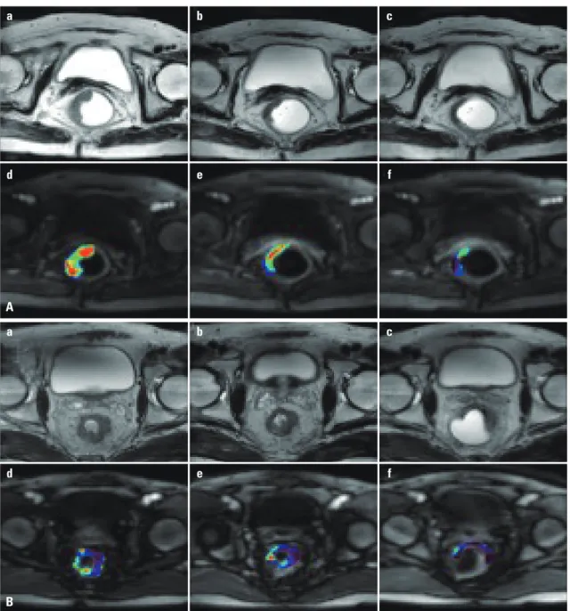
Fig. 5. Perfusion MRI imaging. (A) Good response. (B) Poor response.
A a
d
b
e
c
f
B a
d
b
e
c
f
vascular space, and correlated with the permeability of tumor vasculature. Ktrans is the most popular perfusion parameter in quantitative analysis.
24Gollub, et al.
25evaluated the value of Ktrans before and after preoperative chemotherapy for LARC and found a significantly lower post-treatment Ktrans for pCR, compared to that of non-pCR (p=0.04). George, et al.
26and Lim, et al.
27studied the predictive value of pretreatment Ktrans in rectal cancer patients received preoperative CRT and good re- sponder showed a higher Ktrans than poor responder (Fig. 5).
Oberholzer, et al.
28also observed same findings that the high- er pretreatment Ktrans was correlated with tumor down stag- ing. On the other hand, Devries, et al.
29and Kremser, et al.
30showed that, Ktrans before preoperative CRT was higher in down staging (-) tumors, compared to down staging (+) tumors.
DW-MRI provides qualitative and quantitative information about the basic cellular architecture of the tissue based on dif- ferences in movement (diffusion) of water protons within the various tissues. Tissues with high cellular density like malig- nant tumor show restricted proton movement and an enhanc- ed signal on DW-MRI images. Numbers of studies have report- ed the efficacy of DW-MRI to distinguish the fibrosis from re- mnant tumor tissue after chemoradiaton in rectal cancer. Ac- cording to a meta-analysis, DW-MRI can improve the diagnos- tic value for the assessment of pCR after preoperative CRT in rectal cancer, with an increase in sensitivity from 19% to 84%.
Lambregts, et al.
31reported a effectiveness of DW-MRI for iden- tifying residual tumor and distinguishing non-pCR with a NPV of 90%.
The apparent diffusion coefficient (ADC) is the objective value of DW-MRI. ADC values increase during and after CRT in rectal cancer, which indicate cell death and tumor necrosis.
The higher ADC is generally shown in good tumor responders, and a potential predictive biomarker of chemoradiation.
32-35Significant differences of mean ADC after preoperative CRT have been observed between good versus poor responders;
however, a substantial variation and conflicting results is also found in several studies.
33,34,36Changes in ADC before and after CRT are also different be- tween responders and non-responders. The research of Jung, et al.
34and of Intven, et al.
37found a significantly greater in- crease in ADC in responders than in non-responders.
35The value of pretreatment ADC has been studied as a pre- dictor of response to CRT. Sun, et al.
32found that pretreatment mean ADC of patients with good response was lower than that of patients with poor response. In contrast, Barbaro, et al.
38reported a high pretreatment ADC for good responders. Lam- brecht, et al.
35and Intven, et al.
37found significantly higher pre- treatment ADC values in patients resulting pCR than non-pCR.
In summary, the basis of potential predictive imaging bio- marker in rectal cancer after preop CRT is more evident for DW-MRI than DCE-MRI; however, for both, results are drawn mainly from small, single-center studies. Further investigation through prospective large-scale studies and combined man-
ner are needed to find a promising imaging biomarkers of tr- eatment response in rectal cancer.
Fluorine-18-fluorodeoxyglucose (
18F-FDG) positron emission tomography (PET)
Fluorine-18-fluorodeoxyglucose (
18F-FDG) PET is being in- creasingly used for staging and evaluating treatment response in oncology. Many researchers studied
18F-FDG PET to assess and predict responses to CRT in rectal cancer, and various pa- rameters have been investigated.
39-47Maximum voxel stan- dardized uptake value (SUVmax) is defined as the ratio of ra- dioactivity concentration to the injected activity divided by body weight. ΔSUV was defined as the SUVmax-pre–SUVmax- post difference, and the percentage decrease between the SU- Vmax-pre and the SUVmax-post is presented as the response index (RI)=[ΔSUV/pre-SUV]×100. The percentage change in total lesion glycolysis (TLG) before and after CRT (ΔTLG%) is another semi-quantitative parameter. One qualitative param- eter related to
18F-FDG PET, visual response (VR), was assess- ed for predicting pathologic response to preoperative CRT (Table 1).
39-47The diagnostic performances of the three parameters (RI, SUVmax-post, and VR) related to
18F-FDG PET were similar in predicting pathological response. The parameter ΔTLG% had higher specificity than the other three parameters in predict- ing pathological response.
42-45Most studies have used several parameters but find just one or two to be correlated with CRT response.
40-45,47In our previ- ous study,
40a lower SUVmax-post and a higher RI were shown in good tumor response (T-down staging and TRGs 1 and 2).
The SUVmax-post and RI were also significantly associated with pathological treatment response, especially in pCR (Fig. 6).
Maffione, et al.
46used eight parameters to predict TRG, and found SUVmax, RI, VR, and ΔTLG% to be significantly correlat- ed with pathological treatment response, with SUVmax-post having the highest sensitivity for predicting TRG.
Janssen, et al.
48studied the optimal time point for repeated
18