262 논문접수일 :2012년 07월 1 1일
논문수정일 :2012년 19월 17일 심사완료일 :2012년 10월 23일
교신저자 :정 현, 614-735 부산광역시 부산진구 개금동 633-165 인제대학교 의과대학 부산백병원 이비인후-두 경부외과학교실
전화 :(051) 890-6379·전송:(051) 892-3831 E-mail:[email protected]
서 론
비부비강에서 발생하는 악성종양은 두경부영역에서 발생한 악성종양의 3% 정도 차지하고,1) 이 중 비중격에 원발하는 악성종양은 비강 내 악성종양의 약 9%를 차지 하는 매우 드문 종양이다.2) 비강에 발생하는 악성종양의
90%는 편평상피암이며, 선암종의 경우 매우 드물다.3) 선 암종은 크게 타액선 형(salivary type)과 비타액선형(non- salivary type)으로 나누며, 이 중 비타액선형은 창자형(in- testinal type)과 비창자형(non-intestinal type)으로 나 누고, 각각의 아형으로 고등급와 저등급으로 분류한 다.4) 비중격에서 발생한 저등급 비창자형(low-grade non-intestinal type) 선암종은 지금까지 국내에 보고된 적이 없어 문헌고찰과 함께 보고하는 바이다.
증 례
31세 남자환자가 10년 전부터 발생한 빈번한 비루와 비폐색 증상으로 본원 이비인후과에 방문하였다. 내원시 臨床耳鼻:第 23 卷 第 2 號 2012
• • • • • • • • • • • • • • • • • • • • • • • • • • • • • • • • • • • • • • • • • • • • • • • • • • • • • • • • • • • • • • • • • • • • • • • • • • • • • • • • • • • • • • • • • • • • • • • • • • • • • • • • • • • • • • • • • • • • • • • • • • • • • • • • • • • • • • • • • • • • • • • • • • • • • • • • • • • • • • • • • • • • • • • • • • • • • • • • • • • • • • • • • • • • • • • • • • • • • • • • • • • • • • • • • • • • • • •
J Clinical Otolaryngol 2012;23:262-265
비중격에 발생한 저등급 비창자형 선암종 1예
인제대학교 의과대학 부산백병원 이비인후-두경부외과학교실
정 현
Low-Grade Nonintestinal Sinonasal Adenocarcinoma Originating from Nasal Septum : A Case Report
Hyun Jung, MD
Department of Otorhinolaryngology-Head and Neck Surgery, Inje University, School of Medicine, Busan Paik Hospital, Busan, Korea
-
ABSTRACT -
Adenocarcinoma is the third most common mucosal malignancy found in sinonasal tract, after squamous cell carcinoma and adenoid cystic carcinoma, and represents approximately 15% of all sinonasal cancers. The 2005 World Health Organization (WHO) classification of sinonasal adenocarcinoma (SNAC) includes 2 categories : salivary- and nonsalivary-type SNACs. Nonsalivary types are classified into intestinal type SNAC (ITAC) and non-intestinal type SNAC (non-ITAC), each of which is separated into low- and high-grade categories. Low- grade non-ITAC can arise in the sinonasal tract ; the nasal cavity is the mostly afftected, followed by the ethmoid and maxillary sinuses. Over recent years, little doubt remains that low-grade non-ITAC represents a separate en- tity based on morphology, ultrastructural features, and behavior. There was no report about low-grade non-IT- AC arising from nasal septum in South Korea. We performed successful endoscopic excision of this rare case, and so present it with literature review. (J Clinical Otolaryngol 2012;23:262-265)
KEY WORDS:AdenocarcinomaㆍNasal septumㆍNonintestinalㆍLow grade.
증 례
정 현 : 비중격에 발생한 저등급 비창자형 선암종
263
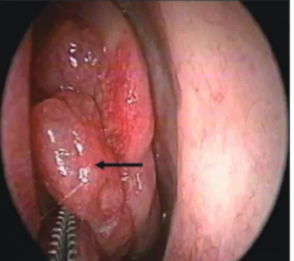
시행한 이학적 검사에서 특이 소견은 없었으며, 비내시경 검사에서 좌측 비강 내 비중격에서 기원하는 과혈관성 의 유두상 종괴가 관찰되었다(Fig. 1). 당뇨로 4년간 투 약하고 있었으며, 직업력 및 과거력에서 중금속이나 방 사선, 분진 등에 노출된 적은 없었고, 가족력상 특이 사항 은 없었다. 비부비동 컴퓨터 전산화 단층 촬영상 좌측 비중격과 좌측 중비갑개 사이에 1.1×2.3×2.9 cm 가량 의 연부조직음영이 보였고, 골파괴, 안와 내 침범, 또는 두개골 기저부 침범 등의 소견은 보이지 않았다(Fig. 2).
일반혈액검사, 간기능 검사, 심전도, 흉부단순촬영 등 에서 특이 소견을 보이지 않았다. 저자는 반전성 유두종 Fig. 1. Nasal endoscopy demonstrated papillomatous
reddish mass (arrow) between nasal septum and left mid- dle turbinate.
Fig. 2. Paranasal sinus computed tomography scans (A : Axial and B : Coronal view) revealed a soft tissue density mass (arrow) between nasal septum and left middle turbinate without destructive bone change.
A B
Fig. 3. Histopathologic photographs. The tumor showed focally preserved surface (A), but focally involved by the tumor tissue (B). The mass consists of compactly arranged, well formed glands and occasional squamous morules (C), and mitotic figures (arrow) are easily found (D)(H&E stain).
A
C
B
D
J Clinical Otolaryngol 2012;23:262-265
264 의심하에 전신마취 하 내시경적 절제술을 시행하였다.
수술 중 좌측 비중격에서 발생한 불규칙한 표면을 가진 쉽게 출혈하는 종괴를 내시경하에 정상 비중격 점막에 절개를 가한 후 종괴와 함께 박리하여 제거하였다. 종 괴는 비중격에서 쉽게 박리가 되었으며, 종괴를 제거 후 정상 점막을 추가로 1 cm 정도 회전식 흡입기(microde- brider)를 이용하여 제거하였다. 환자는 수술 후 2일째 비강패킹을 제거하였고, 수술 후 3일째 퇴원하였다.
병리조직검사상 비강 내 종괴는 점막 아래 위치하고 있으며, 부분적으로 점막과 종양의 연결성을 보였다. 종 양 조직은 불규칙한 덩어리를 형성하며, 빽빽하게 증식 한 선 구조와 군데군데 편평상피가 섞여 있었다. 선 구조 는 대부분 한 층의 세포로 구성되어, 세포학적 이형성은 심하지 않으나, 유사분열상은 쉽게 관찰되었고, 악성 소 견은 뚜렷하지 않았다(Fig. 3). 절제 변연부 침범 소견은 관찰되지 않았다. 이와 같은 조직소견을 바탕으로 저등 급 비창자형 선암종으로 확진되어 술 후 양전자방출단 층촬영을 시행하였으나 원격전이의 소견은 관찰되지 않 았다. 본 증례는 악성종양의 일종인 선암종으로 진단되 었으나, 저등급 비창자형 선암종의 경우 전이가 거의 없 고, 좋은 예후를 보이기 때문에 수술 후 추가적인 항암화 학치료나 방사선 치료를 시행하지 않았으며, 술 후 2년 간 정기적인 외래추적관찰 중이며, 현재까지 재발소견 을 보이고 있지 않다.
고 찰
비부비동에 발생한 선암종은 조직학적으로 표면점막 상피-기원형 또는 점막하선-기원형으로 분류하였으 나,9) 2005년 WHO는 타액선형과 비타액선형으로 분류 하였고, 이중 비타액선형 선암종은 창자형과 비창자형 으로 나누고, 각각 아형으로 고등급와 저등급으로 분류 했다.4) 국내에서는 이러한 WHO 분류에 따른 비부비동 에 발생한 선암종이 아직 보고되지 않았다.
비부비동에 발생한 선암종은 성별과 인종적 차이 없 이 동일한 비율로 발생하며 발생연령은 40대에서 80대 사이에 호발한다.5) 창자형 선암종에서 나무 분진에 대 한 장기적인 노출이 특징적인 위험요인이며, 특히 가구 제작 노동자들에게 70배 높은 발생 위험도가 있다고 보
고되었다.3,6) 그렇지만 비창자형 선암종은 성별과 인종 차이가 없고, 50세 이후에 호발하며, 위험요인, 환경적 인 영향, 선행인자가 없다고 보고되었다.7) 본 증례에서 도 나무분진에 대한 노출 같은 위험요인이나 환경적인 영향을 찾을 수 없었다.
비부비동에 발생한 선암종은 다양한 증상을 보이며, 그 중 비폐색과 비출혈이 가장 흔한 증상으로 나타난다.
하지만 이러한 증상은 많은 비부비동 질환이나 다른 종 양에 있어서도 흔하게 나타나기 때문에 진단까지 많은 시간이 걸릴 수 있다.8) 본 증례에 있어서도 10년동안 빈 번한 비폐색과 비출혈이 있었지만 병원에 늦게 방문하 여 진단이 늦게 되었다.
치료 계획과 예후에 있어서 조직학적 감별은 매우 중 요하다. 저등급 비창자형 선암종은 육안적으로 유두상 또는 꽃양배추 모양이며 대부분 부드러운 양상을 띄며, 크기는 0.3~4.0 cm으로 다양하게 보고되었고, 현미경적 으로 유두(papillary)와 선양 성장양상으로 캡슐에 둘러 싸여 있지 않고, 주변 조직에 침투하는 경향을 보이고 있다. 유사분열이 빈번하지 않고 핵이 명확하지는 않지 만 사립체(psammoma body)를 보일 수 있다. 고등급 비 창자형 선암종을 시사하는 조직학적 소견은 단단한 성장 양상을 보이고, 경계가 불분명하고, 핵이형성증이 중등 도 이상이며, 유사분열률이 높다.8) 본 증례는 섬모 호흡 상피의 창자형 화생(metaplasia)이 없고, 고등급 비창자 형 선암종의 조직학적 소견이 없어서 저등급 비창자형 선암종으로 진단되었다.
면역조직화학적 검사상 창자형 선암종은 CK20, CDX2, villin에 양성을 보이며, CK7에 음성소견을 보이며, 비창 자형 선암종은 이와 반대로 CK20, CDX2, villin에 음성 을 보이며, CK7에 양성소견을 보인다. 이러한 면역조직 화학적 검사는 창자형과 비창자형 선암종을 구별할 수 있 지만 비창자형 선암종과 과오종의 감별진단을 내릴 수 없다.7)
고등급 비창자형 선암종은 나쁜 예후를 보이는 반면, 저등급은 전이가 거의 없고, 아주 좋은 예후를 보인다.5)
비부비동에 발생한 비창자형 선암종의 일반적 치료 는 완전한 수술적 절제이며, 고등급 또는 광범위한 종 양의 경우 수술 후 방사선치료가 필요하다. 저등급 비창 자형 선암종의 경우 완전한 절제가 치료원칙이지만 매
정 현 : 비중격에 발생한 저등급 비창자형 선암종
265 우 드문 종양이어서 아직 정확한 안전 절제 범위는 정립 되지 않았다.10,11) 본 증례에서는 종양 주위의 정상 비중 격점막에 절개하여 종양을 제거하였고, 이후 1 cm의 안 전연을 확보하였다. 저등급 비창자형 선암종은 근치적 수술적 수술보다는 보존적 수술로 충분할 것으로 생각 된다.5,7) 술 후 방사선 치료는 원발성 고등급 선암종 또 는 재발한 저등급 선암종 환자에서 고려될 수 있다.10,11)
결 론
일차적으로 비중격에서 원발하는 악성 종양은 드물 며, 특히 선암종은 매우 드물다. 본 증례와 같이 비중격 에서 발생한 저등급 비창자형 선암종의 경우 악성 종양 이지만 전이가 거의 없고, 아주 좋은 예후를 보이기 때문 에 보전적인 수술로 치료할 수 있으며 수술 후 추가적 인 항암화학치료나 방사선 치료가 필요하지 않을 것으로 사료된다.
중심 단어:선암종・비중격・비창자형・저등급.
REFERENCES
1) Weber AL, Stanton AC. Malignant tumors of the parana- sal sinuses: radiologic, clinical, and histopathologic eval- uation of 200 cases. Head Neck Surg 1984;(3)6:761-76.
2) Echeverria-Zumarraqa M, Kaiser C, Gavilan C. Nasal sep-
tal carcinoma: initial symptom of nasal septal perforation.
J Laryngol Otol 1988;102(9):834-5.
3) Weimert TA, Batsakis JG, Rice DH. Carcinomas of the na- sal septum. J Laryngol Otol 1978;92(3):209-13.
4) Franchi A, Santucci M, Wenig BM. Adenocarcinoma. WHO histological classification of tumors of the nasal cavity and paranasal sinuses. In: Barnes L, Eveson JW, Reichart P, Sidransky D, editors. Pathology and Genetics of Head and Neck Tumors. Lyon: IARC Press;2005. p.22-3.
5) Heffner DK, Hyams VJ, Hauck KW, Lingeman C. Low- grade adenocarcinoma of the nasal cavity and paranasal sinuses. Cancer 1982;50(2):312-22.
6) Luce D, Gerin M, Leclerc A, Morcet JF, Brugere J, Gold- berg M. Sinonasal cancer and occupational exposureto formaldehyde and other substances. Int J Cancer 1993;
53(2):224-31.
7) Bhaijee F, Carron J, Bell D. Low-grade nonintestinal sino- nasal adenocarcinoma: a diagnosis of exclusion. Ann Di- agn Pathol 2011;15(3):181-4.
8) Orvidas LJ, Lewis JE, Weaver AL, Bagniewski SM, Olsen KD. Adenocarcinoma of the nose and paranasal sinuses:
a retrospective study of diagnosis, histologic characteris- tics, and outcomes in 24 patients. Head Neck 2005;27(5):
370-5.
9) Franquemont DW, Fechner RE, Mills SE. Histologic clas- sification of sinonasal intestinal-type adenocarcinoma.
Am J Surg Pathol 1991;15(4):368-75.
10) LeLiever WC, Bailey BJ, Griffiths C. Carcinoma of the na- sal septum. Arch Otolaryngol 1984;110(11):748-51.
11) Beaty CW, Pearson BW, Kern EB. Carcinoma of the nasal septum: experience with 85 cases. Otolaryngol Head Neck Surg 1982;90(1):90-4.