대흉외지 2005;38:510-513 □ 증례보고 □
- 510 - 증 례
종합 검진상 우연히 발견한 좌측 폐 하엽의 종괴를 주 소로 49세 여자가 내원하였다. 환자는 B형 간염이 있었으 며, 6개월 전 우측 무릎 관절경 수술을 받은 것 외에 흡연 이나 결핵으로 치료받은 과거력 등은 없었다.
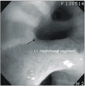
단순 흉부 방사선 촬영상 좌측 폐 하엽의 중앙 부위에 약 3 cm 정도 크기의 음영이 관찰되었으며(Fig. 1), 폐기능 검사상 FEV1 2.41L (94%), FVC 2.99L (89%), FEV1/FVC 81% 등이었으며, 기타 검사실 소견상 이상은 없었다. 기 관지 내시경 검사에서 기관지 내의 종괴 소견은 보이지 않았으나, 좌측 하 폐엽의 내측 기저 분엽 기관지가 바깥 쪽으로부터 눌리는 양상으로 관찰되었으며, 기관지 내시 경 하 조직 생검상으로는 이상 소견이 보이지 않았다(Fig.
2). 흉부 전산화 단층 촬영상 좌측 주 폐동맥이 작아져 있 고, 좌측 하 폐엽이 하행 대동맥으로부터 기시하는 동맥 에 의해 혈액 공급을 받고 있었으며, 좌측 하 폐엽내 혈관
들은 전반적으로 확장되고, 폐 실질은 젖빛 유리 양상 (Ground-glass appearance)을 보였다(Fig. 3).
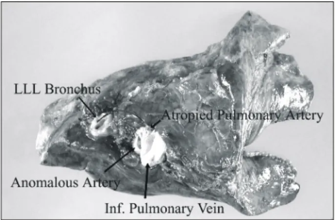
수술은 환자를 전신 마취하 우측와위에서 좌측 후측방 개흉술을 시행하고, 5번째 늑간으로 접근하였으며, 수술 시야를 안전하게 확보하기 위해 6번째 늑골을 절개하였 다. 흉막 유착이나 삼출액 등은 없었으며, 8번째 늑골 높 이에서 하행 대동맥에서 기시하여 좌측 하 폐엽으로 들어 가는 이상 동맥을 발견할 수 있었다(Fig. 4). 좌측 상, 하 폐엽을 각각 분리하고, 하폐 정맥을 먼저 결찰하고, 하폐 동맥을 결찰하여 분리하였다. 하폐 동맥은 위축되어 직경 이 약 3∼4 mm 정도로 작아져 있었으며, 하폐 정맥은 약 간 커져 있었다. 하행 대동맥에서 기시하는 이상 동맥은 수술 후 출혈을 예방하기 위해 3회 결찰 후 절제하였으며, fibrin glue로 도포하였다. 기관지는 TA (Ethicon Endo-Sur- gery, Creek Road Cincinnati, USA) #30을 이용하여 절제하 였으며, 기관지 절제 부위도 fibrin glue로 도포하였다. 공 기 누출 검사에서 상 폐엽이나 기관지 절제 부위에서 공
폐분획증이 없는 좌측 하폐엽의 이상 기시 체혈관
홍성범*․나국주*․박정민*․안병희*․김상형*
Anomalous Systemic Arterial Supply to Normal Basal Segments of Left Lower Lobe without Sequestration
Seong-Beom Hong, M.D.*, Kook-Ju Na, M.D.*, Jung-Min Park, M.D.*, Byung-Hee Ahn, M.D.*, Sang-Hyung Kim, M.D.*
Anomalous systemic arterial supply to the normal basal segments with normal bronchial connection of the lung without sequestration is a rare anomaly. It was classified as a type of sequestration according to Pryce's termi- nology, but whether the term - one of the sequestration is appropriate or not, is controversial because of normal bronchial connection. We describe our experience with surgical treatments for anomalous arterial supply to the normal basal segments of the left lower lobe.
(Korean J Thorac Cardiovasc Surg 2005;38:510-513) ꠏꠏꠏꠏꠏꠏꠏꠏꠏꠏꠏꠏꠏꠏꠏꠏꠏꠏꠏꠏꠏꠏꠏꠏꠏꠏꠏꠏꠏꠏꠏꠏꠏꠏꠏꠏꠏꠏꠏꠏꠏꠏꠏꠏꠏꠏꠏꠏꠏꠏꠏꠏꠏꠏꠏꠏꠏꠏꠏꠏꠏꠏꠏꠏꠏꠏꠏꠏꠏꠏꠏꠏꠏꠏꠏꠏꠏꠏꠏꠏꠏꠏꠏꠏꠏꠏꠏꠏꠏꠏꠏꠏ Key words: 1. Lung sequestration
2. Pulmonary arteries
*전남대학교 의과대학 흉부외과학교실
Department of Thoracic and Cardiovascular Surgery, Chonnam National University Medical School 논문접수일:2005년 4월 8일, 심사통과일:2005년 5월 25일
책임저자:나국주 (501-190) 광주시 동구 학 1동 8번지, 전남대학교 의과대학 흉부외과학교실 (Tel) 062-220-6546 (Fax) 062-227-1636, E-mail: [email protected]
본 논문의 저작권 및 전자매체의 지적소유권은 대한흉부외과학회에 있다.
홍성범 외 Systemic Arterial Supply, Sequestration
- 511 -
기 누출은 없었으며, 하행 대동맥에서의 출혈 소견도 보 이지 않았다. 절단된 좌측 하 폐엽은 11×7.5×5 cm 크기 였으며(Fig. 5), 수술 후 병리 조직 검사상 폐의 이상 소견 은 관찰되지 않았다.환자는 수술 후 1일 흉관 한 개를 제거하였으며, 나머지 한 개는 흉관으로 나오는 배액량이 많아 수술 후 8일째 제거하였다. 이후 합병증 없이 수술 후 15일째 퇴원하였 으며, 환자는 수술 후 3개월이 지난 현재까지 특별한 문제 없이 외래 추적 관찰 중이다.
고 찰
폐분획증은 비정상적인 기관지 연결 관계와 함께 체혈 관에서 혈액 공급을 받는 폐 질환으로, 폐분획증이 없이 정상 폐분엽에 체혈관이 혈액 공급하는 것은 매우 드문 질환이다[1]. 1940년 Harris와 Lewis[2]가 폐 절제 후에 비 정상적인 체혈관으로부터의 출혈로 인한 사망 례를 보고 한 이후, Pryce 등[3]에 의해 폐로 가는 비정상적인 체혈관 에 대해 자세히 기술하였으며, 이에 따르면 폐분획증이 없이 정상 폐분엽에 체혈관이 혈액 공급하는 것은 Pryce type I 폐분획증으로 분류될 수 있다. 최근에는 같은 의미
Fig. 1. Preoperative chest X-ray: About 3 cm nodular density in left
central lower lung field.
Fig. 3. Preoperative chest CT shows
relatively hypoplastic left main pul- monary artery and anomalous sys- temic artery originating from the descending aorta suppling the ba- sal segment of left lower lobe. Nor- mal branching pattern of the left lower lobar bronchus was seen.Additionally, diffuse dilatation of in- trapulmonary peripheral vascula- ture, areas of ground-glass opacity were also noted in the involved ba- sal segments.
Fig. 2. Preoperative bronchoscopic finding: External compression of
mediobasal segment of the left lower lobe.Lt. mediobasal segment
대흉외지 2005;38:510-513
- 512 -
로 ‘Systemic arterial supply .to (the basal segments of) the lung[1]', 'Systemic origin of the sole artery to the basal seg- ments of the lung[4]', 'Systemic arterialization of lung with- out sequestration[5]' 등으로 불리기도 한다.대부분의 환자는 무증상이지만, 이 질환의 주 증상은 객혈이나, 운동시 호흡 곤란이다[1,6,7]. 무증상 환자의 경 우 흉부 방사선 촬영상 발견한 종괴 소견이나, 흉부 청진 상 잡음이 들려서 정밀 검사를 하게 되는 경우가 있으나, 성인에서는 청진 상 잡음은 거의 들리지 않는다[1]. 체혈 관에서의 단락 양이 많을 경우 좌측 심장의 부하 과중으 로 울혈성 심부전에 빠질 수도 있다[1,7].
비정상적인 혈관이 발생하는 원인은 논란이 많으나 배 아기에 대동맥의 후새궁(postbranchial arch)이 주 폐동맥이 발달하기 전에 비정상적으로 잔존한 결과로 보인다[7].
진단은 흉부 방사선 촬영과 기관지 내시경, 흉부 전산 화 단층 촬영 등을 할 수 있으나 가장 정확한 검사법은 혈 관 조영술이다[1]. 혈관 조영술을 통해 병변이 있는 폐엽 의 정상 폐동맥의 분지를 확인하고, 이상 기시 체혈관의 폐엽 내 모세 혈관 상에서 폐정맥으로의 연결 관계를 설 명할 필요가 있다. 본원의 증례에서 혈관 조영술을 시행 하지는 않았으나 CT 혈관 조영술(CT angiography)을 통하 여 비교적 정확한 혈관 관계를 알 수 있었다. Kim 등[7]은 흉부 방사선 촬영상 ① 심장 음영 뒤쪽의 결절, ② 하행 대동맥 경계의 부분적인 소실, ③ 좌측 폐문 하방의 정상 적인 하 폐동맥 음영의 소실, ④ 병변 폐엽의 간질 음영의 증가 등이 나타나는 것으로 기관지 확장증, 폐분획증, 기 관 폐쇄증 등과 감별할 수 있다고 하였다.
이 질환을 가진 모든 환자는 폐동맥 고혈압으로 인한 객혈, 심부전 등의 잠재적인 위험성 때문에 수술의 적응 증이 된다[1,6]. 대부분 폐엽 절제술을 시행하나, 분엽 절 제술이나 이상 기시 체혈관과 폐동맥의 문합술, 이상 기 시 체혈관만을 결찰하는 방법 등이 제시되고 있다[1,6].
Toshihiko 등[6]은 수술 후 폐 기능의 보존을 위해 폐엽 절 제술보다는 분엽 절제술만을 시행하는 것을 권하고 있다.
그러나 분엽 절제술은 이상 기시 체혈관의 정확한 분포를 확인하고 신중하게 고려해야 할 것으로 여겨진다.
국내에서는 폐분획증에 대한 보고가 몇 차례 있었으나, 폐분획증이 없이 폐동맥이 복대동맥에서 기시하는 증례 를 1985년 Kim 등[8]이 보고한 바 있다. 이 증례에서는 복 대동맥에서 기시한 체혈관이 횡격막을 지나 우측 하 폐엽 에 분포하였으며, 우측 하 폐엽 절제술을 시행하였다. 그 러나 대부분의 다른 보고는 이상 기시 체혈관은 좌측 하 폐엽의 기저 분엽에서 발생하였다[1,6,7]. 본원에서 치험한 증례도 좌측 하 폐엽의 내측 기저 분엽에서 발생하였으 며, 좌측 하 폐엽 절제술을 시행하였다.
저자는 폐분획증이 없이 정상적인 기관지 교통과 폐 조 직을 가지고 있으면서, 하행 대동맥에서 기시하는 체혈관 에서 혈액 공급을 받는 증례를 치험 하였기에 문헌 고찰 과 함께 보고하는 바이다.
참 고 문 헌
1. Akira Y, Takashi H, Toshio F, et al. Anomalous systemic
arterial supply to normal basal segments of the left lower lobe. Ann Thorac Surg 1999;68:332-8.
2. Harris HA, Lewis I. Anomalies of lungs with special refer-
Fig. 4. Intraoperative finding. +=Anomalous systemic artery origi-
nating from the descending aorta.
Pericardium
Descending aorta
Lt. lower lobe
Fig. 5. Specimen of the left lower lobe.
홍성범 외 Systemic Arterial Supply, Sequestration
- 513 - ence to danger of abnormal vessels in lobectomy. J Thorac
Surg 1940;9:666-71.3. Pryce DM. Lower accessory pulmonary artery with intralo-
bar sequestration of lung: a report of seven cases. J Pathol
1946;58:457-67.4. Hessel EA II, Boyden EA, Stamm SJ, Sauvage LR. High
systemic origin of the sole artery to the basal segments of the left lung: Findings, surgical treatment, and embryologic interpretation. Surgery 1970;67:624-32.
5. Flisak ME, Chandrasekar AJ, Marsan RE, Ali MM. Systemic
arterialization of lung without sequestration. Am J Radiol
1982;138:751-3.6. Toshihiko I, Yukiko H, Kenzo H, Takehiko F. Systemic ar-
terial supply to the left basal segment without the pulmonary artery: Four consecutive cases. Eur J Cardiothorac Surg
2003;23:847-9.7. Kim TS, Lee KS, Im JG, et al. Systemic arterial supply to
the normal basal segments of the left lower lobe. J Thorac
Img 2002;17:34-9.8. Kim YH, Sun K, Back KJ, Kim HM, Kim IS. Anomalous
systemic arterial supply to the lung without sequestration - A case report. Korean J Thorac Cardiovasc Surg 1985;18:104-
10.=국문 초록=
폐분획증 없는 정상적인 폐분엽에 혈액 공급하는 이상 기시 체혈관은 매우 드문 질환이다. 이것은 Pryce의 용어 정의에 따라 폐분획증의 한 종류로 분류되기도 하지만, 정상적인 기관지 교통 관계 때 문에 아직 논란이 있다. 최근 본원에서 수술로 치료한 정상적인 폐분엽에 혈액 공급하는 이상 기시 체혈관을 치험하였기에 보고하고자 한다.
중심 단어:1. 폐분획증 2. 폐동맥