• 검색 결과가 없습니다.

초음파 유도하 요추 및 제1천추 신경근 차단술의 타당성 연구

N/A
N/A
Protected

Academic year: 2021

Share "초음파 유도하 요추 및 제1천추 신경근 차단술의 타당성 연구"

Copied!
6
0
0

로드 중.... (전체 텍스트 보기)

전체 글

(1)

https://doi.org/10.35827/cp.2019.18.2.59

접수일

: 2019

2

13

,

게재승인일

: 2019

7

29

일 책임저자

:

원선재

,

서울시 영등포구

63

10

󰂕

07345,

가톨릭대학교 의과대학 여의도성모병원 재 활의학과

Tel: 02-3779-1064, Fax: 02-780-9114 E-mail: [email protected]

초음파 유도하 요추 및 제1천추 신경근 차단술의 타당성 연구

가톨릭대학교 의과대학 여의도성모병원 재활의학과

1

, 국립교통재활병원 재활의학과

2

김재원1ㆍ박혜정2ㆍ이원일1ㆍ원선재1

Feasibility of Ultrasound-Guided Lumbar and S1 Nerve Root Block: A Cadaver Study

Jaewon Kim, M.D.

1

, Hye Jung Park, M.D.

2

, Won Ihl Lee, M.D., Ph.D.

1

and Sun Jae Won, M.D., Ph.D.

1

1

Department of Rehabilitation Medicine, Yeouido St. Mary’s Hospital, College of Medicine, The Catholic University of Korea, Seoul,

2

Department of Rehabilitation Medicine, National Traffic Injury Rehabilitation Hospital, Yangpyeong, Korea

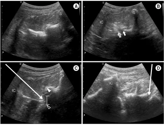
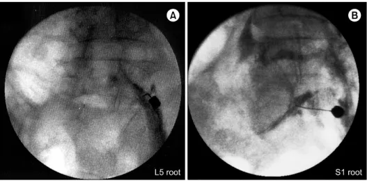
Objective: This study evaluated the feasibility of ultrasound-guided lumbar nerve root block (LNRB) and S1 nerve root block by identifying spread patterns via fluoroscopy in cadavers. Method: A total of 48 ultrasound-guided injections were performed in 4 fresh cadavers from L1 to S1 roots. The target point of LNRB was the midpoint between the lower border of the transverse process and the facet joint at each level. The target point of S1 nerve root block was the S1 foramen, which can be visualized between the median sacral crest and the posterior superior iliac spine, below the L5-S1 facet joint. The injection was performed via an in-plane approach under real-time axial view ultrasound guidance. Fluoroscopic validation was performed after the injection of 2 cc of contrast agent. Results: The needle placements were correct in all injections. Fluoroscopy confirmed an intra-foraminal contrast spreading pattern following 41 of the 48 injections (85.4%). The other 7 injections (14.6%) yielded typical neurograms, but also resulted in extra-foraminal patterns that occurred evenly in each nerve root, including S1. Conclusion: Ultrasound-guided injection may be an option for the delivery of injectate into the S1 nerve root, as well as lumbar nerve root area. (Clinical Pain 2019;18:59-64)

Key Words: Ultrasonography, Spinal injection, Lumbosacral Region, Spinal roots

INTRODUCTION

With advances in technology, imaging tools, such as magnetic resonance imaging, computed tomography (CT), and neuromuscular ultrasound, now provide higher-re- solution and clearer images in the medical field. Ultra- sound-guided injection is now attracting much attention from clinicians managing spinal pain.

1-6

In cervical spinal interventions, ultrasound-guided techniques can ‘prevent’

intravascular injections, whereas fluoroscopy-guided proce- dures can ‘detect’ intravascular injections.

7,8

In addition, ul- trasound-based procedures do not entail radiation exposure and can be performed in a relatively small space, and with relatively little manpower.

9

Using ultrasound, it is more difficult to obtain clear im- ages in the lumbar area compared to cervical area because the spine is located relatively deeply, especially in obese patients. In an ultrasound-guided facet joint injection study, the accuracy of the procedure in an obese group with body mass indexes (BMI) of over 30 kg/m

2

was reportedly re- duced to 62%.

10

However, in non-obese patients, ultra- sound-guided paravertebral procedures are reportedly asso- ciated with good accuracy and efficiency in the spinal area.

1,2,5,9,11-16

Because the lumbar nerve root is located deeper than the target point of the facet and medial bundle branch, it is harder to perform ultrasound-guided injection at that site.

Thus, the technique, and the feasibility and efficacy of ul-

trasound-guided lumbar nerve root block (LNRB) have not

been well characterized, and most relevant studies were re-

ported after 2011.

14,17-19

Notably, the approaches used differ

from study to study, including axial, paramedian sagittal,

and paramedian sagittal oblique approaches and had several

limitations. In a cadaver study reported by Gofeld et al.

2

in 2011, the feasibility was good with a 91.3% intra-fora-

(2)

Fig. 1. Schematic view for needle insertion. (A) At the level of the lower margin of the L5 transverse process. (B) L5∼S1 facet joint was identified between the inferior articular process (filled arrow) and the superior articular process (unfilled arrow). (C) L5 nerve root injection. The target point level was identified as the mid-point be- tween (A) and (B) cephalocau- dally. The target depth was showed as the lower half between the later- al border of the L5 lamina (filled arrowhead) and the most medial border of the vertebral body (unfil- led arrowhead). Arrow shows the needle approaching (D) S1 fora- mens (*) are shown bilaterally.

Arrow shows the needle approa- ching. IC: iliac crest, S: spinous process, T: transverse process.

minal contrast spread pattern, and needle placement in the ultrasound-guided procedure was correct in all cases (100%).

In a prospective randomized clinical trial reported by Yang et al.,

17

ultrasound-guided lumbar transforaminal epidural injections yielded an 85% success rate and shorter oper- ation times than a fluoroscopy-guided procedure (518 s vs.

929 s). In another study, compared with a CT-guided tech- nique, ultrasound-guided pararadicular injections also yielded good accuracy (90%) and shorter operation times (4.0 min vs. 7.6 min), and entailed less radiation.

14

Howev- er, in those studies, the injection point was not clear (just described as the most medially visible shadow of the verte- bral body between two adjacent transverse process)

2

or the injection was performed in limited levels (L3∼4 and L4∼

5).

17

In addition, there has only been one case report of ul- trasound-guided S1 root injection.

20

Therefore, it would be meaningful to confirm the feasi- bility of ultrasound-guided LNRB and S1 root injection in a study incorporating a clear description of the approach utilized. This study evaluated the feasibility of lumbar nerve root and S1 root injection using fluoroscopic confir- mation.

MATERIALS AND METHODS

This study was performed in accordance with the Decla- ration of Helsinki. This study was approved by the institu- tional review board. A total of 48 ultrasound-guided in- jections (L1 to S1, bilateral) were performed on 4 unem- balmed cadavers who had no history of lumbar spine sur- gery (2 males and 2 females, mean age 79.7 years, and BMI 23.8 kg/m

2

). An Accuvix XG ultrasound system (Medison Co, Seoul, Korea) equipped with a 2∼7 MHz curved-array transducer was used for all procedures. The same operator performed all procedures. The cadaver was placed in a prone position with slight lumbar flexion using a soft pillow.

First, the transducer was positioned on the midpoint be-

tween the bilateral iliac crest with a transverse scan to the

lumbar vertebra axis, then moved caudally until the sacrum

and sacral foramens were visualized. Subsequently, the

probe was moved cephalad to find the L5∼S1 facet joint

and L5 transverse process (Supplementary Video 1). The

facet joint and the transverse process at each vertebra level

appear sequentially as the probe was moved to the cephalic

direction. After slightly moving the probe laterally to place

the facet joint in the center of the view, the target level was

(3)

Table 1. Ultrasound-Guided Injection into Lumbar and S1 Roots (n = 48) No. of cases

Total

L1 L2 L3 L4 L5 S1

Extra-foraminal spread 1 2 1 0 2 1 7/48 (14.6%)

Intra-foraminal spread 7 6 7 8 6 7 41/48 (85.4%)

Ventral spread only 5 4 5 8 5 7 34

Ventral and dorsal spread 2 2 2 0 1 0 7

Fig. 2. Fluoroscopic validation of ultrasound-guided lumbar (L5) nerve root injection (A) and S1 root injection (B).

determined as the midpoint between the lower border of the transverse process and the facet joint at each level (Fig.

1-A, 1-B). The depth of the target point was the lower half between the lateral border of the laminar and the uppermost part of the vertebral body (Fig. 1-C). The needle tip was placed as deeply within the target area as possible keeping with the principle that the needle tip should be visible (Supplementary Video 2, 3). In L5 root level injections, clear visualization of the L5 vertebral body and disc was sometimes difficult due to acoustic shadowing by the iliac crest. In such cases, the target depth was presumed to be the same distance from the lateral margin of the L5 lamina, which was estimated from the distance between the target point and the lateral margin of the L4 lamina. The target point of the S1 root was just beyond the posterior S1 foramen. It can be visualized between the median sacral crest and the posterior superior iliac spine, below the L5∼

S1 facet joint. The needle tip was advanced into the S1 foramen while maintaining visualization of the needle tip (Fig. 1-D, Supplementary Video 4). After the needle tip was appropriately located, 2 cc of contrast agent was in-

jected at the target point at each level from L1 to S1, with an in-plane approach under real-time ultrasound guidance using a 22-gauge spinal needle. During the injection, local spreading of contrast agent should be visible. In cases where local spreading was not observed, the operator tried to re-locate the needle tip. Fluoroscopic validation was per- formed after the injection of contrast agent in all roots studied.

RESULTS

Fluoroscopic validation was performed after the in- jection of contrast agent in all roots studied (Fig. 2-A, 2-B).

It suggested that all of the needle tips were well placed at the lower part of the cephalad vertebra in all injections.

The needle tip was located under the pedicle from the ven-

tral to the mid-part of the intervertebral foramen on a later-

al view. An intra-foraminal contrast spreading pattern was

confirmed in 41 of the 48 cases (85.4%). All intra-foramin-

al spreading patterns exhibited ventral spreading, but 7 of

41 ventral spreading patterns also showed spreading to the

(4)

dorsal sides. Seven of 48 cases (14.6%) revealed extra-fo- raminal spreading patterns, and it occurred evenly in each nerve root, including S1 (Table 1). Two of 48 cases (4.2%) showed intravascular injection.

DISCUSSION

This study is the first to show ultrasound-guided S1 nerve root injection procedure with fluoroscopy confir- mation with high success rate (7 of 8, 87.5%). Also, it also validated the feasibility of ultrasound guided lumbar nerve roots (L1 to L5) injections, with similar overall rate of suc- cess (41 of 48, 85.4%) compared to previously reported studies (85∼100%). In this study, all injections yielded typical neurograms. The rate of intra-foraminal contrast agent spreading (85.4%) was similar to that reported in pre- vious studies (85∼100%), including S1 root injection.

2,14,17-19

Also, the procedure was performed using an in-plane ap- proach with axial imaging, similar to that described by Gofeld et al.

2

In contrast, Kim et al.

18

and Loizides et al.

19

have suggested a paramedian approach using sagittal ima- ging. In axial imaging, which is used method in this study, the visualization of structures deeper than the transverse process level is clearer than it is in sagittal imaging. Also, identification of the vertebral body is difficult and visual- ization of the needle tip also becomes harder when the nee- dle tip advances deeper than transverse process level in sagittal imaging. In our opinion, an axial imaging approach can usually provide clearer visualization of the needle tip during the procedure and would be a more appropriate ap- proach to maximize the advantages of the ultrasound-gui- ded procedure with the exception of L5 root injection. In L5 root injection, visualization at vertebral body level is frequently difficult in both an axial and a sagittal view. In such cases in this study, the target depth was estimated from the lateral margin of the L5 lamina to be the same depth from the L4 level, and tried to keep the needle tip visible. To keep the needle tip visible during ultrasound- guided injection, the target point in this study was slightly shallower than that reported in previous studies. Previous studies have targeted the ventral area of the intervertebral foramen.

2,18

The different target depths in this study may have caused a slightly lower intra-foraminal contrast agent spreading rate (85.4% in our study, 87.5% in Kim et al.,

18

91.3% in Gofeld et al.

2

) and higher dorsal spreading pattern

(7 of 48 in our study, 1 of 35 in Kim et al.,

18

0 of 42 in Gofeld et al.

2

). Because sonoanatomy differs from patient to patient, especially in patients with scoliosis or spondylo- listhesis, the target point should be adjusted based on each individual patient’s condition and the operator’s preference.

The results of this study may be helpful when deciding on the injection target point.

Several techniques can be used to perform a spinal in-

jection, such as the blind technique, CT-guided injection,

fluoroscopy-guided injection, and ultrasound-guided injec-

tion. Each technique has associated benefits. With fluoro-

scopy-guided injections, the clinician can confirm the dye

spreading pattern. For example, intra-foraminal, which im-

ply that the contrast spread to epidural space at the level

of posterior longitudinal ligament area, and extra-fora-

minal, spreading along the distal nerve root. This con-

firmation may be important when developing the subse-

quent treatment plan. In our opinion, the most important

benefit of ultrasound-guided injection in spinal procedures

is that it can provide real-time visualization of the tissue

surrounding the needle tip, particularly arteries with the

Doppler mode. Due to this advantage, ultrasound-guided

injection may facilitate the prevention of intra-arterial

injections. In this study, there were two intravascular con-

trast spreading patterns because the present study was per-

formed in unembalmed cadavers; therefore, the vessels

were not examined with the Doppler mode. In living pa-

tients, Doppler imaging can efficiently provide relevant in-

formation about vessels around the lumbosacral spine.

20

Notably, ultrasound-guided injection has the disadvantage

that clearly confirming the cephalocaudal spread pattern of

contrast agent in the epidural space is difficult via the pro-

cedure, while it is possible with fluoroscopy-guided injec-

tion. In a previous study, which investigated ultrasound-

guided cervical root injection, a crescent pattern of contrast

agent spreading in the ultrasound image was related to peri-

radicular contrast spreading in fluoroscopy, and was asso-

ciated with significantly better outcomes with regard to cer-

vical radicular pain reduction.

21

However, in the lumbar

area the resolution and quality of ultrasound imaging is

lower than it is in the cervical area. Thus, the local spread-

ing pattern could not be identified clearly in the lumbar

area. When confirming the spreading pattern is more im-

portant, fluoroscopy-guided injection would be advanta-

geous.

(5)

The visualization of the lumbar structure was clear in this study because the body mass index of the cadaver was within a normal range and there was no definite spinal de- formity (e.g., scoliosis, spondylolisthesis, or sacralization).

The ultrasound guided lumbar medial branch block in obese patients (BMI > 30 kg/m

2

) was successful in only 62% of cases,

10

even though the target point of the medial branch block was located superficially compared to that of ultrasound-guided LNRB. This imply that ultrasound guid- ed lumbar root injection may be more difficult to perform in obese subjects. Also, if the patient undergoing ultra- sound-guided LNRB is believed to have a severe spinal de- formity, the results of other imaging studies, such as X-ray, should be checked to specify the exact target level.

Identifying vessels using Doppler ultrasound is an im- portant benefit in ultrasound-guided injection. High fre- quency ultrasound can detect small arteries sensitively in Doppler mode.

3,8,22

In ultrasound-guided LNRB, a rela- tively lower frequency transducer is used compared with that used during a cervical nerve root injection, which ef- fects the resolution and sensitivity of ultrasound. To our knowledge, there have been no reports of the sensitivity of Doppler ultrasound to detect the vessels surrounding the lumbar vertebra. Since Doppler ultrasound is a valuable tool for estimating the blood flow in the ovarian artery with a 1∼2 mm diameter,

22,23

Doppler ultrasound may have a potential to provide information about vessels in the lumbar vertebral area. However, avoiding vessels during ultra- sound-guided procedures can be affected by many factors (e.g., spinal deformities, clinician’s skill, and the resolution of the ultrasound machine). Therefore, further studies in re- al patients will be necessary.

The accuracy of ultrasound-guided injection is good, and can be similar to that of CT-guided and fluoroscopy-guided procedures. Additionally, the operation time is shorter, there is no radiation exposure to patients and the operator, and it requires less space than CT or fluoroscopy-guided procedures.

2,11,14,17-19

In the present study, the accuracy of ultrasound-guided injection was approximately the same as it was in previously reported studies.

2,14,18

Ultrasound-guid- ed injection may be an option for the delivery of injectate into the S1 root, as well as lumbar nerve root area.

CONCLUSION

Ultrasound-guided S1 nerve root, as well as lumbar nerve root injection, is feasible and valid treatment option considering that it showed similar success rate to fluoro- scopy-guided process with less harm to patients in non- obese cases. Also, in-plane approach using axial imaging can visualize exact needle tip during the procedure, which can enhance treatment accuracy.

DISCLOSURE STATEMENT

The authors declare that there are no conflicts of interest.

SUPPLEMENTARY MATERIALS

Supplementary materials can be found via https://doi.

org/10.35827/cp.2019.18.2.59.

REFERENCES

1. Galiano K, Obwegeser AA, Bodner G, Freund MC, Gruber H, Plonder F, et al. Ultrasound-guided periradicular in- jections in the middle to lower cervical spine: an imaging study of a new approach. Reg Anesth Pain Med 2005; 30:

391-396

2. Gofeld M, Bristow SJ, Chiu SC, McQueen CK, Bollag L.

Ultrasound-Guided Lumbar Transforaminal Injections:

Feasibility and Validation Study. Spine (Phila Pa 1976) 2011; 37: 808-812

3. Narouze SN, Vydyanathan A, Kapural L, Sessler DI, Mekhail N. Ultrasound-guided cervical selective nerve root block: a fluoroscopy-controlled feasibility study. Reg Anesth Pain Med 2009; 34: 343-348

4. Pekkafahli MZ, Kiralp MZ, Basekim CC, Silit E, Mutlu H, Drusun H, et al. Sacroiliac joint injections performed with sonographic guidance. J Ultrasound Med 2003; 22: 553- 559

5. Shim JK, Moon JC, Yoon KB, Kim WO, Yoon DM.

Ultrasound-guided lumbar medial-branch block: a clinical study with fluoroscopy control. Reg Anesth Pain Med 2006; 31: 451-454

6. Won SJ, Rhee WI, Yoon JS, Lee UY, Ko YJ. Ultra- sound-guided lower cervical nerve root injectate volumes associated with dorsal root ganglion and epidural spread.

J Ultrasound Med 2016; 35: 305-310

7. Narouze SN, Galiano K, Obwegeser AA, Bodner G,

(6)

Gruber H. Ultrasound-guided cervical periradicular in- jection: cautious optimism. Reg Anesth Pain Med 2006;

31: 87

8. Narouze SN. Ultrasound-guided cervical spine injections:

ultrasound “prevents” whereas contrast fluoroscopy “de- tects” intravascular injections. Reg Anesth Pain Med 2012;

37: 127-130

9. Galiano K, Obwegeser AA, Walch C, Schatzer R, Ploner F, Gruber H. Ultrasound-guided versus computed tomog- raphy-controlled facet joint injections in the lumbar spine:

a prospective randomized clinical trial. Reg Anesth Pain Med 2007; 32: 317-322

10. Rauch S, Kasuya Y, Turan A, Neamtu A, Vinayakan A, Sessler DI. Ultrasound-guided lumbar medial branch block in obese patients: a fluoroscopically confirmed clinical fea- sibility study. Reg Anesth Pain Med 2009; 34: 340-342 11. Jung H, Jeon S, Ahn S, Kim M, Choi Y. The validation

of ultrasound-guided lumbar facet nerve blocks as con- firmed by fluoroscopy. Asian Spine J 2012; 6: 163-167 12. Greher M, Scharbert G, Kamolz LP, Beck H, Gustorff B,

Kapral S, et al. Ultrasound-guided lumbar facet nerve block: a sonoanatomic study of a new methodologic approach. Anesthesiology 2004; 100: 1242-1248

13. Darrieutort-Laffite C, Hamel O, Glemarec J, Maugars Y, Le Goff B. Ultrasonography of the lumbar spine: sonoanatomy and practical applications. Joint Bone Spine 2014; 81:

130-136

14. Loizides A, Gruber H, Peer S, Galiano K, Bale R, Obernauer J. Ultrasound guided versus CT-controlled para- radicular injections in the lumbar spine: a prospective randomized clinical trial. AJNR Am J Neuroradiol 2013;

34: 466-470

15. Won SJ, Lee UY, Cho SU, Rhee WI. Feasibility of ultra- sound-guided atlanto-occipital joint injection. Ann Rehabil Med 2012; 36: 627-632

16. Galiano K, Obwegeser AA, Bale R, Harlander C, Schatzer R, Gruber H, et al. Ultrasound-guided and CT-navigation- assisted periradicular and facet joint injections in the lum- bar and cervical spine: a new teaching tool to recognize the sonoanatomic pattern. Reg Anesth Pain Med 2007; 32:

254-257

17. Yang G, Liu J, Ma L, Cai Z, Meng C, Zhou H, et al.

Ultrasound-guided versus fluoroscopy-controlled lumbar transforaminal epidural injections: a prospective random- ized clinical trial. Clin J Pain 2016; 32: 103-108 18. Kim YH, Park HJ, Moon DE. Ultrasound-guided para-

radicular injection in the lumbar spine: a comparative study of the paramedian sagittal and paramedian sagittal oblique approaches. Pain Pract 2015; 15: 693-700

19. Loizides A, Gruber H, Peer S, Brenner E, Galiano K, Obernauer J. A new simplified sonographic approach for pararadicular injections in the lumbar spine: a CT-con- trolled cadaver study. AJNR Am J Neuroradiol 2011; 32:

828-831

20. Park D. Ultrasound-guided S1 transforaminal epidural in- jection using the in-plane approach and color Doppler imaging. Am J Phys Med Rehabil 2018; 97: e14-e16 21. Lee SH, Kim JM, Chan V, Kim HJ, Kim HI. Ultrasound-

guided cervical periradicular steroid injection for cervical radicular pain: relevance of spread pattern and degree of penetration of contrast medium. Pain Med 2013; 14: 5-13 22. Guzel AI, Erkılınc S, Ozer I, Tokmak A, Kurt Sahin A,

Ugur M. Are uterine and ovarian artery Doppler velocim- etry values good pregnancy predictors in clomiphene cit- rate cycles? Int J Fertil Steril 2015; 9: 41-46

23. Pelage JP, Walker WJ, LeDref O, Rymer R. Ovarian artery:

angiographic appearance, embolization and relevance to

uterine fibroid embolization. Cardiovasc Intervent Radiol

2003; 26: 227-233

수치

Fig. 1. Schematic view for needle  insertion. (A) At the level of the  lower margin of the L5 transverse  process
Table 1. Ultrasound-Guided Injection into Lumbar and S1 Roots (n = 48) No. of cases Total L1 L2 L3 L4 L5 S1 Extra-foraminal spread 1 2 1 0 2 1 7/48 (14.6%) Intra-foraminal spread 7 6 7 8 6 7 41/48 (85.4%)

참조

관련 문서