1178
Growth Factor-β
1의 발현에 미치는 영향
Effect of 5alpha-Reductase Inhibitor in Expression of Transforming Growth Factor-β
1in Benign Prostatic Hyperplasia Patients
Hong Wook Kim, Je Soo Lim1, Young Seop Chang, Ki Hak Song From the Department of Urology, 1Myeonggok Clinical Institute, Konyang University College of Medicine, Daejeon, Korea
Purpose: Transforming growth factor (TGF)-beta is a member of the super- family of polypeptides, which control cell cycle progression and a variety of other cellular activities. TGF-β1 has been implicated as an effector of the induction of apoptosis in response to 5alpha-reductase inhibitor (5ARI) and; therefore, causes a decrease in the prostate volume. We investigated the effect of 5ARI in the expression of TGF-β1 in benign prostatic hyper- plasia (BPH).
Materials and Methods: 50 patients diagnosed with BPH were divided into two groups. The control group (n=30), in which a transurethral resection of the prostate (TURP) was performed without medication, and the 5ARI group (n=20), who were administrated with 5 mg of 5ARI daily for at least 3 months, followed by TURP. The resected specimens were stained with anti-rabbit TGF-β1 polyclonal antibody using immuno- fluoroscent staining. The expression of TGF-β1 was analyzed with a confocal laser scanning microscope and an image analyzer. The mRNA level of TGF-β1 was determined by reverse transcriptase-polymerase chain reaction (RT-PCR).
Results: There were no statistical differences in the patient characteristics, including age, serum prostate-specific antigen (PSA) level and prostate volume, between the two groups. The expression of TGF-β1 was demon- strated in the luminal epithelium and smooth muscle cells in BPH. TGF-β1
was more strongly expressed in the luminal epithelium of both groups, and in the 5ARI group than the control (p<0.001).
Conclusions: These results suggest that 5ARI up-regulates the expression of TGF-β1 in BPH patients, and may a play role as an inhibitor in the proliferation of BPH through the TGF-β1 signal pathway. (Korean J Urol 2006;47:1178-1184)
Key Words: Benign prostatic hyperplasia, 5alpha-reductase, Transforming growth factor-β1
대한비뇨기과학회지 제 47 권 제 11 호 2006
건양대학교 의과대학 비뇨기과학교실,
1명곡임상연구소
김홍욱․임지수1․장영섭․송기학
접수일자:2006년 5월 15일 채택일자:2006년 10월 9일
교신저자: 송기학
건양대학교병원 비뇨기과 대전시 서구 가수원동 685번지
}320-718 TEL: 042-600-8835 FAX: 042-542-3790 E-mail: urosong@kyuh.
co.kr
이 논문은 2004년도 명곡임상 교수 연구비 의 일부 지원에 의하여 이루어진 것임.
서 론
전립선비대증은 다양한 배뇨증상과 전립선 크기의 증가, 방광출구폐색 등이 복합된 질환이고 50세 이상의 남자에서 가장 흔한 질환이며 전립선의 간질세포에서 분비된 여러
가지 성장인자들이 측분비 (paracrine) 또는 자가분비 (auto- crine) 작용을 통해 상피세포 또는 간질세포의 증식을 촉진 시키거나 세포고사 (apoptosis)의 결함으로 인한 상대적인 증식, 그리고 세포노화 (senescence)로 발생한다.1
전립선비대증에 의한 폐색은 두 가지로 설명하는데 첫 번째는 동적 요소인 전립선 평활근의 긴장이며 두 번째는
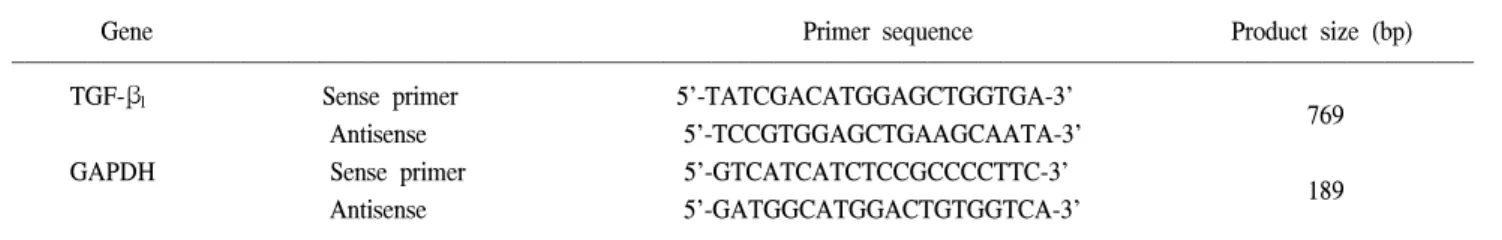
Table 1. Primer sequence for RT-PCR
Gene Primer sequence Product size (bp)
TGF-β1 Sense primer 5’-TATCGACATGGAGCTGGTGA-3’
Antisense 5’-TCCGTGGAGCTGAAGCAATA-3’ 769
GAPDH Sense primer 5’-GTCATCATCTCCGCCCCTTC-3’
Antisense 5’-GATGGCATGGACTGTGGTCA-3’ 189
RT-PCR: reverse transcriptase-polymerase chain reaction, TGF-β1: transforming growth factor-beta 1 정적 요소인 전립선 요도의 물리적 폐쇄이다.2 이중 전립선
요도의 물리적인 폐쇄는 전립선의 상피세포 및 간질세포의 증식으로 유발되는데 이 과정에서 꼭 필요한 것이 dehydro- testosterone (DHT)이다. DHT는 1형과 2형 5알파환원효소에 의해 테스토스테론 (testosterone)으로부터 전환되어 생성된 다. 전립선비대증을 유발하는 전립선 내 DHT 농도는 전립 선에 주로 분포하는 2형 5알파환원효소에 의해 결정된다.3 5알파환원효소억제제는 이 2형 5알파환원효소를 억제하여 전립선 내 DHT 농도를 낮추어 전립선비대증을 치료하는 약물이다. 그러나 DHT의 저하가 어떤 경로로 전립선비대 증을 치료하는지에 대한 분자생물학적 기전은 아직 정확히 밝혀지지 않았다.
전립선비대증의 성장에는 많은 성장인자가 관련되어 있 다. 그 중 세포의 성장, 분화 등을 조절하는 TGF-β1은 다른 의견도 있으나 대체로 전립선비대증에서 발현과 분비가 증 가되며 저농도의 경우는 성장을 촉진하지만 고농도의 TGF-β1는 전립선 조직의 성장을 억제시키는 것으로 알려 져 있다.4-7 현재 전립선비대증 치료제와 여러 성장인자의 관련성에 대한 많은 연구가 진행되고 있으며 5알파환원효 소억제제가 TGF-β1의 발현을 증가시켜 전립선비대증에 작 용하리라는 연구결과가 발표되었다.8,9
이에 저자들은 전립선비대증의 치료 목적으로 투여된 5 알파환원효소억제제가 인체 전립선비대증 조직에서 TGF- β1의 발현에 어떠한 영향을 주는지 알아보고자 하였다.
대상 및 방법
1. 대상
2003년 1월부터 2005년 12월까지 전립선비대증으로 본원 에서 경요도전립선절제술을 시행 받은 135명 중 5알파환원 효소억제제의 사용 유무에 따라 50명을 대상으로 5알파환 원효소억제제를 복용하지 않은 군 (n=30)과, 5알파환원효소 억제제를 3개월 이상 복용한 군 (n=20)으로 나누어 실험하 였다. 5알파환원효소억제제를 3개월 이상 복용 후 경요도
전립선절제술을 받은 군은 Speakman 등10이 하부요로증상 을 가진 70세 이상의 남자, 국제 전립선 증상 점수가 7 이상 인 경우, 요속이 12ml/s 미만인 경우, 전립선 부피가 30ml를 넘거나 전립선특이항원치가 1.4ng/ml를 넘는 경우, 잔뇨가 100ml를 넘어 5알파환원효소억제제를 사용하도록 권고한 환자들 중에서 3개월 이상 복용한 환자로 정하였다.
2. 방법
1) 면역 형광 염색: 절제된 전립선 조직을 10% 중성 포 르말린에 하루 동안 고정시키고 흐르는 물에 수세하여 고 정제를 제거하고 탈수하였다. 투명화 과정 후에 paraffin에 포매하였으며 조직블럭은 slide glass에 4μm두께로 절편을 제작하여 부착시켰다. 탈파라핀화 및 함수화를 위해 Xylene I, II에 각 10분씩, 100%, 95%, 90%, 80%, 70% 알코올에 순 차적으로 각각 5분씩 반응시켰다. 항원을 노출시키기 위하 여 1x sodium citrate buffer 용액에 담가 10분간 극초단파오 븐으로 전처리하고, 0.01M (pH 7.4) PBS로 10분씩 3번 세척 후 비특이적인 반응을 없애고자 10% 정상 염소 혈청 (nor- mal goat serum, Dakocytomation, Denmark)으로 1시간 동안 37oC에서 항온처리하였다. 그리고 1차 항체인 rabbit TGF- β1 polyclonal antibody (1:100, Santa Cruz Biotechnology, Inc., Santa Cruz, USA)로 4oC에서 16시간 반응시킨 뒤, 0.01M (pH 7.4) PBS로 10분씩 3번 세척하였다. 2차 항체로 37oC의 암실 에서 1시간 동안 항온처리 하였고, 2차 항체로는 goat anti- rabbit IgG-FITC (1:200, Santa Cruz Biotechnology, Inc., Santa Cruz, USA)를 사용하였다. 0.1% Triton X-100 (Sigma-Aldrich Corp., St. Louis, USA)가 함유된 0.01M PBT로 30분씩 2회 세척한 후 PBS로 20분씩 15회 세척하였다. 염색이 끝난 슬 라이드는 mounting media (Dako, Carpinteria, USA)를 이용하 여 봉입하였다.
2) RT-PCR: 두 군 간에 생성된 TGF-β1의 양을 정량적으 로 비교해 보고자 PCR을 시행하였다. RNA 분리는 조직을 채취하여 액체질소로 냉동시키고, 냉동상태가 유지된 상태 에서 분쇄하고 TRIzol (In vitrogen, USA)을 이용하여 total
A B C
D E F
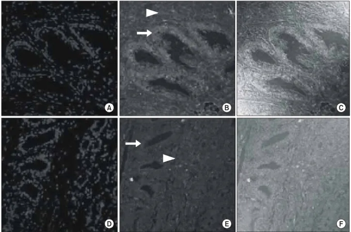
Fig. 1. Immunofluoroscent staining of TGF-β1 in the control and 5ARI groups (x400). (A), (D): DAPI staining of the nucleus in BPH specimens. (B), (E): the 5ARI group shows stronger expression of TGF-β1 than the control group. The gland area (arrow) expresses stronger TGF-β1 immunoactivity than the stromal area (arrowhead) in the 5ARI group. (C), (F): merged image of TGF-β1(A-C: 5ARI group.
D-F: control group.). 5ARI: 5alpha-reductase inhibitor, TGF: transforming growth factor, BPH: benign prostatic hyperplasia.
Table 2. Summary of patient characteristics
Control group 5ARI group
p-value
(n=30) (n=20)
Age (years) 68.9±1.2 70.1±1.6 0.87
S-PSA (ng/ml) 5.77±1.26 4.98±0.93 0.80 Prostate vol. (ml) 70.59±4.56 69.68±5.47 0.17
5ARI: 5alpha-reductase inhibitor, S-PSA: serum-prostate specific antigen
RNA를 추출하였다. 추출된 RNA의 농도와 질을 평가하기 위해서, 100배 희석된 RNA 용액으로 UV spectrophotometer 를 이용해 260nm와 280nm에서 흡광도를 측정, 비교 확인하 였다. 역전사 중합효소 연쇄반응법은 2㎍의 total RNA를 정 량하여 oligo-dT primer에 의한 cDNA 합성을 시행하였다 (TaKaRa RNA PCR kit, TaKaRa, JAPAN). 합성된 cDNA 용 액은 PCR을 실시할 때까지 -20oC에서 보관하였다. 시발체 (primer)는 조사대상인 TGF-β1 유전자와 내부표준유전자 (internal standard)로서 GAPDH 유전자를 사용하였다 (Table 1). PCR reagent system (TaKaRa, JAPAN)을 사용하여 PCR을 실시하였으며, PCR에 사용된 반응 혼합액은 다음과 같다.
TGF-β1 혼합액은 cDNA 2μl, 각 TGF-β1 시발체 0.32mM, 각 GAPDH 시발체 0.1125mM였고, Taq polymerase는 0.5unit 첨가하였다. TGF-β1의 경우 95oC 5분간 변성 후, 95oC 30초, 62oC 30초, 72oC 30초의 반응 조건으로 반응 횟수는 35회 시행하였다. 72oC에서 5분간 연장 반응 후 PCR 반응을 종 결시켰다. PCR 산물에 대한 전기영동은 TGF-β1의 경우
2%의 ethidium bromide가 포함된 한천 겔 상에서 100V의 전압 하에 30분간 실시하고 자외선 투사기 위에서 mRNA 의 발현 유무를 확인하였다. 상기 시발체들에 의한 PCR 산물의 크기는 TGF-β1, GAPDH 각각 769bp, 189bp였다 (Table 1).
3) 영상분석: 전립선비대증 조직에서 TGF-β1의 면역염색 강도의 평가는 한 장의 슬라이드에서 최소한 5군데 이상
Gland
Control group 5ARI group
Gray scale
0 50 100 150 200
Total *
Stroma
Fig. 2. The intensity of TGF-β1 in the immunofluoroscent stain- ing. The glandular area expresses stronger TGF-β1 immunoac- tivity than the stromal area in both the control and 5ARI groups (p<0.05). The expression of TGF-β1 is not significantly stronger in the stroma of the control group (p>0.05). There is a signi- ficantly stronger expression of TGF-β1 in the gland of the 5ARI group (p<0.05). The total intensity of TGF-β1 expression in the 5ARI group is stronger than in the control group (p<0.05). *,†: p<0.05. 5ARI: 5alpha-reductase inhibitor, TGF-β1: transforming growth factor-β1.
1 2 3 4 M 5 6 7 8
TGF-β1
GAPDH
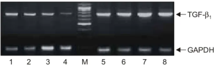
Fig. 3. Expression of TGF-β1 mRNA is increased in the 5ARI compared to the control group (M: marker, 1-4: control group, 5-8:
5ARI group). 5ARI: 5alpha-reductase inhibitor, TGF-β1: trans- forming growth factor-β1.
전립선 상피세포와 간질의 TGF-β1의 면역염색 강도를 측 정하였으며 그 평균값을 구하여 환자의 TGF-β1의 면역염 색 강도로 하였고 공초점 레이저 주사현미경 (Zeiss LSM, Jena, Germany)과 영상분석장치를 이용하여 측정하였다.
3. 통계
모든 실험성적은 평균±표준편차 및 표준오차로 표시하였 으며, 두 군 간의 통계적인 분석은 Student's t-test를 사용하 여 분석하였다. 통계처리는 SPSS for windows (Version 11.0, SPSS Inc., Chicago, USA) 프로그램을 이용하였으며 p값이 0.05 미만인 경우에 통계학적으로 유의한 것으로 판정하였다.
결 과
경요도전립선절제술만 받은 군의 평균연령은 68.9±1.2 세, 전립선 특이항원치는 5.77±1.26ng/ml, 전립선의 부피는 70.59±4.56ml였고 5알파환원효소억제제를 3개월 이상 복 용한 군의 평균연령은 70.1±1.6세, 전립선 특이항원치는 4.98±0.93ng/ml, 전립선 부피는 69.68±5.47ml로 두 군의 연 령, 전립선 특이항원치 및 전립선의 부피에 유의한 차이는 없었다 (p>0.05) (Table 2). 5ARI군에서 5알파환원효소억제 제를 복용한 기간은 평균 8.0±6.7개월 (3-24)이었다.
TGF-β1는 대조군과 5알파환원효소억제제 복용군의 전립 선비대증 검체 중 상피세포와 간질의 평활근에서 발현 되
었으며 전립선 간질보다는 상피세포에서 강하게 발현되었 다 (Fig. 1). TGF-β1의 발현 강도는 상피세포를 비교하였을 때 대조군에서 144.74±0.95, 5알파환원효소억제제 복용군 에서 153.04±1.53으로 5알파환원효소억제제 복용군에서 더 높게 발현되었다 (p=0.000) (Fig. 2). 간질을 비교하였을 때는 대조군에서 137.87±0.92, 5알파환원효소억제제 복용 군에서 135.20±1.12으로 대조군에서 더 높게 발현되었지만 통계적인 유의성은 없었다 (p=0.066) (Fig. 2). 상피세포와 간 질을 합하여 두 군을 비교하였을 때 TGF-β1의 발현 강도는 대조군에서 141.31±0.72, 5알파환원효소억제제 복용군에서 144.12±1.29로 5알파환원효소억제제 복용군에서 더 높게 발현되었다 (p=0.049). RT-PCR을 시행하여 TGF-β1의 mRNA 발현 정도를 비교하였을 때 대조군보다 5알파환원효소억 제제 복용군에서 높게 발현되었다 (Fig. 3).
고 찰
전립선비대증은 병태생리학적으로 basic fibroblastic growth factor (bFGF), epidermal growth factor (EGF), platelet derived growth factor (PDGF)와 insulin-like growth factor (IGF) 같은 성장자극요소와1 DHT같은 남성 호르몬 등이 관여하며, B cell lymphoma-2 (bcl-2) 유전자는 세포고사를 억제하여 증식 이 일어나게 하고 TGF-β1는 전립선 상피 세포 부근의 섬유 모세포를 통해 전립선 상피의 성장을 억제하는 것으로 알 려져 있다.11,12
TGF-β는 세포분화와 세포주기진행을 조절하는 다기능 폴리펩타이드이며 25,000MW의 homodimer로 인간혈소판 과 태반, 돼지의 신장에서 처음으로 분리되었다. TGF-β는 TGF-β1 TGF-β2 TGF-β3의 아형이 있으며 112개의 아미노 산으로 되어 있다.13,14 TGF-β1은 세포의 성장 억제, 세포의 이동, 분화, 고사 등을 조절하며 전립선 조직에서도 세포의 성장과 억제를 조절하고 전립선 성장에 필요한 상피-간질 상호 작용에 중요한 역할을 한다.15,16
전립선에서는 3가지 아형 모두가 발현되며 전립선 간질 과 상피 조직에 따라서 TGF-β 발현 정도와 subtype에 차이 가 있는데 Story 등17은 정상 전립선과 전립선비대증 조직에 서 각각 간질과 상피 조직을 배양하여 비교하였을 때 전립 선의 간질에서는 TGF-β1이 주로 발현되고 전립선의 상피 조직에서는 TGF-β2가 TGF-β1보다 많이 발현되나 통계학 적인 유의성은 없다고 하였다. Timme 등18은 전립선의 간질 세포에서 TGF-β1이 많이 발현된다고 하였으나 Kyprianou 등19은 경요도전립선절제술 또는 근치적 전립선절제술로 얻은 전립선비대증 조직의 상피세포에서 간질세포보다 TGF-β1가 많이 발현된다고 보고하였고 Lucia 등9은 세포 배양을 통한 실험에서 5알파환원효소억제제를 사용하였을 때 상피세포에서 농도에 비례하여 세포수는 감소하지만 세 포당 분비되는 TGF-β1의 양은 증가한다고 하였다. 본 연구 에서도 상피세포와 간질 세포에서 TGF-β1이 모두 발현되 었으나 면역염색 강도를 비교하였을 때 상피세포에서 간질 세포보다 TGF-β1이 통계적으로 유의하게 높은 발현을 보 였다 (Fig. 2). 5알파환원효소억제제 복용군에서 복용 기간 이 길수록 간질에서의 TGF-β1 발현이 유의하게 감소하는 경향을 보이나 실험군의 수가 적어 보다 많은 실험군을 대 상으로 한 결과가 필요할 것으로 보인다.
병리학적으로 전립선 비대 결절은 주로 상피 선조직의 발아 및 분기 (branching)에 의해 야기되고 적은 정도에서 선 단위 비대 혹은 전립선 기질 요소의 증식에 의해 발생한
다.20,21 상피의 증식이 주가 되는 전립선비대증에서 상피 세
포의 증식에는 여러 가지 성장 인자 및 호르몬이 필요하지 만 그 중에서 세포 내 DHT 대사 산물을 필요로 한다. DHT 는 전립선에 존재하는 5알파환원효소 2형에 의해 주로 생 성되며 이 효소는 간질 섬유아세포와 기저상피세포에 존재 하고 정상 조직에 비해 전립선비대증 조직에서 활성도가 높다고 한다.3 5알파환원효소억제제는 이 5알파환원효소 2 형에 작용하여 남성호르몬의 변환을 억제하게 된다. 이 결 과로 간질 조직에서는 세포고사가 관찰되지 않으나 상피 세포에서는 세포의 고사, 관 위축 및 혈관 감소 등에 의하여 전립선 용적을 20-30% 감소시켜 주는 것으로 알려져 있다.3 5알파환원효소억제제는 전립선비대증과 관련된 증상을 호 전시켜 주고 요속을 1-2ml/s 향상시켜 준다.22 Lowe 등23은 전립선비대증 환자에서 6년 동안의 5알파환원효소억제제 를 사용한 결과 증상점수를 4점 호전시켰고, 전립선 부피를 24% 감소시켰으며 최대요속을 2.9ml/s 향상시켰다고 하였 다. 이런 기전에 의해 5알파환원효소억제제는 전립선비대 증 환자의 급성 요폐와 전립선비대증과 관련된 수술의 위 험성을 유의하게 감소시켜 주는 효과가 있는 것으로 밝혀 져 전립선비대증 환자의 약물치료에 많이 사용되고 있다.24
또한 5환원효소억제제는 전립선비대증과 관련된 혈뇨를 가진 환자에서 사용하였을 때 혈뇨의 정도를 감소시키거나 사라지게 하였으며 경요도전립선절제술을 시행하기 전에 3개월 이상 복용하였을 경우 수술 중 실혈의 양을 감소시켜 주었다고 하였다.25,26 Speakman 등10은 하부요로증상을 가진 70세 이상의 남자, 국제 전립선 증상 점수가 7 이상인 경우, 요속이 12ml/s 미만인 경우, 전립선 부피가 30ml를 넘거나 전립선특이항원치가 1.4ng/ml를 넘는 경우, 잔뇨가 100ml를 넘는 경우 등 전립선비대증이 진행되고 급성 요폐의 위험 성이 있는 남성들에게 5알파환원효소억제제를 복용할 것 을 권유하였고 3-6개월 이상 사용하였을 때 전립선의 부피 가 가장 많이 작아진다고 하였다. 5알파환원효소억제제가 증상의 호전, 요속증가, 전립선비대증과 관련된 수술 및 급 성 요폐의 방지 등의 효과를 갖지만 남성호르몬의 저하가 어떠한 경로를 거쳐 세포 수준에 영향을 주는지는 아직 정 확하게 밝혀지지 않았다. Zhu와 Kyprianou27은 남성 호르몬 이 TGF-β1 리간드, 수용체 발현, TGF-β 신호의 기본적인 세포 내 구성요소인 Smad의 발현 및 활성화를 억제하고 특 히 DHT가 남성호르몬 수용체와 Smad3의 상호작용을 통해 전립선 상피세포에서 TGF-β의 신호를 억제한다고 하였다.28 Saez 등8은 5알파환원효소억제제로 치료 받은 군과 치료 받지 않은 군을 비교하였을 때 치료받은 군의 상피세포에 서 TGF-β1이 결합하는 TGF-β 수용기 II가 많이 발현되며 또한 상피세포에서 간질세포보다 전립선 샘의 위축이 많이 관찰되는 것을 보고 5알파환원효소억제제가 TGF-β 신호 체계를 통해 전립선 샘의 위축에 영향을 주었을 것이라고 주장하였다. 또한 Glassman 등29은 전립선비대증 환자에서 약물 치료 후 전립선의 용적이 줄어드는 세포고사는 TGF- β신호체계가 중요한 역할을 한다고 주장하였고 알파교감 신경억제제 단독투여, 5알파환원효소억제제 단독투여, 복 합요법 순으로 더욱 TGF-β1이 증가하는 경향을 보였다고 하였다. 본 실험에서도 5알파환원효소억제제 복용군의 상 피세포에서 TGF-β1의 발현이 강하게 나타난 것을 알 수 있 었다 (Fig. 1). 또한 5알파환원효소억제제 복용군과 대조군 을 비교하였을 때 전반적으로 5알파환원효소억제제 복용 군에서 TGF-β1의 발현이 강하게 나타났다 (Fig. 2) (Fig. 3).
이것은 Glassman 등29의 실험과 같이 TGF-β1의 발현이 간 질세포에 비해 상피세포에서 증가되며 상피와 간질을 합하 여 전반적인 발현을 비교하였을 때 상피세포에서의 5알파 환원효소억제제 영향으로 전체적인 증가를 나타낸다. 이런 점들을 종합하면 5알파환원효소억제제가 전립선 내의 DHT 수치를 낮추고 이것이 TGF-β1의 신호를 억제하지 않 게 되어 TGF-β1의 발현이 증가하는 것으로 보인다.
본 실험의 결과로 보았을 때 5알파환원효소억제제는 전
립선 내의 DHT에 영향을 주며 전립선의 상피세포에서 TGF-β1의 발현을 증가시키는 것으로 생각한다.
결 론
5알파환원효소억제제 복용군과 대조군에 대해 TGF-β1
에 대한 면역조직화학염색을 시행한 결과 5알파환원효소 억제제 복용군의 상피세포에서 TGF-β1의 발현이 통계학적 으로 유의하게 증가하였다. 전립선비대증 환자에서 5알파 환원효소억제제의 사용은 전립선비대증과 관련된 임상지 표를 향상시키며 분자 생물학적으로 전립선 상피세포에서 TGF-β1의 발현을 증가시켜 전립선의 성장에 영향을 줄 것 으로 생각되지만 아직까지 명확한 기전에 대해서 밝혀지지 않았다. 그러나 본 연구에서 5알파환원효소억제제가 전립 선 상피세포에서 TGF-β1의 발현을 증가시킴을 보여줌으로 써 5알파환원효소억제제가 TGF-β1을 통해 전립선비대증 의 성장을 억제하는 한 가지 방법을 제시하고 있다.
앞으로 좀 더 많은 실험군으로 전립선비대증과 관련된 여러 성장인자들에 대한 실험을 통해 5알파환원효소억제 제가 전립선의 성장에 미치는 영향을 규명해야 할 것으로 생각한다.
REFERENCES
1. Lee KL, Peehl DM. Molecular and cellular pathogenesis of benign prostatic hyperplasia. J Urol 2004;172:1784-91 2. Chapple CR. Pharmacological therapy of benign prostatic
hyperplasia/lower urinary tract symptoms: an overview for the practising clinician. BJU Int 2004;94:738-44
3. Steers WD. 5alpha-reductase activity in the prostate. Urology 2001;58(6 Suppl 1):17-24
4. Djonov V, Ball RK, Graf S, Mottaz AE, Arnold AM, Flanders K, et al. Transforming growth factor-beta 3 is expressed in nondividing basal epithelial cells in normal human prostate and benign prostatic hyperplasia, and is no longer detectable in prostate carcinoma. Prostate 1997;31:103-9
5. Hong SJ. Benign prostatic hyperplasia: multiple factors for prostate tissue change with aging. Korean J Urol 2005;46:
547-54
6. Reynolds AR, Kyprianou N. Growth factor signalling in prostatic growth: significance in tumour development and therapeutic targeting. Br J Pharmacol 2006;147(Suppl 2):
S144-52
7. Untergasser G, Madersbacher S, Berger P. Benign prostatic hyperplasia: age-related tissue-remodeling. Exp Gerontol 2005;
40:121-8
8. Saez C, Gonzalez-Baena AC, Japon MA, Giraldez J, Segura DI, Miranda G, et al. Regressive changes in finasteride-treated
human hyperplastic prostates correlate with an upregulation of TGF-b receptor expression. Prostate 1998;37:84-90
9. Lucia MS, Sporn MB, Roberts AB, Stewart LV, Danielpour D. The role of transforming growth factor-beta1, -beta2, and -beta3 in androgen-responsive growth of NRP-152 rat prostatic epithelial cells. J Cell Physiol 1998;175:184-92
10. Speakman MJ, Kirby RS, Joyce A, Abrams P, Pocock R.
Guideline for the primary care management of male lower urinary tract symptoms. BJU Int 2004;93:985-90
11. Alan WP, Ronald R. The molecular biology, endocrinology, and physiology of the prostate and seminal vesicles. In: Walsh PC, Retik AB, Vaughan ED Jr, Wein AJ, editors. Campbell's urology. 8th ed. Philadelphia: Saunders; 2002;1237-96 12. Danielpour D. Functions and regulation of transforming
growth factor-beta (TGF-b) in the prostate. Eur J Cancer 2005;
41:846-57
13. Sporn MB, Roberts AB. Interactions of retinoids and transforming growth factor-β in regulation of cell differen- tiation and proliferation. Mol Endocrinol 1991;5:3-7 14. Sporn MB, Roberts AB. TGF-β: problems and prospects. Cell
Regul 1990;1:875-82
15. Zhou W, Park I, Pins M, Kozlowski JM, Jovanovic B, Zhang J, et al. Dual regulation of proliferation and growth arrest in prostatic stromal cells by transforming growth factor-beta1.
Endocrinology 2003;144:4280-4
16. Itoh N, Patel U, Cupp AS, Skinner MK. Developmental and hormonal regulation of transforming growth factor-beta1 (TGF beta1), -2, and -3 gene expression in isolated prostatic epi- thelial and stromal cells: epidermal growth factor and TGF beta interactions. Endocrinology 1998;139:1378-88
17. Story MT, Hopp KA, Molter M. Expression of transforming growth factor beta 1 (TGF-β1), -β2, and - β3 by cultured human prostate cells. J Cell Physiol 1996;169:97-107 18. Timme TL, Truong LD, Merz VW, Krebs T, Kadmon D,
Flanders KC, et al. Mesenchymal-epithelial interactions and transforming growth factor-beta expression during mouse prostate morphogenesis. Endocrinology 1994; 134:1039-45 19. Kyprianou N, Tu H, Jacobs SC. Apoptotic versus proliferative
activities in human benign prostatic hyperplasia. Hum Pathol 1996;27:668-75
20. McNeal J. Pathology of benign prostatic hyperplasia. Insight into etiology. Urol Clin North Am 1990;17:477-86
21. Price H, McNeal JE, Stamey TA. Evolving patterns of tissue composition in benign prostatic hyperplasia as a function of specimen size. Hum Pathol 1990;21:578-85
22. Kaplan SA. 5a-reductase inhibitor: What role should they play? Urology 2001;58(6 Suppl 1):65-70
23. Lowe FC, McConnell JD, Hudson PB, Romas NA, Boake R, Lieber M, et al. Long-term 6-year experience with finasteride in patients with benign prostatic hyperplasia. Urology 2003;61:791-6
24. Boyle P, Roehrborn C, Harkaway R, Logie J, de la Rosette
J, Emberton M. 5-Alpha reductase inhibition provides superior benefits to alpha blockade by preventing AUR and BPH- related surgery. Eur Urol 2004;45:620-7
25. Miller MI, Puchner PJ. Effects of finasteride on hematuria associated with benign prostatic hyperplasia: long-term follow- up. Urology 1998;51:237-40
26. Sandfeldt L, Bailey DM, Hahn RG. Blood loss during transurethral resection of the prostate after 3 months of treat- ment with finasteride. Urology 2001;58:972-6
27. Zhu B, Kyprianou N. Transforming growth factor beta and prostate cancer. Cancer Treat Res 2005;126:157-73
28. Kaminska B, Wesolowska A, Danilkiewicz M. TGF beta signalling and its role in tumour pathogenesis. Acta Biochim Pol 2005;52:329-37
29. Glassman DT, Chon JK, Borkowski A, Jacobs SC, Kyprianou N. Combined effect of terazosin and finasteride on apoptosis, cell proliferation, and transforming growth factor-beta expres- sion in benign prostatic hyperplasia. Prostate 2001;46:45-51