고관절 인공관절 수술 후 비구 삽입물 주위 골절의 치료 원칙
박찬우ㆍ오형근*ㆍ이우석
†ㆍ박윤수ㆍ임승재
삼성서울병원 정형외과, 인제대학교 일산백병원 정형외과*, 강남세브란스병원 정형외과†
Principles for Management of Periprosthetic Acetabular Fractures after Hip Arthroplasty
Chan-Woo Park, M.D., Hyoung-Keun Oh, M.D.*, Woo-Suk Lee, M.D.†, Youn-Soo Park, M.D., Seung-Jae Lim, M.D.
Department of Orthopedic Surgery, Samsung Medical Center, Seoul, Department of Orthopedic Surgery, Inje University Ilsan Paik Hospital*, Goyang, Department of Orthopedic Surgery, Gangnam Severance Hospital†, Seoul, Korea
Received May 8, 2019 Revised May 15, 2019 Accepted May 15, 2019 Correspondence to:
Seung-Jae Lim, M.D.
Department of Orthopedic Surgery, Samsung Medical Center, 81 Irwon-ro, Gangnam-gu, Seoul 06351, Korea Tel: +82-2-3410-1226
Fax: +82-2-3410-0061 E-mail: [email protected] Financial support: None.
Conflict of interests: None.
Periprosthetic acetabular fracture (PAF) is an uncommon complication following hip arthroplasty.
However, as the number of people needing hip prostheses continues to rise, the absolute number of PAF is expected to increase as well. These fractures may occur either intraoperatively or postoperatively.
Postoperative fractures can be caused by traumatic events or by pathologic conditions related to peri- acetabular osteolysis. The management of PAF usually depends on the degree of displacement and the stability of the acetabular component. While most of non-displaced fractures can be managed non- operatively by protected weight bearing, displaced fractures with unstable implants require surgical intervention, which is often technically challenging. This review summarized the latest findings on the epidemiology, the diagnosis, the classification, and the treatment of PAF.
Key Words: Periprosthetic fractures, Acetabulum, Hip, Arthroplasty, Classification, Therapeutics
Copyright © 2019 The Korean Fracture Society. All rights reserved.
This is an Open Access article distributed under the terms of the Creative Commons Attribution Non-Commercial License (http://creativecommons.org/licenses/by-nc/4.0) which permits unrestricted non-commercial use, distribution, and reproduction in any medium, provided the original work is properly cited.
서 론
인공 고관절 치환술 후 발생하는 비구 삽입물 주위 골절 은 상당히 드문 것으로 알려져 있으나 수술 받는 환자수가 꾸준히 증가함에 따라 그 발생 건수 또한 점차 증가할 것으 로 예상된다.1-3) 비구 주위 골절은 크게 수술 중 발생하는 골 절과 수술 후 발생하는 골절로 나눌 수 있다. 최근 고관절 전 치환술(total hip arthroplasty)에서 주로 비구컵을 압박고정 (press-fit)하는 경향을 보이면서 수술 중 발생하는 비구 골절 또한 과거에 비해 발생률이 증가하고 있다.4-7) 수술 후 골절은
주로 외상에 의해 발생하거나 폴리에틸렌(polyethylene) 마모 입자(wear particle)에 의한 골반골의 골융해(osteolysis)와 같 은 병적인 상태에서 발생한다.8,9) 전치환술 후 뿐만 아니라 반 치환술(hemiarthroplasty) 후에도 비구 주위 골절이 발생할 수 있으며 종종 수술적 치료를 요한다.
비구 주위 골절의 치료 방침은 골절의 전위 정도 및 비구 삽입물의 안정성 여부에 따라 결정된다.10) 전위가 적은 안정 형 골절에서는 보존적인 방법으로 치료가 가능하지만 전위 가 심한 골절의 경우 대부분 비구 삽입물의 불안정성이 동반 되어 있어 수술적 치료가 필요하다. 치료의 목표는 비구의 전
방 골주(anterior column)와 후방 골주(posterior column)의 정확한 복원과 적절한 골질(bone stock)의 회복, 그리고 비구 컵 또는 반치환 삽입물의 안정성 회복이다.2)
본 종설에서는 기존의 문헌들을 바탕으로 비구 삽입물 주 위 골절의 역학, 진단 및 분류 방법에 대해 정리해보고 각 상 황 별로 구체적인 치료 방법에 대해 논의하도록 하겠다.
본 론
1. 역학 및 위험요인
고관절 치환술 후 비구 삽입물 주위 골절은 1972년 Mill- er11)에 의해 처음 보고되었다. 과거 시멘트형 비구컵이 주로 사용될 때는 발생 빈도가 약 0.02%에 불과할 정도로 드물었 으나,12) 점차 무시멘트형 비구컵이 대세를 이룸에 따라 이전 보다 발생률이 증가하는 추세이다. Peterson과 Lewallen13)은 주로 무시멘트형 비구컵으로 시행한 23,850예의 인공관절 전치환술에서 약 0.07%의 발생률을 보고하였다. Haiduke- wych 등7)은 7,121예의 전치환술 중 시멘트형 비구컵을 사용 한 경우에는 1건의 비구 골절도 발생하지 않은 반면 무시멘트 형 비구컵에서는 약 0.4%의 비구 골절이 발생하였다고 보고 하였다.
비구 주위 골절의 가장 흔한 원인들로는 외상, 무균성 해 리 및 골융해 등이 있다. 수술 중 무시멘트형 비구컵을 비구 에 단단히 감입(impaction)시키기 위해 가해지는 힘에 의해 골절이 발생할 수 있다. Haidukewych 등7)은 비구 주위 골절 은 비구컵의 디자인에 의해 영향을 받으며 구형 일체형 비구 컵이나 원형 조립형 비구컵에 비해 타원형 일체형 비구컵에 서 골절 발생률이 높았다고 보고하였다. 특히 비구 크기에 비 해 지나치게 크게 확공(underreaming)을 시행하거나 삽입 되는 비구컵의 직경보다 2 mm 이상 작게 확공하였을 때 비 구컵 삽입 중 골절 발생 위험이 증가하는 것으로 알려져 있
다.5,14) 재치환 수술 시 이전 삽입물을 비구로부터 제거하는
과정에서도 골절이 발생할 수 있다.9,15) 그밖에 위험 인자로는 여성, 골다공증, 류마티스 관절염, 발달성 이형성증, 감염, 재 수술, 골반 방사선 치료 이력 등이 있다.2,13,16-19)
2. 방사선적 검사
골절의 전위 정도 및 비구 삽입물의 안정성 여부를 파악 하기 위해 일반적인 비구 골절과 마찬가지로 단순 방사선 검
사를 우선적으로 시행한다. 골반 및 비구는 골구조가 단순하 지 않기 때문에 골반의 전후면이나 측면 방사선 사진만으로 는 부족하며, 장골익 영상(iliac oblique view) 및 폐쇄골 영상 (obturator oblique view) 등 45도 측면 촬영 영상이 필수적
이다.2,20) 장골익 영상은 후방 골주의 골절을 판단하는 데 효
과적이며 폐쇄골 영상은 전방 골주를 확인하는 데 효과적이 다. 또한 골절 발생 전과 후의 전후면 방사선 사진을 비교함으 로써 비구 삽입물의 안정성 여부를 평가할 수 있다. 비구컵이 눈물방울(teardrop)을 기준으로 2 mm 이상 이동하였거나 무 시멘트형 비구컵 주위 전역에 걸친 방사선 투과성선(radiolu- cent line)이 관찰되는 경우, 시멘트형 비구컵 주위 시멘트 멘 틀의 갈라짐(crack)이 확인되는 경우 비구컵이 해리된 것으로 판단할 수 있다.21-23)
일반적인 비구 골절과는 달리 비구의 전연과 후연은 주로 비구 삽입물에 의해 가려져 있기 때문에 단순 방사선 촬영만 으로는 골절을 놓칠 수 있고 골절에 대한 충분한 정보를 얻기 어려운 경우가 많다. 따라서 골절 패턴을 정확히 이해하고 적 절한 치료 방침을 세우기 위해서는 컴퓨터 단층 촬영(com- puted tomography, CT)이 필요하다.24) 최근 단층 촬영 기술 이 발전을 거듭함에 따라 인공관절을 시행한 경우에도 금속 인공물(metal artifact)에 의한 왜곡을 최소화하면서 고해상 도의 영상을 얻을 수 있게 되었다.25) 특히 3차원적으로 재구 성된 영상은 골절의 양상 및 비구 삽입물과의 관계를 정확히 파악하는 데 큰 도움이 된다. 강한 충격에 의해 발생한 골절 의 경우 골반 내부로 전위된 골편 및 삽입물로 인해 주요 혈 관이 손상되었을 가능성이 있기 때문에 수술 전 컴퓨터 단층 혈관조영검사(CT angiography)를 시행하여 혈관 손상 여부 를 확인해야 한다.9,13,26)
3. 분류
비구 삽입물 주위 골절은 1996년 Peterson과 Lewallen13) 에 의해 처음 분류되었다. Type 1은 비구컵이 방사선적으 로 안정된 경우, type 2는 비구컵이 명백히 전위되었거나 방 사선적으로 불안정한 경우이다. 1999년 Callaghan 등27)은 비구 주위 골절의 해부학적인 위치 및 방향에 따라 전방벽 (anterior wall), 횡방향(transverse), 하연(inferior lip), 후방벽 (posterior wall) 골절로 구분하였다.
2003년 Della Valle 등15)은 골절의 발생 시점, 기전, 임상 양상 등에 따라 크게 다섯 가지로 구분한 뒤 비구 삽입물의 안정성 및 골소실의 정도에 따라 각각을 세분화하였다. 이 분
류법은 최근까지 가장 널리 이용되던 분류법 중 하나로 각 환 자의 치료 방침을 결정하는 데 있어 유용한 정보를 제공한다.
그러나 지나치게 세분화되어 복잡한 면이 없지 않아 2008년 Davidson 등1)은 이를 간소화하여 크게 3가지 타입으로 분류 하였다(Table 1).
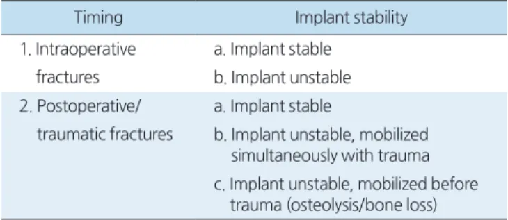
2014년 Duncan과 Haddad28)는 모든 관절 삽입물 주위 골 절에 공통적으로 적용할 수 있는 Unified Classification Sys- tem 분류법을 제시하였다. 이에 따라 비구 주위 골절 역시 해 부학적 위치에 따라 type A에서 F까지 분류되었으며, 특히 비 구에 바로 인접한 type B 골절은 삽입물의 안정성 및 주변 골 질에 따라 B1에서 B3로 세분화되었다. 이 분류법은 이후 AO foundation에 의해 채택되어 현재 가장 정확한 분류법으로 인정되고 있다. 하지만 이 분류법 또한 상당히 복잡하여 최근 Pascarella 등29)은 좀 더 간소화된 분류법을 새롭게 제시하기 도 하였다(Table 2).
4. 치료 원칙
비구 주위 골절은 골절의 전위 여부 및 삽입물의 안정성 여부에 따라 보존적 치료 또는 수술적 치료를 시행할 수 있 다. 수술적 치료의 원칙은 전위된 전후방 골주, 특히 후방 골 주의 안정적인 복원과 관절 중심의 회복, 그에 따른 비구 삽 입물의 안정성 회복이다. 전치환 비구컵의 경우 골절로 인해 한 번 해리가 일어나게 되면 반드시 재치환술을 시행해야 하 는 반면 반치환 삽입물의 경우에는 골절의 정복 및 내고정을 통해 비구의 안정성 및 관절내 조화(congruency)가 회복되면 삽입물을 유지할 수 있다.
1) 수술 중 발생한 골절
수술 중 골절은 대부분 작게 확공된 비구에 삽입물을 강 하게 감입시킬 때, 주로 경화된 후방 골주에서 발생하게 된다.
비전위 골절의 경우에는 골절 자체에 대해서는 보존적인 치 료를 시행할 수 있다. 그러나 아무리 삽입물이 압박 고정력을 얻은 것으로 보이더라도 비구컵의 돔 나사못(dome screw)을
통해 추가적인 안정성을 확보하는 것이 중요하다. 수술 후에 는 주기적인 방사선 검사를 통한 추시 관찰로 비구 삽입물의 위치 변동 여부를 확인하여야 한다.2)
전위된 골절의 경우, 특히 삽입물이 충분한 고정력을 얻을 수 없는 상황이라면 반드시 플레이트나 나사못 등을 통해서 전위된 골주를 안정화시켜야 한다. 비전위성 골절과 마찬가지 로 삽입물에 돔 나사못을 사용해야 하며 필요시 동종 골이식 등을 통해 고정력을 최대한 확보해야 한다. 간혹 골소실이 심 하거나 골편을 제대로 정복할 수 없을 때에는 탄탈륨 보강물 (tantalum augmentation) 또는 항돌출 케이지(antiprotrusio cage)를 사용해야 하는 경우도 있다.30)
2) 수술 후 발생한 골절
(1) 비전위성 골절: 외상 등에 의해 발생한 비전위성 골절
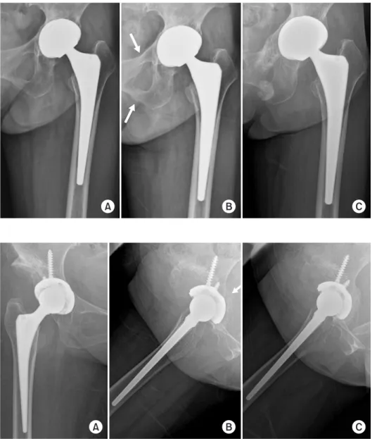
의 경우 비구 삽입물이 안정적이면 보존적 치료를 시행할 수 있다. 삽입물의 잠재적인 불안정성 여부를 평가하기 위하여 CT 등을 통해 골절의 정확한 위치와 패턴을 확인하는 것이 중요하다. 상치골지(superior pubic ramus)와 하치골지(infe- rior pubic ramus) 골절은 대부분 삽입물의 이완를 초래하지 않으며 특별한 치료 없이도 만족할 만한 골유합을 얻을 수 있 다(Fig. 1). 후방 골주를 포함하는 횡골절(transverse fracture) 의 경우에도 전위가 없고 삽입물이 안정적이라면 보존적 치 료가 가능하다(Fig. 2). Springer 등31)은 고관절 재치환술 후 발생한 7명의 횡골절 환자 중 2명의 비전위성 골절 환자에서 보존적 치료로 골유합을 얻을 수 있었다고 보고하였다. 이 경 우 약 6-8주간의 체중 부하 제한과 고관절의 과도한 굴곡 제 한이 필요할 수 있다.2) 보존적 치료를 시행하는 경우에는 반 드시 주기적인 방사선 검사를 시행하여 이차적인 삽입물 이 완의 발생 여부를 확인하여야 한다.13) 골절이 치유된 후에도 삽입물의 이완이 발생할 수 있으며, 이 경우 비구 재치환술이 불가피하다.Table 1. Classification of Periprosthetic Acetabular Fractures by David- son et al.1)
Type I Nondisplaced fracture not compromising the stability of the implant
Type II Nondisplaced fracture that may compromise the stability of the implant
Type III Displaced fracture
Table 2. Classification of Periprosthetic Acetabular Fractures by Pas- carella et al.29)
Timing Implant stability
1. Intraoperative fractures
a. Implant stable b. Implant unstable 2. Postoperative/
traumatic fractures
a. Implant stable
b. Implant unstable, mobilized simultaneously with trauma c. Implant unstable, mobilized before
trauma (osteolysis/bone loss)
(2) 삽입물의 불안정성을 동반한 비전위성 골절: 비전위성
골절이라고 하더라도 삽입물이 안정적이지 않은 경우나 잠재 적으로 불안정성을 초래할 수 있는 경우에는 수술적 치료가 필요하다. 이러한 유형의 골절에는 전방 골주와 후방 골주를 모두 침범하는 횡골절이나 전방 골주와 돔을 후방 골주로부 터 분리시키는 사선 골절(oblique fracture)이 포함된다. 이는 비구의 상방과 하방이 완전히 분리되어 전방 골주와 후방 골 주가 모두 단정된 것을 의미하며 흔히 골반 불연속성(pelvic discontinuity)이라는 용어로 표현한다.16) 정상적인 골반골이 라면 골반 불연속성은 주로 강한 충격에 의해 급성으로 발생 하지만 만성적인 골융해로 이미 병적인 상태에 있는 골반골 에서는 가벼운 외상 또는 외상 없이 서서히 발생할 수 있다.만성 골반 불연속성의 경우에는 급성 불연속성의 경우보
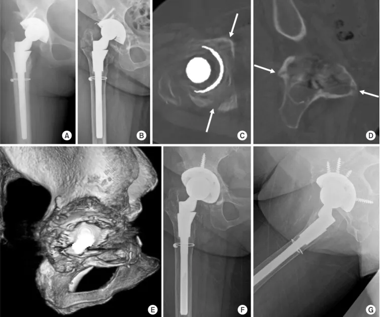
다 상대적으로 전위가 심하지 않으며 전후방 골주가 안정적 인 경우가 많기 때문에 골절에 대한 고정 없이도 재치환술이 가능하다.10) 특히 골결손의 정도가 심하지 않고 환자의 골질 이 나쁘지 않은 경우라면 비구 내 자가 해면골 또는 동종 해 면골 이식을 시행한 뒤 점보컵(jumbo cup)을 이용하여 어느 정도의 압박 고정력을 얻을 수 있다(Fig. 3).32) 하지만 이 경우 반드시 여러 방향으로 돔 나사못을 삽입하여 비구컵에 추가 적인 고정력을 부여해야 한다. 비구내 골결손 정도가 심하거 나 전후방 골주가 불안정한 경우에는 구조적 동종 골이식, 금 속 보강물, 항돌출 비구 케이지, 컵-케이지 구조 등 다양한 보조물의 도움을 받아 최대한 고정력을 얻어야 한다.33)
(3) 전위성 골절: 외상에 의한 급성 골반 불연속성은 주로
횡골절 또는 T형 골절을 유발하며 골편의 심한 전위를 동반Fig. 2. (A) Postoperative radiograph of a 52-year-old female with developmental dysplasia of the hip. (B) Non-displaced fracture of the posterior column was observed in the Lowenstein lateral ra- diograph at 2 months postoperatively.
An arrow indicates posterior column fracture. (C) The fracture was healed with callus formation after 4 months of protected weight-bearing.
Fig. 1. (A) A 55-year-old female un- derwent total hip arthroplasty due to rheumatoid arthritis on her left hip. (B) Fractures on the superior and inferior ramus were found after the patient suf- fered a minor trauma at 8 months post- operatively. Arrows indicate pubic rami fractures. (C) The fractures were healed after 6 months with conservative man- agement. The acetabular component was stable without pain.
할 수 있다. 상당한 정도의 전위가 동반되어 있을 때에는 골 편을 정복 및 고정하지 않는 한 새로운 비구컵을 안정적으로 고정할 수 없다. 특히 후방 골주의 전위가 있는 경우 대부분 플레이트나 나사못을 통해 골편을 안정화시켜야 한다. Rog- ers 등34)은 주로 외상에 의해 발생한 9명의 급성 골반 불연속 성 환자 중 8명에서 후방 골주의 내고정이 필요했다고 기술하 였다. 만약 이전 수술에서 전방 접근법이나 외측 접근법을 사 용하였다면 추가적인 후방 접근법을 통해 골편을 정확하게 정복하고 견고하게 고정해야 한다. 골절부의 적절한 시야 확
보를 위해 전자부 활주 절골술(trochanteric slide osteotomy) 이나 광범위 대전자 절골술(extended throchanteric oste- otomy) 등이 필요할 수 있다. 골편을 고정한 후에는 빠른 골 유합 및 삽입물의 안정성을 위해 남아 있는 골절 틈을 골이식 을 통해 메워주는 것이 권장된다.2)
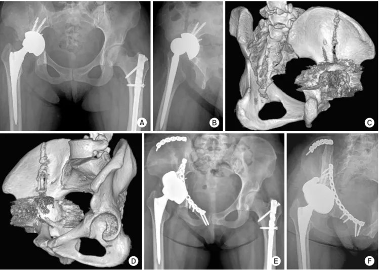
전방 및 후방 골주가 적절히 고정되었다면 일반적인 재치 환용 비구컵 및 여러 방향의 돔 나사못을 이용하여 비구컵을 새롭게 고정시킬 수 있다. 골절의 정복이 만족스럽지 못하거 나 골소실이 있는 경우, 골질이 좋지 않은 경우에는 금속 보
Fig. 3. (A) A 55-year-old female with secondary osteoarthritis due to tuberculosis sequelae underwent total hip arthroplasty. (B) Eighteen-year postoperative radiograph showed severe osteolytic lesions around the acetabular cup and proximal stem. A minor traumatic event caused peri- prosthetic acetabular and greater trochanteric fractures. (C-E) The corresponding computed tomography scan indicated pelvic discontinuity caused by a transverse acetabular fracture. Arrows indicate fracture lines causing pelvic discontinuity. (F, G) Massive impaction bone grafting was per- formed, followed by acetabular revision using a 64-mm jumbo cup with multiple screw holes.
강물이나 항돌출 케이지 등을 사용하여 최대한 견고하게 비 구컵을 고정해야 한다(Fig. 4).15) 만약 이러한 방법으로도 비 구컵의 안정적인 고정력을 얻을 수 없다면 일차 수술로 골절 을 고정하여 먼저 골유합을 이룬 뒤 이차 수술을 통해 새로 운 비구컵을 고정하는 방법도 고려할 수 있다.10) 전치환 삽입 물과 달리 반치환 삽입물 주위 골절에서는 전후방 골주가 비 교적 유지된 채로 삽입물이 골반 내로 돌출(protrusion)되는 경우가 발생할 수 있다. 이 경우 내측으로 전위된 골편이 적절 히 정복되어 조화로운 관절면이 유지될 수 있다면 골절 부위 를 견고하게 고정한 채 반치환물을 그대로 유지하는 방법도 가능하다(Fig. 5).
결 론
비구 삽입물 주위 골절의 치료 방침은 골절의 전위 정도 및 삽입물의 안정성 여부에 따라 신중히 결정되어야 한다. 골 편의 전위가 거의 없고 비구 삽입물이 안정적인 경우 대부분 보존적 치료가 가능하다. 그러나 이 경우에도 주기적인 추적 검사를 통해 삽입물의 전위 여부를 재평가해야 하며 삽입물 이 해리된 것으로 판단되는 경우 언제든 재치환술을 시행해 야 한다. 외상으로 인해 전방 또는 후방 골주가 전위된 경우 전후방 골주의 정확한 정복 및 고정력이 재치환물의 운명을 결정하기 때문에 보다 정교한 수술적 술기가 요구된다. 비구 를 재치환할 시에는 비구 주위의 골소실 및 골주의 안정성 정 도에 따라 적절한 보강물을 선택하여 강한 초기 고정력을 얻 어야 한다.
Fig. 4. (A, B) The acetabular prosthesis became unstable due to a traumatic periprosthetic acetabular fracture. (C, D) The three-dimensional recon- struction images showed a displaced acetabular fracture involving both columns and the iliac wing. (E, F) After removing the previous prosthesis, the fracture was treated by open reduction and internal fixation with two reconstruction plates. The acetabular component was revised with the help of tantalum metal block and additional screws. Three-month postoperative radiographs showed stable implant fixations.
요 약
비구 삽입물 주위 골절은 고관절 치환술 후 발생하는 흔하 지 않은 합병증이다. 하지만 고관절 치환술을 시행받은 인구 가 지속적으로 증가함에 따라 비구 삽입물 주위 골절의 발생 건수 역시 앞으로 점차 증가할 것으로 예상된다. 비구 삽입물 주위 골절은 크게 수술 중 발생하는 골절과 수술 후 발생하 는 골절로 나눌 수 있다. 수술 후 발생하는 골절은 주로 외상
또는 비구 주위 골융해와 같은 병적인 상태에서 발생할 수 있 다. 비구 주위 골절의 치료 방향은 골편의 전위 정도 및 비구 삽입물의 안정성 여부에 따라 결정된다. 대부분의 비전위 골 절은 체중 부하 제한과 같은 비수술적 방법으로 치료될 수 있 는 반면 전위된 골절의 경우 주로 삽입물의 불안정성을 동반 하기 때문에 종종 고난도의 수술적 술기를 요한다. 이 종설에 서는 현재까지의 문헌들을 바탕으로 비구 주위 골절의 역학, 진단, 분류 및 치료 원칙에 대해 고찰하였다.
Fig. 5. (A, B) Preoperative radiographs showing a displaced acetabular fracture with the protrusion of the bipolar cup into the pelvic cavity. (C, D) Computed tomography images showed comminuted fracture of the medial wall of the acetabulum. (E) The displaced quadrilateral surface was visualized through the modified Stoppa approach. (F-H) After open reduction of depressed articular surface of the acetabulum, buttress plating was performed using reconstruction plates and screws. (I, J) Five-month postoperative radiographs showed the stable bipolar component in the congruent hip joint.
색인 단어: 삽입물 주위 골절, 비구, 고관절, 인공관절 치환술,
분류, 치료법ORCID
박찬우, https://orcid.org/0000-0002-4332-5863 오형근, https://orcid.org/0000-0001-5388-7096 이우석, https://orcid.org/0000-0002-0798-1660 박윤수, https://orcid.org/0000-0001-9605-9055 임승재, https://orcid.org/0000-0002-7209-9690
References
1. Davidson D, Pike J, Garbuz D, Duncan CP, Masri BA: Intra- operative periprosthetic fractures during total hip arthroplasty.
Evaluation and management. J Bone Joint Surg Am, 90: 2000- 2012, 2008.
2. Simon P, von Roth P, Perka C: Treatment algorithm of acetabu- lar periprosthetic fractures. Int Orthop, 39: 1995-2003, 2015.
3. Helfet DL, Ali A: Periprosthetic fractures of the acetabulum. In- str Course Lect, 53: 93-98, 2004.
4. Kim YS, Callaghan JJ, Ahn PB, Brown TD: Fracture of the ac- etabulum during insertion of an oversized hemispherical compo- nent. J Bone Joint Surg Am, 77: 111-117, 1995.
5. Sharkey PF, Hozack WJ, Callaghan JJ, et al: Acetabular fracture associated with cementless acetabular component insertion: a report of 13 cases. J Arthroplasty, 14: 426-431, 1999.
6. Curtis MJ, Jinnah RH, Wilson VD, Hungerford DS: The initial stability of uncemented acetabular components. J Bone Joint Surg Br, 74: 372-376, 1992.
7. Haidukewych GJ, Jacofsky DJ, Hanssen AD, Lewallen DG: In- traoperative fractures of the acetabulum during primary total hip arthroplasty. J Bone Joint Surg Am, 88: 1952-1956, 2006.
8. Chatoo M, Parfitt J, Pearse MF: Periprosthetic acetabular frac- ture associated with extensive osteolysis. J Arthroplasty, 13:
843-845, 1998.
9. Chitre A, Wynn Jones H, Shah N, Clayson A: Complications of total hip arthroplasty: periprosthetic fractures of the acetabulum.
Curr Rev Musculoskelet Med, 6: 357-363, 2013.
10. Benazzo F, Formagnana M, Bargagliotti M, Perticarini L: Peri- prosthetic acetabular fractures. Int Orthop, 39: 1959-1963, 2015.
11. Miller AJ: Late fracture of the acetabulum after total hip re- placement. J Bone Joint Surg Br, 54: 600-606, 1972.
12. McElfresh EC, Coventry MB: Femoral and pelvic fractures after total hip arthroplasty. J Bone Joint Surg Am, 56: 483-492, 1974.
13. Peterson CA, Lewallen DG: Periprosthetic fracture of the ac-
etabulum after total hip arthroplasty. J Bone Joint Surg Am, 78:
1206-1213, 1996.
14. Takigami I, Ito Y, Mizoguchi T, Shimizu K: Pelvic discontinuity caused by acetabular overreaming during primary total hip ar- throplasty. Case Rep Orthop, 2011: 939202, 2011.
15. Della Valle CJ, Momberger NG, Paprosky WG: Periprosthetic fractures of the acetabulum associated with a total hip arthro- plasty. Instr Course Lect, 52: 281-290, 2003.
16. Berry DJ, Lewallen DG, Hanssen AD, Cabanela ME: Pelvic discontinuity in revision total hip arthroplasty. J Bone Joint Surg Am, 81: 1692-1702, 1999.
17. Zwartelé RE, Witjes S, Doets HC, Stijnen T, Pöll RG: Cement- less total hip arthroplasty in rheumatoid arthritis: a systematic review of the literature. Arch Orthop Trauma Surg, 132: 535- 546, 2012.
18. McGrory BJ: Periprosthetic fracture of the acetabulum during total hip arthroplasty in a patient with Paget’s disease. Am J Or- thop (Belle Mead NJ), 28: 248-250, 1999.
19. Desai G, Ries MD: Early postoperative acetabular discontinu- ity after total hip arthroplasty. J Arthroplasty, 26: 1570.e17-e19, 2011.
20. Martin JR, Barrett IJ, Sierra RJ, Lewallen DG, Berry DJ: Preop- erative radiographic evaluation of patients with pelvic disconti- nuity. J Arthroplasty, 31: 1053-1056, 2016.
21. Goodman SB, Adler SJ, Fyhrie DP, Schurman DJ: The acetabu- lar teardrop and its relevance to acetabular migration. Clin Or- thop Relat Res, (236): 199-204, 1988.
22. Onsten I, Carlsson AS, Ohlin A, Nilsson JA: Migration of ac- etabular components, inserted with and without cement, in one- stage bilateral hip arthroplasty. A controlled, randomized study using roentgenstereophotogrammetric analysis. J Bone Joint Surg Am, 76: 185-194, 1994.
23. Udomkiat P, Wan Z, Dorr LD: Comparison of preoperative ra- diographs and intraoperative findings of fixation of hemispheric porous-coated sockets. J Bone Joint Surg Am, 83: 1865-1870, 2001.
24. Fehring KA, Howe BM, Martin JR, Taunton MJ, Berry DJ:
Preoperative evaluation for pelvic discontinuity using a new re- formatted computed tomography scan protocol. J Arthroplasty, 31: 2247-2251, 2016.
25. Gelalis ID, Politis AN, Arnaoutoglou CM, Georgakopoulos N, Mitsiou D, Xenakis TA: Traumatic periprosthetic acetabular fracture treated by acute one-stage revision arthroplasty. A case report and review of the literature. Injury, 41: 421-424, 2010.
26. Harvie P, Gundle R, Willett K: Traumatic periprosthetic acetab- ular fracture: life threatening haemorrhage and a novel method of acetabular reconstruction. Injury, 35: 819-822, 2004.
27. Callaghan JJ, Kim YS, Pederson DR, Brown TD: Periprosthetic fractures of the acetabulum. Orthop Clin North Am, 30: 221- 234, 1999.
28. Duncan CP, Haddad FS: The Unified Classification System (UCS): improving our understanding of periprosthetic fractures.
Bone Joint J, 96: 713-716, 2014.
29. Pascarella R, Sangiovanni P, Cerbasi S, et al: Periprosthetic ac- etabular fractures: a new classification proposal. Injury, 49 Suppl 3: S65-S73, 2018.
30. Ko PS, Chan WF, Wong MK, Leung MF, Lee OB, Lam JJ:
Fixation using acetabular reconstruction cage and cancellous allografts for intraoperative acetabular fractures associated with cementless acetabular component insertion. J Arthroplasty, 19:
643-646, 2004.
31. Springer BD, Berry DJ, Cabanela ME, Hanssen AD, Lewallen
DG: Early postoperative transverse pelvic fracture: a new com- plication related to revision arthroplasty with an uncemented cup. J Bone Joint Surg Am, 87: 2626-2631, 2005.
32. Whaley AL, Berry DJ, Harmsen WS: Extra-large uncemented hemispherical acetabular components for revision total hip ar- throplasty. J Bone Joint Surg Am, 83: 1352-1357, 2001.
33. Paprosky W, Sporer S, O’Rourke MR: The treatment of pelvic discontinuity with acetabular cages. Clin Orthop Relat Res, 453:
183-187, 2006.
34. Rogers BA, Whittingham-Jones PM, Mitchell PA, Safir OA, Bircher MD, Gross AE: The reconstruction of periprosthetic pelvic discontinuity. J Arthroplasty, 27: 1499-1506.e1, 2012.