INTRODUCTION
In the last decade, preimplantation genetic diagnosis (PGD) has become an important alternative to prenatal diagnosis for couples at high risk of transmitting inherited disorders to their children. Fluorescence in situ hybridization (FISH) has been offered for gender selection in X-linked diseases (1, 2), chromosomal translocations (3-5), aneuploidy, and recur- rent implantation failure (6, 7). Procedures using polymerase chain reaction (PCR) has been applied for single gene defects including X-linked diseases (8-11).
Ornithine transcarbamylase (OTC; EC 2.1.3.3) is placed in the mitochondrial matrix where it contributes to the detox- ification of nitrogenous wastes through the urea cycle, and catalyzes the synthesis of citruline from carbamyl phosphate and ornithine. The OTC gene is located on the short arm of the X chromosome within band Xp21.1 (12). It spans 73 kb, has an open reading frame of 1,062 nucleotides, and con- tains 10 exons and 9 introns (13, 14). OTC deficiency (MIM-
#311250) is an X-linked and co-dominant metabolic disor-
der with partial penetrance in females (15). The phenotype of OTC deficiency is extremely heterogeneous, ranging from asymptomatic adult hemizygous males to acute neonatal hyperammonemic coma in the first week of affected male babies. Approximately 80% of heterozygous females are asy- mptomatic and remaining 20% show clinical severity simi- lar to males with partial deficiencies (16, 17). There are more than 341 mutations known to cause OTC deficiency, all of which are specific for the individual families. Mutations have been found in all exons and introns with a relative paucity of mutations in the sequence encoding the leader peptide (exon 1 and a part of exon 2) and in exon 7 (17, 18).
To our knowledge, only two cases have been reported where specific PGD for OTC deficiency were carried out using mul- tiplex or duplex-nested PCR assay and healthy babies devoid of the OTC mutation were born (19, 20). In this study, we describe the successful pregnancy and birth after PGD for OTC deficiency with simultaneous analyses of FISH and du- plex-nested PCR in Korea. We have applied a duplex-nested PCR for the amplification of both the causative mutation and
Hyoung-Song Lee, Jin Hyun Jun, Hye Won Choi, Chun Kyu Lim, Han-Wook Yoo�, Mi Kyoung Koong*, Inn Soo Kang*
Laboratory of Reproductive Biology and Infertility, Department of Obstetrics and Gynecology*, Cheil General Hospital & Women’s Healthcare Center, Kwandong University College of Medicine, Seoul;
Medical Genetics Clinic & Laboratory�, Department of Pediatrics, Asan Medical Center, University of Ulsan College of Medicine, Seoul, Korea
Address for correspondence Jin Hyun Jun, Ph.D.
Laboratory of Reproductive Biology & Infertility, Cheil General Hospital & Women’s Healthcare Center, Kwandong University College of Medicine, 1-19 Mukjeong-dong, Jung-gu, Seoul 100-380, Korea Tel : +82.2-2000-7590, Fax : +82.2-2265-5621 E-mail : junjh55@hanmail.net
572
Preimplantation Genetic Diagnosis for Ornithine Transcarbamylase Deficiency by Simultaneous Analysis of Duplex-nested PCR and Fluorescence In Situ Hybridization
: A Case Report
Ornithine transcarbamylase (OTC) deficiency is an X-linked co-dominant disorder.
A couple, with a previous history of a neonatal death and a therapeutical termination due to OTC deficiency, was referred to our center for preimplantation genetic diag- nosis (PGD). The female partner has a nonsense mutation in the exon 9 of the OTC gene (R320X). We carried out nested polymerase chain reaction (PCR) for R320X mutation and fluorescence in situ hybridization (FISH) for aneuploidy screening.
Among a total of 11 embryos, two blastomeres per embryo from 9 embryos were biopsied and analyzed by duplex-nested PCR and FISH, and one blastomere per embryo from 2 embryos by only duplex-nested PCR. As a result of PCR and restric- tion fragment length polymorphism analysis, four embryos were diagnosed as una- ffected embryos having the normal OTC gene. Among these embryos, only one embryo was confirmed as euploidy for chromosome X, Y and 18 by FISH analy- sis. A single normal embryo was transferred to the mother, yielding an unaffected pregnancy and birth of a healthy boy. Based on our results, PCR for mutation loci and FISH for aneuploidy screening with two blastomeres from an embryo could provide higher accuracy for the selection of genetically and chromosomally normal embryos in the PGD for single gene defects.
Key Words : Preimplantation Genetic Diagnosis (PGD); Ornithine Transcarbamylase (OTC) Deficiency; Nest- ed PCR; FISH; Single Gene Defects
Received : 18 September 2006 Accepted : 6 December 2006
the Y chromosome specific Sry gene, and FISH for aneuploidy screening of chromosome X, Y and 18. A healthy boy was born by the transfer of only one unaffected normal embryo.
CASE REPORT
A couple was referred to our center after therapeutic ter- mination of the second pregnancy following prenatal diag- nosis for OTC deficiency. In the first pregnancy, a male baby was born but died at 10 days after birth due to liver dysfunc- tion accompanying hyperammonemia. Molecular genetic analysis of the couple’s OTC gene was performed by PCR and direct sequencing at Seoul Asan Hospital and revealed a sin- gle base substitution (R320X) in the exon 9 of the female partner. In the second pregnancy, the fetus was identified to be an affected boy by PCR and direct sequencing on a chori- onic villus sample, and the pregnancy was therapeutically terminated. In order to avoid a pregnancy with an affected baby, the couple was counseled for PGD in Cheil General Hospital.
In order to determine the efficiency of single cell PCR and allele drop-out (ADO) rate, primers used for the detection of the causative mutation and Y chromosome specific Sry gene were tested using single lymphocytes (Table 1). Ovarian stim-
ulation, in vitro fertilization, and blastomere biopsy were done as previously described (4, 5). After the blastomere biopsy procedure, the embryos were washed several times and trans- ferred to the G2.2 medium (Vitrolife Sweden AB, Kungs- backa, Sweden). Each blastomere was washed twice through two drops of G2.2 medium and transferred into sterile 0.2 mL PCR tubes containing 5 L of alkaline lysis buffer. For each embryo biopsied, blank negative controls were prepared from the wash drops. The nested-duplex PCR analysis was performed by previously described protocol (6). Mutation analysis by restriction fragment length polynorphism (RFLP) with BclI restriction enzyme was carried out only for positively amplified PCR products of blastomeres. Another blastomere from the same embryo was prepared for FISH. The FISH anal- ysis for chromosome X, Y and 18 was done as previously des- cribed and the manufacture’s protocol (4, 5). The embryos were cultured under standard culture condition until the diagno- sis was established. The embryos with the normal genotype were selected and transferred on day 4 after fertilization. Am- niocentesis was performed at 16 weeks of gestation to con- firm the results of PGD. Postnatal genetic and physiologi- cal diagnosis was also conducted.
All 10 exons and exon-intron boundaries of the OTC gene
Annealing temp. (°C)
Name Sequences (5′→3′)
Product sizes
(bp) Ornithine trans- CACTCTGCTCCTTTGTCTCT 62 319
carbamylase GTTGGAACCACACAAAGAAC Exon 9
Ornithine trans- GGCCATGTGTGTTTTTAGAT 64 218 carbamylase GTCCACTTTCTGTTTTCTGC
Exon 9-nested
Sry GAATATTCCCGCTCTCCGGA 64 472
GCTGGTGCTCCATTCTTGAG
Table 1.Sequences of oligonucleotide primers and PCR con- ditions for duplex-nested PCR for OTC deficiency
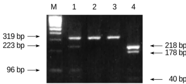
M 1 2 3 4
319 bp
218 bp 178 bp 223 bp
96 bp
40 bp Fig. 1.Identification of the maternal mutation (heterozygote type) in exon 9 of the OTC gene. After digesting the PCR product with BclI, the 319 or 218 bp PCR products of the affected mother were digested into 223 and 96 bp (lane 1) and 178 and 40 bp products (lane 4), respectively. The 40 bp product was hardly seen on an agarose gel. Lane 1 and 4; the first and nested PCR products of the affected mother, respectively, lane 2 and 3; BclI-digested pro- ducts from the first PCR products of the father. M: molecular weight marker.
Fig. 2.Results of agarose gel electrophoresis of the restriction fragment length polymorphism (RFLP) analysis with BclI restriction enzyme in the preimplantation genetic diagnosis for ornithine transcarbamylase deficiency. In normal embryos (No. 1, 2, 3 and 8), the 218-bp PCR products were not digested with BclI.
Em 1 2 3 4 6 7 8 9 10 11
218 bp 178 bp Mother Control Mother Control PCR, polymerase chain reaction; OTC, ornithine transcarbamylase.
were screened by PCR followed by direct DNA sequencing analysis. The heterozygous OTC gene with R320X nonsense mutation (CGA→ TGA) and wild type was detected in the exon 9 of the female partner by RFLP (Fig. 1) and direct sequ- encing (data not shown). This R320X nonsense mutation was associated with acute neonatal hyperammonemia in their first pregnancy and reduced enzyme activity by 10-15% (21).
Male partner has a normal OTC gene.
The efficiency and accuracy of the duplex-nested PCR anal- ysis were tested on single lymphocytes from the heterozygous female partner and normal male partner. PCR on single lym- phocytes resulted in 90.6% (58/64) amplification rate, where- as none of the negative controls showed a positive band. ADO was detected in 4 out of 30 lymphocytes (13.3%).
After ovarian hyperstimulation, a total of 18 oocytes were retrieved and 17 oocytes were injected using intracytoplas- mic sperm injection in order to prevent DNA contamination from inseminated sperm. Among a total of 11 embryos, two blastomeres per embryo from 9 embryos were biopsied and simultaneously analyzed by duplex-nested PCR and FISH, and one blastomere per embryo from 2 embryos by only du- plex-nested PCR. As a result of duplex-nested PCR and RFLP analysis with the BclI restriction enzyme, ten showed success- ful amplification for the exon 9 of the OTC gene, and the amplification rate was 90.9%, almost same as that of the sin- gle lymphocyte test. There were two unaffected males, five affected males, two unaffected females, and one heterozygous female (Fig. 2 and Table 2). No amplification was detected in any of the negative controls. On the other hand, out of nine blastomeres analyzed by FISH, only six blastomeres were identified as chromosomally normal embryos (Fig. 3, Table 2).
However, among total four unaffected embryos (embryo 1, 2, 3, and 8) diagnosed by PCR, only two embryos (embryo 3 and 8) were identified as chromosomally normal by FISH.
The other two embryos (embryo 1 and 2) were shown to have trisomy 18 and monosomy 18, respectively. Only one male embryo (embryo 8) was transferred, and another female unaf- fected embryo (embryo 3) was cryopreserved at blastocyst stage on day 5 after fertilization. This PGD cycle resulted in a singleton pregnancy and the genotype of the fetus was con- firmed to be a normal male by amniocentesis at 16 weeks of gestation. A healthy boy of 2.8 kg was delivered at 36 weeks.
The genotype of the baby for the exon 9 of OTC gene and level of ammonia were confirmed as normal.
DISCUSSION
Most PGD cycles for X-linked disorders such as Duchenne muscular dystrophy and fragile X syndrome involved the selection of female embryos which were diagnosed as normal or heterozygous. These gender selections were supported by couples at risk of X-linked recessive conditions. However, OTC deficiency is an X-linked and co-dominant disorder.
Some heterozygous females represent severe phenotypic abnor- malities such as nausea, vomiting and ataxia. ADO for affect-
Plus (+) and minus (-) signs indicate whether a band was detected cor- responding to the Sry gene. N.A. (not assayed) indicates that blastomere was not available for FISH. In the table, transferred embryo is presented with bold and italic characters.
PGD, preimplantation genetic diagnosis; OTC, ornithine transcarbamy- lase; PCR, polymerase chain reaction; FISH, fluorescence in situ hybri- dization.
No. of embryo (stage)
FISH
(X;Y;18) Diagnosis PCR
OTC exon
9-RFLP Sry
1 (8 cells) Normal - 3;0;3 Trisomy
2 (12 cells) Normal + 1;1;1 Monosomy 18 3 (8 cells) Normal - 2;0;2 Female unaffected 4 (8 cells) Mutant + 1;1;2 Male affected 5 (8 cells) Amplification failure + 1;0;1 No result 6 (8 cells) Mutant + 1;1;2 Male affected 7 (8 cells) Heterozygote - 2;0;2 Female heterozygote 8 (8 cells) Normal + 1;1;2 Male unaffected 9 (8 cells) Mutant + 1;1;2 Male affected 10 (5 cells) Mutant + N.A. Male affected 11 (4 cells) Mutant + N.A. Male affected Table 2.Summary of the results of the PGD for OTC deficiency with simultaneous analysis of duplex nested PCR and FISH
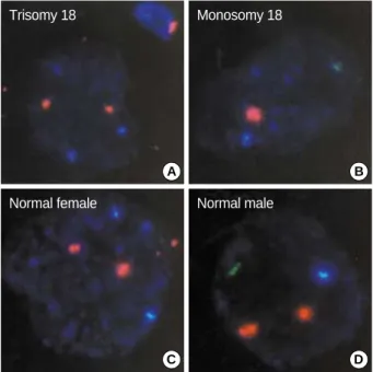
Fig. 3.Results of the fluorescence in situ hybridization (FISH) anal- ysis on the embryos diagnosed to be normal by PCR in the PGD for OTC deficiency. Panels A-Dshow the photographs for the results of FISH on the embryos No. 1, 2, 3, and 8 in Table II, respectively.
Aqua, green and orange signals indicate the presence of the chro- mosome X, Y and 18 in the nucleus of the blastomeres, respec- tively.
PGD, preimplantation genetic diagnosis; OTC, ornithine transcar- bamylase; PCR, polymerase chain reaction
A B
C D
Trisomy 18 Monosomy 18
Normal female Normal male
ed allele could lead to yielding an affected heterozygous baby, even if the female embryo was diagnosed as normal by PGD for OTC deficiency. Gender selection with FISH for Y chro- mosome-specific loci is more reliable because amplification failure could occasionally occur in single cell PCR. Thus, our protocol with PCR and FISH in this study provides higher accuracy for the selection of genetically and chromosomally normal embryos in the PGD for X-linked co-dominant de- fects.
We applied duplex-nested PCR and FISH analysis in two blastomeres from each embryo, which were more than 6-cell stage. Biopsy of more than a quarter of blastomeres from each embryo could interfere with developmental potential of em- bryos. Thus, single blastomere biopsy was applied to less than 6-cell stage embryos in this study. Sequential PCR and FISH analysis in one blastomere (cell recycling) was reported and could be used to detect the aneuploidy, but the rate of ADO after PCR seems to be higher using this method (22, 23).
Therefore, we carried out two methods separately on two blastomeres. A duplex-nested PCR analysis was developed for the simultaneous diagnosis of a R320X mutation and a gender selection, and FISH analysis was used to detect ane- uploidy for chromosome X, Y and 18. As a result of duplex nested PCR and FISH analysis, concordant results have been obtained from two blastomeres at the sex locus, and the am- plification efficiency was 100% (9/9 except two blastomeres, that were not analyzed by FISH analysis) for Sry. And the amplification rate was 90.9% (10/11) for exon 9 of the OTC gene.
Several strategies have been developed to decrease ampli- fication failure and ADO in single cell analysis of PGD. These included the development of sequential first and second polar body analysis (24), improvement of the PCR condition by increasing the denaturation temperature and time (19), using a more powerful lysis method (25-28) and the use of fluores- cent PCR for mutations or linked markers and multiplex PCR (29-32). As mentioned above, we have also endeavored to optimize single cell PCR conditions, which included longer initial denaturation time, higher denaturation temperature, and the selection of Taq polymerase, the concentration of MgCl2, and lysis methods. The incidence of ADO was 5- 15% in many PGD laboratories (27, 28). At a preliminary experiment on single lymphocytes in this study, the ADO rate for exon 9 of the OTC gene was about 13.0% when using duplex-nested PCR. We could not absolutely exclude the possibility of ADO in the diagnosis of unaffected female em- bryo owing to the relatively higher ADO. Thus, FISH was simultaneously applied for the aneuploidy screening and sex selection to prevent the pregnancy of heterozygous female.
In this study, we report the successful PGD and delivery of a healthy boy in a couple at risk of transmitting the OTC deficiency. This was carried out by the simultaneous analy- sis of the duplex-nested PCR for a R320X mutation in the OTC gene underlying the OTC deficiency and Y chromo-
some-specific loci, and FISH for aneuploidy screening of chro- mosome X, Y and 18. This strategy could provide higher accuracy for the selection of genetically and chromosomally normal embryos in PGD for single gene defects.
REFERENCES
1. Munne S, Sultan KM, Weier HU, Grifo JA, Cohen J, Rosenwaks Z.
Assessment of numerical abnormalities of X, Y, 18 and 16 chromo- somes in preimplantation embryos before transfer. Am J Obstet Gy- necol 1995; 172: 1191-201.
2. Verlinsky Y. Preimplantation genetic diagnosis. J Assist Reprod Genet 1996; 13: 87-9.
3. Delhanty JD. Chromosome analysis by FISH in human preimplan- tation genetics. Hum Reprod 1997; 12 (11 Suppl): 153-5.
4. Lim CK, Jun JH, Min DM, Lee HS, Kim JY, Koong MK, Kang IS.
Efficacy and clinical outcome of preimplantation genetic diagnosis using FISH for couples of reciprocal and Robertsonian transloca- tions: the Korean experience. Prenat Diagn 2004; 24: 556-61.
5. Lim CK, Min DM, Lee HS, Byun HK, Park SY, Ryu HM, Kim JY, Koong MK, Kang IS, Jun JH. Improvement of pregnancy rate in pre- implantation genetic diagnosis with FISH procedure by the labora- tory optimization and experiences. Korean J Fertil Steril 2004; 31:
29-39.
6. Gianaroli L, Magli MC, Ferraretti AP, Munne S. Preimplantation diagnosis for aneuploidies in patients undergoing in vitro fertiliza- tion with a poor prognosis: identification of the categories for which it should be proposed. Fertil Steril 1999; 72: 837-44.
7. Munne S, Magli C, Cohen J, Morton P, Sadowy S, Gianaroli L, Tuck- er M, Marquez C, Sable D, Ferraretti AP, Massey JB, Scott R. Posi- tive outcome after preimplantation diagnosis of aneuploidy in human embryos. Hum Reprod 1999; 14: 2191-9.
8. Lissens W, Sermon K. Preimplantation genetic diagnosis: current status and new development. Hum Reprod 1997; 12: 1756-61.
9. Ray PF, Handyside AH. Increasing the denaturation temperature during the first cycles of amplification reduces allele dropout from single cells for preimplantation genetic diagnosis. Mol Hum Reprod 1996; 2: 213-8.
10. Malcov M, Schwartz T, Mei-Raz N, Yosef DB, Amit A, Lessing JB, Shomrat R, Orr-Urtreger A, Yaron Y. Multiplex nested PCR for preim- plantation genetic diagnosis of spinal muscular atrophy. Fetal Diagn Ther 2004; 19: 199-206.
11. Lee HS, Choi HW, Lim CK, Koong MK, Kang IS, Yoo HW, Choi JH, Jun JH. Identification of a novel single nucleotide polymorphism of HADHA gene at a referred primer-binding site during pre-diag- nostic tests for preimplantation genetic diagnosis. J Korean Med Sci 2006; 21: 794-9.
12. Lindgren V, de Martinville B, Horwich AL, Rosenberg LE, Francke U. Human ornithine transcarbamylase locus mapped to band Xp21.1 near the Duchenne muscular dystrophy locus. Science 1984; 226:
698-700.
13. Horwich AL, Fenton WA, William KR, Kalousek F, Kraus JP, Doo- little RF, Konigsberg W, Rogenberg LE. Structure and expression
of a complementary DNA for the nuclear coded precursor of human mitochondrial ornithine transcarbamylase. Science 1984; 224: 1068- 74.
14. Hata A, Tsuzuki T, Shimada K, Takiguchi M, Mori M, Matsuda I.
Structure of the human ornithine transcarbamylase deficiency. Am J Hum Genet 1988; 48: 212-22.
15. Pelet A, Rotig A, Bonaiti-Pellie C, Rabier D, Cormier V, Toumas E, Hentzen D, Saudubray JM, Munnich A. Carrier detection in a par- tially dominant X-linked disease: ornithine transcarbamylase defi- ciency. Hum Genet 1990; 84: 167-71.
16. Hudak ML, Douglas Jones M, Brusilow SW. Differentiation of tran- sient hyperammonemia of the newborn and urea cycle enzyme defects by clinical presentation. J Pediatr 1985; 107: 712-9.
17. Tuchman M, Jaleel N, Morizono H, Sheehy L, Lynch MG. Muta- tions and polymorphisms in the human ornithine transcarbamylase gene. Hum Mutat 2002; 19: 93-107.
18. Yamaguchi S, Brailey LL, Morizono H, Bale AE, Tuchman M. Muta- tion and polymorphisms in the human ornithine transcarbamylase (OTC) gene. Hum Mutat 2006; 27: 626-32.
19. Ray PF, Gigarel N, Bonnefont JP, Attie T, Hamamah S, Frydman N, Vekemans M, Frydman R, Munnich A. First specific preimplan- tation genetic diagnosis for ornithine transcarbamylase deficiency.
Prenat Diagn 2000; 20: 1048-54.
20. Verlinsky Y, Rechitsky S, Verlinsky O, Strom C, Kuliev A. Preim- plantation diagnosis for ornithine transcarbamylase deficiency. Re- prod Biomed Online 2000; 1: 45-7.
21. Yoo HW, Kim GH, Lee DH. Identification of new mutations in the ornithine transcarbamylase (OTC) gene in Korean families. J Inherit Metab Dis 1996; 19: 31-42.
22. Rechitsky S, Freidine M, Verlinsky Y, Strom CM. Allele dropout in sequential PCR and FISH analysis of single cells (cell recycling). J Assist Reprod Genet 1996; 13: 115-24.
23. Thornhill AR, Monk M. Cell recycling of a single human cell for preimplantation diagnosis of X-linked disease and dual sex determi- nation. Mol Hum Reprod 1996; 2: 285-9.
24. Strom CM, Ginsberg N, Rechitsky S, Cieslak J, Ivakhenko V, Wolf
G, Lifchez A, Moise J, Valle J, Kaplan B, White M, Barton J, Kuliev A, Verlinsky Y. Three births after preimplantation genetic diagnosis for cystic fibrosis with sequential first and second polar body analy- sis. Am J Obstet Gynecol 1998; 178: 1298-306.
25. Wu K, Cuppens H, Buyse I, Decorte R, Marynen P, Gordts S, Cas- siman JJ. Co-amplification of the cystic fibrosis delta F508 mutation with the HLA DQA1 sequence in single cell PCR: implications for improved assessment of polar bodies and blastomeres in preimplan- tation diagnosis. Prenat Diagn 1993; 13: 1111-2.
26. Thornhill AR, McGrath JA, Eady RA, Braude PR, Handyside AH.
A comparison of different lysis buffers to assess allele dropout from single cells for preimplantation genetic diagnosis. Prenat Diagn 2001;
21: 490-7.
27. Piyamongkol W, Bermudez MG, Harper JC, Wells D. Detailed inves- tigation of factors influencing amplification efficiency and allele drop- out in single cell PCR: implications for preimplantation genetic diag- nosis. Mol Hum Reprod 2003; 9: 411-20.
28. Choi HW, Lee HS, Lim CK, Koong MK, Kang IS, Jun JH. Reliability of the single cell PCR analysis for preimplantation genetic diagnosis of single gene disorders. Korean J Fertil Steril 2005; 32: 293-300.
29. Dreesen J, Jacobs L, Bras M, Herberg J, Dumoulin JC, Geraedts JP, Evers JL, Smeets HJ. Multiplex PCR of polymorphic markers flank- ing the CFTR gene: a general approach for preimplantation genetic diagnosis of cystic fibrosis. Mol Hum Reprod 2000; 6: 881-5.
30. Eftedal I, Schwartz M, Bendtsen H, Andersen A, Zieve S. Single intra- genic microsatellite preimplantation enetic diagnosis for cystic fibro- sis provides positive allele identification of all CFTR genotypes for informative couples. Mol Hum Reprod 2001; 7: 307-12.
31. Goossens V, Sermon K, Lissens W, Vandervorst M, Vanderfaeillie A, De Rijcke M, De Vos A, Henderix P, Van De Velde H, Van Steir- teghem A, Liebaers I. Clinical application of preimplantation genetic diagnosis for cystic fibrosis. Prenat Diag 2000; 20: 571-81.
32. Lee HS, Choi HW, Lim CK, Park SY, Kim JY, Koong MK, Jun JH, Kang IS. Efficacy of duplex-nested PCR and fluorescent PCR in the preimplantation genetic diagnosis for Duchenne muscular dystrophy.
Korean J Fertil Steril 2005; 32: 17-26.