Prevalence and Associated Factors of Diabetic Retinopathy in Rural Korea: The Chungju Metabolic Disease Cohort Study
This study was aimed to investigate the prevalence of diabetic retinopathy and its associated factors in rural Korean patients with type 2 diabetes. A population-based, cross- sectional diabetic retinopathy survey was conducted from 2005 to 2006 in 1,298 eligible participants aged over 40 yr with type 2 diabetes identified in a rural area of Chungju, Korea. Diabetic retinopathy was diagnosed by a practicing ophthalmologist using
funduscopy. The overall prevalence of diabetic retinopathy in the population was 18% and proliferative or severe non-proliferative form was found in 5.0% of the study subjects. The prevalence of retinopathy was 6.2% among those with newly diagnosed type 2 diabetes and 2.4% of them had a proliferative or severe non-proliferative diabetic retinopathy. The odds ratio of diabetic retinopathy increased with the duration of diabetes mellitus (5-10 yr:
5.2- fold; > 10 yr: 10-fold), postprandial glucose levels (> 180 mg/dL: 2.5-fold), and HbA1c levels (every 1% elevation: 1.34-fold). The overall prevalence of diabetic retinopathy in rural Korean patients was similar to or less than that of other Asian group studies. However, the number of patients with proliferative or severe non-proliferative diabetic retinopathy was still high and identified more frequently at the time of diagnosis.
This emphasizes that regular screening for diabetic retinopathy and more aggressive management of glycemia can reduce the number of people who develop diabetic retinopathy.
Key Words: Diabetic Retinopathy; Prevalence; Risk Factors Ji-Hyun Kim1, Hyuk-Sang Kwon1,
Yong-Moon Park2, Jin-Hee Lee4, Man-Soo Kim3, Kun-Ho Yoon1, Won Chul Lee2, Bong-Yun Cha1 and Ho-Young Son1
Departments of 1Internal Medicine, 2Preventive Medicine and 3Ophthalmology, College of Medicine, The Catholic University of Korea, Seoul; 4The Catholic Institute of Ubiquitous Healthcare, Seoul, Korea Received: 18 February 2011
Accepted: 21 June 2011 Address for Correspondence:
Ho-Young Son, MD
Division of Endocrinology and Metabolism, Department of Internal Medicine, The Catholic University of Korea, Seoul St. Mary’s Hospital, 641 Banpo-ro, Seocho-gu, Seoul 137-701, Korea
Tel: +82.2-2258-1026, Fax: +82.2-599-3589 E-mail: [email protected]
DOI: 10.3346/jkms.2011.26.8.1068 • J Korean Med Sci 2011; 26: 1068-1073 Endocrinology, Nutrition & Metabolism
INTRODUCTION
The number of people with type 2 diabetes mellitus has increased worldwide (1). This epidemic is pronounced in the Asia-Pacific region, and the increase in type 2 diabetes has been more rapid in Asia than in other regions (2). Data showed that during the last 25 yr, the prevalence of diabetes has doubled in the USA and mul- tiplied by three to five times in India, Indonesia, China, Korea, and Thailand (3). Consequently, diabetic retinopathy, the ma- jor ocular complication of diabetes, is the leading cause of visu- al impairment and blindness in working-age people in the Asia- Pacific region (4). Its contribution to vision impairment in pa- tients with diabetes is of great interest. Because microvascular complications are directly related to the duration of diabetes mellitus, early detection of retinopathy is an important preven- tive strategy (5). Furthermore, type 2 diabetes usually has an as- ymptomatic phase between the actual onset of diabetic hyper- glycemia and clinical diagnosis; diabetic retinopathy may be pres- ent at the time of clinical diagnosis (6). Diabetic retinopathy can be treated effectively if it is detected early, and blindness can be prevented in the majority of cases by good glycemic control and timely laser treatment (7). Therefore, a correct, reliable evalua-
tion of the population prevalence and severity of diabetic reti- nopathy is important for public health planning and treatment services in the individuals with type 2 diabetes.
The prevalence of diabetic retinopathy varies widely among populations and the rate has increased considerably worldwide in recent decades (8-11). However, a few Asian group studies have been conducted, but a paucity of recent population-based data exist on the prevalence of diabetes-related eye diseases in Asian countries such as Korea, which in fact, has rapidly increased in the number of individuals with diabetes (3). The current study determined the prevalence and associated factors of diabetic retinopathy in a cohort of rural Korean type 2 diabetes patients.
Particular emphasis was placed on the group of patients already affected by retinopathy shortly after the onset of diabetes.
MATERIALS AND METHODS
This study was based on the Chungju Metabolic Disease Cohort Study (CMC study), a community-based ongoing prospective cohort study of rural Korean adults, aged 40 yr or older living in Chungju, South Korea, since 2003. At baseline, subjects were selected and investigated using random cluster sampling be-
tween 2003 and 2006 after being stratified by the residential ar- eas of 13 health subcenters and 16 community health clinics on the rural area of Chungju city. Details of recruitment methods have been described previously (12). The eligibility criteria in- cluded age of 40 yr or older, sufficient mental and physical abil- ity to participate.
A population-based, cross sectional diabetic retinopathy sur- vey was conducted from 2005 to 2006. It was based on the data from participants with type 2 diabetes identified in the CMC study between 2003 and 2006. The eligibility criteria included known or newly diagnosed type 2 diabetes, age of 40 yr or older and sufficient mental and physical ability to participate. A total of 1,713 adults with type 2 diabetes were invited to take part in this study, of whom 1,510 participated (88.1%). Among them, 212 individuals with missing value of diabetes related clinical parameters or ophthalmologic test were excluded from the anal- ysis. In total, 1,298 individuals (505 men and 793 women) (75.7%) participated in this diabetic retinopathy survey, of whom 291 patients (116 men and 175 women) were designated as having newly diagnosed type 2 diabetes. Known diabetes was defined as a self-reported history of diabetes and the current use of dia- betic medication using the information from participants who had been asked if they had ever been diagnosed with type 2 di- abetes at a clinic. For people without a history of diabetes, a fast- ing plasma glucose (FPG) ≥ 126 mg/dL on two separate occa- sions comprised the criteria for diagnosis of diabetes, according to the World Health Organization diabetes classification (14). The duration of diabetes from the time of diagnosis was recorded and those who presented within a year of onset were classified as newly diagnosed.
Clinical measurement
The questionnaires were taken and a physical examination was performed by trained investigators using standard protocols. The questionnaires included information about medical history; du- ration of diabetes; family history; medication; lifestyle factors such as diet, exercise, and smoking; history of cardiovascular dis- ease; diabetic foot; and peripheral neuropathy. The question- naire used in this study simply included the presence of diabet- ic neuropathy symptom, and the history of diagnosis or current treatment of diabetic neuropathy in clinics or hospital. The symp- tom modalities for diabetic neuropathy were classified into burn- ing, numbness, tingling, fatigue, aching or cramping in the feet, calves or elsewhere. From the results of questionnaire, the pa- tients with those symptoms or history of diagnosis and/or treat- ment were defined to have diabetic peripheral neuropathy.
Postprandial glucose data was obtained from self measured blood glucose levels of study subject. They were required self measurement of capillary postprandial glucose for a week be- fore their visit for study. Postprandial glucose measurements were made 2 hr after the beginning of the meal, generally peak
levels in patients with diabetes. They checked more than 2 times of postprandial glucose levels daily for a week. And investigators confirmed that levels by record of the subjects, and individuals without values more than 180 mg/dL totally were classified as a group of postprandial glucose ≤ 180 mg/dL and the others as a group of > 180 mg/dL. Authors assessed that levels of postpran- dial glucose levels of 180 mg/dL was according to the glycemic recommendations for peak postprandial capillary plasma glu- cose of the American Diabetes Association guideline.
A physical examination was performed by measuring height, weight, and waist and hip circumference according to the stan- dardized method. Body mass index was calculated as weight (kg)/height (m)2. Blood pressure was measured after participants had been seated for at least 5 min using a standard mercury Bau- momanometer according to the World Health Organization- International Society of Hypertension guidelines (15). The blood pressure on the right upper arm was measured twice, 2 min apart and if the difference in diastolic blood pressure was less than 5 mmHg, the average of two measurements was obtained.
Laboratory test
All blood samples were drawn after an overnight 12-hr fast and centrifuged to obtain serum within 30 min. After being frozen, the samples were shipped on dry ice to the Seoul St. Mary’s Hos- pital and stored at -70°C until analysis. All blood analyses were performed in a central laboratory (Samkwang Medical Labora- tories, Seoul, Korea) for accuracy and consistency. Plasma glu- cose concentrations were assessed using a glucose hexokinase method. HbA1c (Hemoglobin A1c) was determined by ion ex- change high-performance liquid chromatography (Variant II turbo; Bio-Rad Laboratories, Inc., Hercules, CA, USA). Total se- rum cholesterol and triglycerides were measured using an en- zymatic calorimetric test, high density lipoprotein (HDL) cho- lesterol was measured by a selective inhibition method, and low density lipoprotein (LDL) cholesterol was calculated using the Friedewald formula (16).
Diabetic retinopathy
The presence of retinopathy was assessed by one experienced ophthalmologist with indirect funduscopy after dilating the pu- pils. Diabetic retinopathy was clinically graded according to the new diabetic retinopathy disease severity scale (17). The results were defined as no apparent retinopathy, mild, moderate or se- vere non-proliferative diabetic retinopathy (NPDR), and prolif- erative diabetic retinopathy (PDR). The ophthalmologist de- scribed each category of the funduscopic findings as follows: 1) No apparent retinopathy, no abnormalities; 2) mild NPDR, mi- croaneury only; 3) moderate NPDR, more than just microaneu- rysms but less than severe nonproliferative diabetic retinopa- thy; 4) severe NPDR, any of the following: more than 20 intraret- inal hemorrhages in each of 4 quadrants; definite venous bead-
ing in 2 quadrants; prominent intraretinal microvascular abnor- malities in 1 quadrant and no signs of proliferative reinopathy;
and 5) PDR, one or more of the following: neovascularization, vitreous/preretinal hemorrhage.
Analysis and statistics
First, the prevalence of diabetic retinopathy was analyzed in our study population. And then, the relationship between diabetic retinopathy and various parameters was assessed to detect risk factors. The results are expressed as the mean ± SD. For the uni- variate analysis, an independent t-test was used to compare con- tinuous variables, and cross-tab analysis with the chi-squared test or Fisher’s exact test was used to compare proportions among groups. To identify correlates of retinal disease progression, a multivariate logistic regression analysis was conducted using the identified significant variables for all cases with complete data. The odds ratios and 95% confidence limits were calculat- ed to determine the association between diabetic retinopathy and the various parameters. The level of significance was con-
sidered to be P < 0.05. The data were analyzed using SPSS ver- sion 11.0 for Windows (SPSS Inc., Chicago, IL, USA).
Ethics statement
This study was performed in accordance with the revised Dec- laration of Helsinki guidelines for biomedical research involv- ing human subjects (13) and was approved by the institutional review board of the Catholic University of Korea and informed consent was obtained from all participants.
RESULTS
The overall prevalence of diabetic retinopathy was 18.0% in rural Korean patients with type 2 diabetes, including NPDR in 16.7%
and PDR in 1.3%. The prevalence of mild, moderate, and severe NPDR was 9.7%, 3.2%, and 3.7%, respectively (Table 1). Among the 291 newly diagnosed patients, 6.2% had a diabetic retinopa- thy already present and 2.4% suffered from vision threatening form, 1.7% of severe NPDR and 0.7% of PDR.
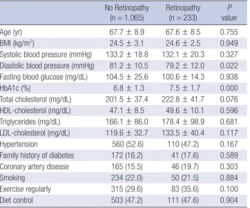
The characteristics of the study population are summarized in Table 2. No difference in the mean age was observed between the retinopathy and no-retinopathy groups, but significant dif- ferences were recorded for diastolic blood pressure and HbA1c between the groups. The mean HbA1c of the patients without retinopathy was 6.8%, which was much less than the 7.5% among patients with retinopathy.
When all patients were considered together, the relative fre- quency of diabetes related parameters differed according to the presence of diabetic retinopathy (Table 3). The clinical parame- ters associated with the incidence of retinopathy in the univari- ate analysis included the duration of diabetes, postprandial plas- ma glucose, and presence of diabetic foot. A longer duration of diabetes and higher postprandial blood glucose levels above 180 mg/dL indicated higher incidences of diabetic retinopathy Table 1. Prevalence of diabetic retinopathy among study subjects
Total number of
diabetes Newly diagnosed diabetes
n 1,298 291
Age (yr) 67.7 ± 8.8 65.9 ± 9.4
Sex (M/F) 505/793 116/175
No retinopathy 1,065 (82.0) 273 (93.8)
Retinopathy Mild NPDR Mod NPDR Severe NPDR PDR
126 (9.7) 42 (3.2) 48 (3.7) 17 (1.3)
7 (2.4) 4 (1.4) 5 (1.7) 2 (0.7) Data are presented as the mean ± SD or n (%). NPDR, non-proliferative diabetic reti- nopathy; PDR, proliferative diabetic retinopathy.
Table 2. Baseline clinical characteristics according to the presence of retinopathy No Retinopathy
(n = 1,065) Retinopathy
(n = 233) P
value
Age (yr) 67.7 ± 8.9 67.6 ± 8.5 0.755
BMI (kg/m2) 24.5 ± 3.1 24.6 ± 2.5 0.949
Systolic blood pressure (mmHg) 133.2 ± 18.8 132.1 ± 20.3 0.327 Diastolic blood pressure (mmHg) 81.2 ± 10.5 79.2 ± 12.0 0.022 Fasting blood glucose (mg/dL) 104.5 ± 25.6 100.6 ± 14.3 0.938
HbA1c (%) 6.8 ± 1.3 7.5 ± 1.7 0.000
Total cholesterol (mg/dL) 201.5 ± 37.4 222.8 ± 41.7 0.076 HDL-cholesterol (mg/dL) 47.1 ± 8.5 49.6 ± 10.1 0.596 Triglycerides (mg/dL) 166.1 ± 86.0 178.4 ± 98.9 0.681 LDL-cholesterol (mg/dL) 119.6 ± 32.7 133.5 ± 40.4 0.117
Hypertension 560 (52.6) 110 (47.2) 0.167
Family history of diabetes 172 (16.2) 41 (17.6) 0.589 Coronary artery disease 165 (15.5) 46 (19.7) 0.303
Smoking 234 (22.0) 50 (21.5) 0.884
Exercise regularly 315 (29.6) 83 (35.6) 0.100
Diet control 503 (47.2) 111 (47.6) 0.904
Data are presented as the mean ± SD or n(%). BMI, body mass index; HDL-choles- terol, high-density lipoprotein cholesterol; LDL-cholesterol; Low-density lipoprotein cholesterol.
Table 3. Relative frequency of diabetes related parameters according to presence of diabetic retinopathy
Parameters No Retinopathy
(n = 1,065) Retinopathy
(n = 233) P value Duration of diabetes (yr)
< 1 1-5 5-10 > 10
208 (19.5) 451 (42.4) 222 (20.8) 184 (17.3)
10 (4.3) 47 (20.2) 54 (23.2) 122 (52.3)
< 0.001
Postprandial glucose ≤ 180 mg/dL
> 180 mg/dL 513 (48.2)
552 (51.8) 43 (18.5) 190 (81.5)
< 0.001
Peripheral neuropathy 494 (46.4) 126 (54.1) 0.104
Diabetic foot 29 (2.7) 19 (8.2) 0.002
Treatment of diabetes Insulin
Oral medication Diet and exercise
293 (27.5) 724 (68.0) 48 (4.5)
50 (21.5) 168 (72.1) 15 (6.4)
0.101
Coronary artery disease 165 (15.5) 46 (19.7) 0.303 Data are presented as n (%).
in patients with type 2 diabetes. A tendency for a history of dia- betic foot was observed in the retinopathy group.
In logistic regression model, diabetic retinopathy was signifi- cantly associated with the duration of diabetes, postprandial glu- cose level and HbA1c. The diabetic retinopathy was increased with the long duration of diabetes mellitus (5-10 yr: OR = 5.19;
> 10 yr: OR = 10.03, < 1 yr as a reference), and was 2.5-fold great- er in patients with postprandial blood glucose levels exceeding 180 mg/dL compared to that of patients with postprandial blood glucose levels below 180 mg/dL with significance. For every 1%
elevation of HbA1c, the risk for diabetic retinopathy increased by a factor of 1.34 (95% CI: 1.545-1.980) (Table 4).
DISCUSSION
This study showed that diabetic retinopathy is a common com- plication among rural Korean patients with type 2 diabetes, and many patients were threatened by visual impairment and re- quired laser treatment. The overall prevalence of retinopathy of 18.0% in our study was less than the 34% to 48% reported in pre- vious hospital-based studies conducted on Korean patients (18- 20). Proliferative retinopathy of 1.3% was also significantly less prevalent than that reported previously (3.8% to 19.9%). A recent nationwide survey from 13 tertiary hospitals in Korea reported a 38.3% prevalence of diabetic retinopathy (21). However, com- munity-based studies assessing diabetic retinopathy are very limited in Korea. As expected, the prevalence of diabetic retinop- athy was lower among those who were examined in the popula- tion-based screening compared to those in the diabetic clinics.
As diabetes is highly prevalent and has increased more rapidly in the Asia-Pacific region (2), diabetic retinopathy is the leading cause of visual impairment and blindness in this region (4). Es- pecially in developing Asian countries, the lack of health care fa- cilities for diabetes management remains a serious public health problem. Consequently, the burden imposed by delayed diag- nosis of diabetes and its complications could be more common and massive than in developed countries. Therefore, understand- ing the actual prevalence and progression of diabetic retinopa- thy is very important for Asian people and worldwide health care planning. In Asian population-based studies conducted prior
to 2000, the prevalence of diabetic retinopathy was and 45.2%
in Taiwan (22) and 27.3% in Chinese hospital (23). Our result of 18% was lower than these epidemiologic data and similar to that of a population study examining urban and rural India after 2000, in which the overall prevalence of diabetic retinopathy was 19.2%
and 17.6%, respectively (24, 25). The causes of this lower overall prevalence of diabetic retinopathy are likely to include the be- havioral and nutritional habits of rural Koreans; typically, they eat vegetable-centered diets and have a relatively higher level of physical activity such as farmwork. In addition, differences in susceptibility to diabetic retinopathy may exist among different ethnic groups.
Notably, the prevalence of diabetic retinopathy in recent Ko- rean and Indian studies was less than that observed in other ep- idemiologic Asian studies conducted before 2000. Although the rate of type 2 diabetes has increased during the past three de- cades in Asian countries, the prevalence of diabetic retinopathy has not increased. In the Blue Mountain Eye Study of suburban Australians comparing the age-specific prevalence of diabetic retinopathy over 6 yr, although the prevalence of diabetic reti- nopathy increased from 29.4% to 33.4%, prevalent diabetic reti- nopathy had become principally mild and the prevalence of more severe diabetic retinopathy levels had decreased (26). In addition, a recent 21.9% prevalence of diabetic retinopathy in Australian population was similar with the result of our study (27).
Another important fact is that usually, type 2 diabetes has an asymptomatic phase with actual diabetic hyperglycemia before clinical diagnosis. This phase has been estimated to last at least 4-7 yr (6). Therefore, identifying diabetic retinopathy from new- ly diagnosed diabetes is valuable to the prevention and appropri- ate treatment of diabetic retinopathy in the early stage. Our study observed 6.2% diabetic retinopathy in newly diagnosed diabe- tes. Compared to the prevalence rate among Asian populations, this rate was lower than that of 30.5% in the Da Qing Study (28), 28.3% in Taiwan (22) and 21.9% in Hong Kong Chinese (29). Re- cent population-based data from India were similar to our data (6.35% and 5.1%, respectively) (24, 30). This is likely to have oc- curred following the introduction of new diagnostic criteria for diabetes, which are now less stringent, a better control of diabet- ic patients by general practitioners and endocrinologists, and more widespread home glucose monitoring. Nevertheless, note that among the newly diagnosed group of patients, proliferative or severe non-proliferative diabetic retinopathy was already pres- ent in 2.4% of subjects in our study and 4.6% in the Indian group (30). An earlier diagnosis and more aggressive control of treat- ment of diabetic retinopathy may be warranted.
Identification and early treatment have a critical role in dia- betic retinopathy because the disease is usually progressive and laser treatment is rarely effective in restoring vision. The recog- nition of modifiable risk factors that have a large potential for Table 4. Multivariate analysis* of the diabetic retinopathy related parameters
Parameter Odds ratio CI (95%) P value
Duration of diabetes (yr) < 1
1-5 5-10 > 10
1.00 2.239 5.192 10.034
0.493-10.159 1.138-23.684 2.284-44.075
0.296 0.033 0.002
HbA1c (increase 1%) 1.344 1.116-1.619 0.002
Postprandial glucose (> 180 mg/dL) 2.496 1.340-4.647 0.004
*Multivariate analysis was performed using a logistic regression model with the for- ward method.CI, confidence interval.
affecting health outcomes is very important. The results of pro- spective, population-based studies have strongly and consistent- ly implicated a longer duration of diabetes and poor glycemic control with diabetic retinopathy (5, 7, 8). Large randomized clin- ical trial has reported that intensive glycemic control results in clinically significant reductions in the incidence and progres- sion of retinopathy and loss of vision (8).
In this study, the duration of diabetes, HbA1c, and postpran- dial glucose levels were associated with diabetic retinopathy in the multivariate analysis. In particular, the duration of diabetes showed a significant association with diabetic retinopathy. This concurs with findings that the duration of diabetes is a key risk factor for diabetic retinopathy (5, 7, 8). The duration of diabetes is considered to be a marker for long-term exposure to hypergly- cemia. Other studies have consistently identified glycemic con- trol as an independent risk factor for retinopathy (26, 27). In this study population, HbA1c and postprandial glucose levels were other factors independently associated with diabetic retinopa- thy. Glycemic control was good with a mean HbA1c of 7.5% and a fasting blood glucose level of 100.6 mg/dL in the retinopathy group; therefore, the postprandial glucose level can be consid- ered to be a determinant of glycemic control.
Although the strength of our study is that it was the first pop- ulation-based study to assess the prevalence of diabetic retinop- athy in Korea, this study has some limitations. First, because it was a cross-sectional study, causality was not evaluated. Second, there is a difference between prevalence in rural area and prev- alence in big cities in Korea. Therefore, the findings of this study can not be regarded as a nationally representative data. Because diabetic retinopathy is a progressive disorder, a one-time cross- sectional screening would not be sufficient to evaluate and al- leviate the burden of diabetic retinopathy. Regularly repeated surveys for the prevalence of diabetic retinopathy and prospec- tive study of progression need to be performed to reduce the vi- sual morbidity of type 2 diabetes. Furthermore, one should not neglect the detection and treatment of diabetic retinopathy just because risk-factor control is expected to minimize its preva- lence. An earlier diagnosis and more aggressive control of blood glucose can decrease the duration-adjusted prevalence of reti- nopathy and sight-threatening complications.
In conclusion, the present study suggests that the prevalence of diabetic retinopathy in rural Korea is lower than that report- ed in other previous Asian groups. Nevertheless, proliferative or severe non-proliferative diabetic retinopathy still exists and pro- gresses in many patients with diabetes due to increased diabe- tes burden, and it remains a public health and economic bur- den in Korea. This emphasizes that regular screening for diabet- ic retinopathy and more aggressive management of glycemia can reduce the number of people who develop diabetic reti- nopathy.
REFERENCES
1. King H, Aubert RE, Herman WH. Global burden of diabetes, 1995-2025:
prevalence, numerical estimates, and projections. Diabetes Care 1998;
21: 1414-31.
2. Chan JC, Malik V, Jia W, Kadowaki T, Yajnik CS, Yoon KH, Hu FB. Dia- betes in Asia: epidemiology, risk factors, and pathophysiology. JAMA 2009; 301: 2129-40.
3. Yoon KH, Lee JH, Kim JW, Cho JH, Choi YH, Ko SH, Zimmet P, Son HY.
Epidemic obesity and type 2 diabetes in Asia. Lancet 2006; 368: 1681-8.
4. Porta M, Bandello F. Diabetic retinopathy: a clinical update. Diabetolo- gia 2002; 45: 1617-34.
5. Klein R, Klein BE, Moss SE, Davis MD, DeMets DL. The Wisconsin Epi- demiologic Study of Diabetic Retinopathy. III: prevalence and risk of di- abetic retinopathy when age at diagnosis is 30 or more years. Arch Oph- thalmol 1984; 102: 527-32.
6. Harris MI, Klein R, Welborn TA, Knuiman MW. Onset of NIDDM occurs at least 4-7 yr before clinical diagnosis. Diabetes Care 1992; 15: 815-9.
7. UK Prospective Diabetes Study (UKPDS) Group. Intensive blood-glucose control with sulphonylureas or insulin compared with conventional treat- ment and risk of complications in patients with type 2 diabetes (UKPDS 33). Lancet 1998; 352: 837-53.
8. Looker HC, Krakoff J, Knowler WC, Bennett PH, Klein R, Hanson RL.
Longitudinal studies of incidence and progression of diabetic retinopa- thy assessed by retinal photography in pima indians. Diabetes Care 2003;
26: 320-6.
9. Leske MC, Wu SY, Hennis A, Nemesure B, Hyman L, Schachat A. Inci- dence of diabetic retinopathy in the Barbados Eye Studies. Ophthalmol- ogy 2003; 110: 941-7.
10. McCarty DJ, Fu CL, Harper CA, Taylor HR, McCarty CA. Five-year inci- dence of diabetic retinopathy in the Melbourne visual impairment proj- ect. Clin Experiment Ophthalmol 2003; 31: 397-402.
11. van Leiden HA, Dekker JM, Moll AC, Nijpels G, Heine RJ, Bouter LM, Stehouwer CD, Polak BC. Risk factors for incident retinopathy in a dia- betic and nondiabetic population: the Hoorn study. Arch Ophthalmol 2003; 121: 245-51.
12. Kwon HS, Park YM, Lee HJ, Lee JH, Choi YH, Ko SH, Lee JM, Kim SR, Kang SY, Lee WC, Ahn MS, Noh JH, Kang JM, Kim DS, Yoon KH, Cha BY, Lee KW, Kang SK, Son HY. Prevalence and clinical characteristics of the metabolic syndrome in middle-aged Korean adults. Korean J Intern Med 2005; 20: 310-6.
13. World Medical Association. Declaration of Helsinki. Ethical Principles for Medical Research Involving Human Subjects. 52nd WMA General Assembly. Edinburgh: 2000.
14. World Health Organization. Definition, diagnosis and classification of diabetes mellitus and its complications part 1: diagnosis and classifica- tion of diabetes mellitus. World Health Organization, 1999.
15. Chalmers J, MacMahon S, Mancia G, Whitworth J, Beilin L, Hansson L, Neal B, Rodgers A, Ni Mhurchu C, Clark T. 1999 World Health Organi- zation-International Society of Hypertension Guidelines for the manage- ment of hypertension. Guidelines sub-committee of the World Health Or- ganization. Clin Exp Hypertens 1999; 21: 1009-60.
16. Friedewald WT, Levy RI, Fredrickson DS. Estimation of the concentra- tion of low-density lipoprotein cholesterol in plasma, without use of the preparative ultracentrifuge. Clin Chem 1972; 18: 499-502.
17. Wilkinson CP, Ferris FL 3rd, Klein RE, Lee PP, Agardh CD, Davis M, Dills D, Kampik A, Pararajasegaram R, Verdaguer JT; Global Diabetic Reti- nopathy Project Group. Proposed international clinical diabetic retinop- athy and diabetic macular edema disease severity scales. Ophthalmolo- gy 2003; 110: 1677-82.
18. Lee HC, Yang JY, Lim SK, Hong CS, Huh KB, Lee SY. A prospective study of diabetic complications. J Korean Diabetes Assoc 1984; 8: 47-53.
19. Nam JH, Lee SH, Lee HJ, Han JH, Ha SW, Kim BW. The prevalence of chronic complications in non-insulin dependent diabetic patients. J Ko- rean Diabetes Assoc 1999; 23: 702-14.
20. Park JY, Kim SW, Cho GY, Lee MH, Je SJ, Lee KU, Kim GS. The prevalence of micro- and macrovascular complications of Korean NIDDM patients.
J Korean Diabetes Assoc 1993; 17: 377-85.
21. Lim S, Kim DJ, Jeong IK, Son HS, Chung CH, Koh G, Lee DH, Won KC, Park JH, Park TS, Ahn J, Kim J, Park KG, Ko SH, Ahn YB, Lee I. A nation- wide survey about the current status of glycemic control and complica- tions in diabetic patients in 2006: The Committee of the Korean Diabetes Association on the Epidemiology of Diabetes Mellitus. Korean Diabetes J 2009; 33: 48-57.
22. Chang C, Lu F, Yang YC, Wu JS, Wu TJ, Chen MS, Chuang LM, Tai TY.
Epidemiologic study of type 2 diabetes in Taiwan. Diabetes Res Clin Pract 2000; 50(Suppl 2): S49-59.
23. Liu DP, Molyneaux L, Chua E, Wang YZ, Wu CR, Jing H, Hu LN, Liu YJ, Xu ZR, Yue DK. Retinopathy in a Chinese population with type 2 diabe- tes: factors affecting the presence of this complication at diagnosis of dia- betes. Diabetes Res Clin Pract 2002; 56: 125-31.
24. Rema M, Premkumar S, Anitha B, Deepa R, Pradeepa R, Mohan V. Prev- alence of diabetic retinopathy in urban India: the Chennai Urban Rural Epidemiology Study (CURES) eye study, I. Invest Ophthalmol Vis Sci 2005;
46: 2328-33.
25. Rani PK, Raman R, Chandrakantan A, Pal SS, Perumal GM, Sharma T.
Risk factors for diabetic retinopathy in self-reported rural population with diabetes. J Postgrad Med 2009; 55: 92-6.
26. Cugati S, Kifley A, Mitchell P, Wang JJ. Temporal trends in the age-specif- ic prevalence of diabetes and diabetic retinopathy in older persons: pop- ulation-based survey findings. Diabetes Res Clin Pract 2006; 74: 301-8.
27. Tapp RJ, Shaw JE, Harper CA, de Courten MP, Balkau B, McCarty DJ, Taylor HR, Welborn TA, Zimmet PZ; AusDiab Study Group. The preva- lence of and factors associated with diabetic retinopathy in the Austra- lian population. Diabetes Care 2003; 26: 1731-7.
28. Hu YH, Pan XR, Liu PA, Li GW, Howard BV, Bennett PH. Coronary heart disease and diabetic retinopathy in newly diagnosed diabetes in Da Qing, China: the Da Qing IGT and Diabetes Study. Acta Diabetol 1991; 28: 169- 73.
29. Wang WQ, Ip TP, Lam KS. Changing prevalence of retinopathy in newly diagnosed non-insulin dependent diabetes mellitus patients in Hong Kong. Diabetes Res Clin Pract 1998; 39: 185-91.
30. Agarwal S, Raman R, Kumari RP, Deshmukh H, Paul PG, Gnanamoor- thy P, Kumaramanickavel G, Sharma T. Diabetic retinopathy in type II diabetics detected by targeted screening versus newly diagnosed in gen- eral practice. Ann Acad Med Singapore 2006; 35: 531-5.
AUTHOR SUMMARY
Prevalence and Associated factors of Diabetic Retinopathy in Rural Korea: The Chungju Metabolic Disease Cohort Study
Ji-Hyun Kim, Hyuk-Sang Kwon, Yong-Moon Park, Jin-Hee Lee, Man-Soo Kim, Kun-Ho Yoon, Won Chul Lee, Bong-Yun Cha and Ho-Young Son
Diabetic retinopathy is the leading cause of blindness in working-age people in the Asia-Pacific region. According to our present study, in 40 ≥ rural Korean patients with type 2 diabetes, the overall prevalence of diabetic retinopathy was 18% and proliferative or severe non-proliferative form was found in 5.0% of the study subjects. Factors associated with retinopathy included duration of diabetes, hemoglobin A1c, and postprandial blood glucose levels. The number of patients with proliferative or severe non- proliferative diabetic retinopathy was comparable with the overall rate in Asian countries, and identified more frequently at the time of diagnosis. More aggressive identification and early treatment are necessary for diabetic retinopathy.