Background and PurposezzCarotid artery stenting (CAS) is emerging as an alternative to ca- rotid endarterectomy for the treatment of carotid artery stenosis (CS), but the effect of CAS on the cognitive function of patients with severe CS has not been fully investigated. The aim of this study was to use comprehensive neuropsychological tests to determine the effect of CAS on cognitive function from baseline to 3 months postprocedure in patients with severe CS.
MethodszzThirty-one patients due to undergo CAS due to high-grade CS (≥70%) and 11 control subjects who were diagnosed with CS, but who did not undergo CAS, and who visited the clinic or emergency room between February 2009 and February 2012 were recruited con- secutively at baseline (i.e., pre-CAS). Follow-up neuropsychological evaluations after 3 months were completed by 23 of the 31 patients who underwent CAS, and by 10 of the 11 control sub- jects. The primary cognitive outcome was assessed using a neuropsychological test containing subcategories designed to test general cognitive function, attention, visuospatial function, lan- guage and related functions, memory, and frontal lobe/executive function.
ResultszzOf the 23 patients undergoing CAS who completed the 3-month follow-up tests, 12 had asymptomatic CS. During the 3-month follow-up period, the patients who underwent CAS and those with asymptomatic CS achieved similar results to the control group on all cognitive tests. However, symptomatic CS patients (n=11) who underwent CAS exhibited improve- ments in visuospatial function (p=0.046) and total Seoul Neuropsychological Screening Bat- tery-Dementia Version scores (p=0.010) in comparison with both the asymptomatic CS pa- tients and the control group.
ConclusionszzThe findings of this study suggest that CAS has a positive effect on cognitive function in patients with symptomatic CS over a 3-month follow-up period. A long-term, mul- ticenter, prospective case-control study would be helpful to predict quality of life and prognoses for patients undergoing CAS.
Key Wordszz carotid artery, stenosis, stenting, cognition, visuospatial, prospective.
Effect of Carotid Artery Stenting on Cognitive Function in Patients with Carotid Artery Stenosis:
A Prospective, 3-Month-Follow-Up Study
INTRODUCTION
Carotid artery stenosis (CS) is one of the most significant risk factors for ischemic stroke.1 Chronic cortical hypoperfusion arising from CS is currently being investigated as an im- portant source of cognitive impairments, regardless of whether it accompanies stroke.2 The artery-to-artery embolism caused by CS can result in multiple infarcts, leading to vascu- lar cognitive impairments and vascular dementia. Furthermore, in cases of severe CS, per- fusion defects can arise in the affected areas of the cerebral cortex, which may result in cog- nitive impairments associated with those defects.3-6 Therefore, when carotid artery stenting Byeol-A Yoona
Sang Wuk Sohna Sang-Myung Cheona Dae-Hyun Kima,b Jae-Kwan Chaa,b SoJeong Yic Kyung Won Parka,d
a Department of Neurology,
b Busan-Ulsan Regional
Cardio-Cerebrovascular Center, and
d Dong-A Anti-aging Research Institute, Dong-A University College of Medicine, Busan, Korea
c Department of Clinical Pharmacology and Therapeutics,
Seoul National University College of Medicine, Seoul, Korea
pISSN 1738-6586 / eISSN 2005-5013 / J Clin Neurol 2015;11(2):149-156 / http://dx.doi.org/10.3988/jcn.2015.11.2.149
Received September 16, 2014 Revised December 3, 2014 Accepted December 3, 2014 Correspondence Kyung Won Park, MD, PhD Department of Neurology,
Dong-A Anti-aging Research Institute, Dong-A University College of Medicine, 32 Daesingongwon-ro, Seo-gu, Busan 602-714, Korea Tel +82-51-240-5266 Fax +82-51-244-8338 E-mail neuropark@dau.ac.kr
cc This is an Open Access article distributed under the terms of the Creative Commons Attribution Non-Com- mercial License (http://creativecommons.org/licenses/by-nc/3.0) which permits unrestricted non-commercial use, distribution, and reproduction in any medium, provided the original work is properly cited.
JCN
Open Access ORIGINAL ARTICLEEffect of Carotid Artery Stenting on Cognition
JCN
(CAS) procedures are undertaken to widen a narrowed area, it is predicted that the improved perfusion in the stenosed area of the cortex will lead to a recovery of cognitive func- tions and eventually help to reduce the risk of vascular cog- nitive impairments and vascular dementia.7,8 However, it has also been reported that cognitive function can be negatively affected due to microembolisms caused during the CAS pro- cedure itself, or temporary perfusion defects that may take place during balloon dilatation. Thus, studies are needed to determine whether CAS has any impact on the cognitive function of patients with CS; such studies should involve de- tailed neuropsychological tests before and after the CAS pro- cedure. Little research has been done in this area to date.9,10
The present study compared the cognitive performance of patients with symptomatic CS and asymptomatic severe CS (≥70%) by testing their cognitive function prior to CAS and 3 months thereafter. Their neuropsychological test results were also compared with those of a control group consisting of CS patients who had not received the CAS procedure, in order to assess whether there are any meaningful differenc- es between the two groups.
METHODS
This prospective, single-center study was conducted at Dong- A University Hospital for 3 months from February 2009 un- til December 2012. The study was approved by the Institu- tional Review Board of Dong-A University Hospital with respect to the clinical protocol, informed consent form (ICF), and patient information sheet (PIS). All subjects voluntarily consented to participate in the study by submitting their ICF after being provided with all relevant information relat- ed to the study through the PIS, including their right to free- ly withdraw from the study at any point.
Subjects
This study involved both symptomatic and asymptomatic CS patients who visited the Department of Neurology or the Stroke Center of Dong-A University Hospital as outpatients or emergency patients. The symptomatic CS patients had within the previous 6 months experienced at least one tran- sient or mild symptomatic ischemic vascular event in the distribution of one or both internal carotid arteries. Patients who met any of the following criteria were excluded from the study: 1) severe language disorders or motor weaknesses caused by acute cerebral infarction; 2) acute confused state lasting for at least several days; 3) brain tumor, infection, or other diseases as diagnosed by brain imaging; or 4) a histo- ry of depression, head injury, drug abuse, or similar trauma that could impact their cognitive functioning. The control
group consisted of patients who had been diagnosed with CS by diagnostic neuroangiography, but who did not under- go CAS.
Neuropsychological screening
The subjects’ overall cognitive performance was evaluated using the Korean version of the Mini Mental State Examina- tion (K-MMSE),11,12 and more detailed tests were performed based on the Seoul Neuropsychological Screening Battery- Dementia Version (SNSB-D).13 The neuropsychological tests were performed within 14 days after the onset of stroke symp- toms or signs. The SNSB-D comprises five test subcategories with a total possible score of 300: attention, 6% (17 points);
language function, 9% (27 points); visuospatial function, 12%
(36 points); memory function, 50% (150 points); and fron- tal lobe executive function, 23% (70 points). Attention was evaluated using the digit span test—forward and digit span test—backward, and visuospatial function was assessed using the Rey-Osterrieth Complex Figure Test (ROCFT). Memory was tested by evaluating both verbal memory and nonverbal memory. Verbal memory was assessed based on the capacity for immediate recall, delayed recall, and the recognition do- main of the Seoul Verbal Learning Test (SVLT). Nonverbal memory was measured based on the capacity for immedi- ate recall, delayed recall, and the recognition domain of the ROCFT (ROCFT-recognition). Frontal executive function was evaluated by conducting contrasting-program, go-no- go, fist-edge-palm, word fluency—animal, word fluency—
phonemic, and Stroop tests. Finally, language and related functions were assessed using the Korean version of the Bos- ton Naming Test,14 and calculation and apraxia tests.
Carotid ultrasonography brain imaging, and operative procedures
Brain magnetic resonance imaging (MRI)/magnetic reso- nance angiography imaging (1.5-tesla GE MRI scanner, GE Healthcare, Piscataway, NJ, USA) was performed on all pa- tients, as were carotid ultrasonography and neuroangiogra- phy. The existence and severity of a CS in the participating patients were evaluated by a single neuroradiologist who conducted neuroangiography based on the North American Symptomatic Carotid Endarterectomy Trial.15 Measurements of the site, severity, and length of the stenosis, the plaque characteristics, and the vessel were performed in order to select an appropriately sized the balloon and stent in cases submitting to the CAS intervention. CAS was performed in a dedicated operating room equipped for endovascular pro- cedures, under local anesthesia, and through a femoral ap- proach. Cerebral protection devices were used to prevent mi- croembolisms during the procedure.
Yoon BA et al.
JCN
Statistical analysis
Statistical analyses were performed using SPSS version 22 (SPSS Inc., Chicago, IL, USA), and in principle the two- tailed test was performed when p≤0.05. The demographic data and neuropsychological test results of the subjects were reviewed using SPSS descriptive statistical analysis. With re- spect to the subjects’ demographic data and baseline scores, the average values were compared between those patients who underwent CAS (the stent treatment group) and the un- stented control group using the independent-samples t-test.
For categorical variables, frequencies were compared using the chi-square test.
Since the neuropsychological tests were conducted for both the stent treatment group and the control group, base- line scores and scores achieved at the 3-month follow-up were compared within each group for the respective cogni- tive test categories. In the analysis, the covariates included subject age, for which meaningful differences were found between the stent treatment group and the control group, and baseline scores. Analysis of covariance (ANCOVA) was conducted to analyze changes in the neuropsychological test scores between baseline and the 3-month follow-up. Fur- thermore, by applying the same method, symptomatic and asymptomatic CS patients were separately compared with the control group to determine the effects of reperfusion fol- lowing the CAS procedure. In addition, patients in the stent treatment group were assigned to the following subgroups according to the presence of factors that were expected to affect cognitive function: 1) patients with microembolism based on brain diffusion-weighted MRI performed after the CAS procedure, 2) patients with perfusion defects based on brain perfusion MRI, and 3) patients with CS (≥70%) in the left carotid artery. The neuropsychological test score changes between baseline and the 3-month follow-up of these three subsets of patients were each compared with those of the control group.
Except where stated otherwise, the data are presented as mean±SD values.
RESULTS
Twenty-three of the 31 patients in the stent treatment group and 10 of the 11 patients in the control group completed the 3-month follow-up tests. The reasons for loss to follow-up in the stent treatment group were withdrawal of informed con- sent (n=6) and no 3-month follow-up test (n=2), while one patient in the control group did not receive the 3-month fol- low-up test. These nine patients were treated as losses to fol- low-up and were excluded from the analyses; thus data from 23 patients in the stent treatment group and 10 control sub-
jects were analyzed.
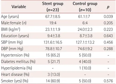
The demographic data and medical characteristics of the stent treatment and control subjects are listed in Table 1. The age differed significantly (p=0.039) between the stent treat- ment group (67.7±8.5 years) and the control group (61.1±7.0 years), but none of the other characteristics differed signifi- cantly between the two groups. Therefore, all of the analyses were conducted after adjusting for age based on ANCOVA.
The stent treatment group comprised 19 males (82.6%) and had an average of 9.4 years of education; the corresponding values for the control group were 6 males (60.0%) and 8.7 years. The most common vascular risk factor was high blood pressure in both the stent treatment group and the control group (frequencies of 65.2% and 50.0%, respectively), fol- Table 1. Demographic findings of carotid artery stenosis patients who were treated with carotid artery stenting (stent group) and those who were not stented (control group)
Variable Stent group (n=23)
Control group
(n=10) p
Age (years) 67.7±8.5 61.1±7 0.039
Male:female (n) 19:4 6:4 0.205
BMI (kg/m2) 23.1±1.9 24.0±2.3 0.223
Education (years) 9.4±3.8 8.7±3.8 0.643
SBP (mm Hg) 131.6±16.5 127.1±13.2 0.456
DBP (mm Hg) 78.8±10.7 74.6±9.2 0.288
Hypertension (%) 15 (65.2) 5 (50.0) -
Diabetes mellitus (%) 5 (21.7) 4 (40.0) -
Hyperlipidemia (%) - 1 (10.0) -
Heart disease (%) 3 (13.0) - -
Smoker (yes) (%) 14 (60.9) 5 (50.0) 0.576 Except where indicated otherwise, data are mean±SD or n (%) values.
BMI: body mass index, DBP: diastolic blood pressure, SBP: systolic blood pressure.
Table 2. Characteristics of the CSs and magnetic resonance imaging (MRI) findings in CS patients with a stent
Characteristic n (%)
Symptomatology
Symptomatic 11 (48)
Asymptomatic 12 (52)
Location
Right 14 (60)
Left 8 (34)
Both 1 (6)
Microembolism
Present 5 (56)
Absent 4 (44)
Perfusion MRI findings at baseline
Perfusion defect (+), abnormal 5 (26) Perfusion defect (–), normal 14 (74) CS: carotid artery stenosis.
Effect of Carotid Artery Stenting on Cognition
JCN
lowed by smoking, diabetes, heart diseases, and hyperlipid- emia. The stent treatment group contained 11 symptomatic CS patients and 12 asymptomatic CS patients. Nine of these 23 patients submitted to MRI (including diffusion-weighted MRI), which revealed that 5 patients had developed a mi- croembolism due to the CAS procedure. Five of the 19 pa- tients for whom brain perfusion MRI was performed were found to have perfusion defects at a meaningful level. Fi- nally, the CS was on the right side in 14 patients, on the left side in 8, and bilateral in 1 (Table 2).
The changes in cognitive performance of the patients in the stent treatment and control groups between baseline and the 3-month follow-up are given in Table 3. The stent treat- ment group had average baseline K-MMSE and SNSB-D scores of 25.0 and 152.5 points, respectively; these scores had increased by 1.3 and 17.6 points, respectively, at the 3-month follow-up. However, the scores in the control group had also increased, by 1.1 and 17.3 points, respectively, in the follow- up test compared to baseline. These findings indicated that there was no statistically significant improvement in test scores between the patients and control group. From a clini- cal point of view, the participating patients exhibited im-
provements in all test categories of neuropsychological as- sessment except for the drawing of a Luria loop. However, the level of improvement in the stent treatment group was not statistically meaningful when compared with the con- trol group. In fact, in the test of SVLT-recognition, the control group achieved a more statistically meaningful improve- ment than the stent treatment group.
Symptomatic CS patients had average K-MMSE and SNSB- D scores of 23.5 and 142.5 points, respectively; the corre- sponding values for the asymptomatic CS patients were 26.3 and 161.7 points. At the 3-month follow-up test the K-MMSE scores of the two groups were 1.8 and 0.3 points higher, re- spectively, than their baseline scores, and their total SNSB-D scores had increased by 32.3 and 4.2 points. When compared to both the asymptomatic CS patients in the stent treatment group and the control group, symptomatic CS patients ex- hibited statistically meaningful improvements in ROCFT, ROCFT-recognition, and total SNSB-D scores (Table 4). Con- versely, asymptomatic CS patients exhibited declines in sev- eral test results, including the ROCFT, orientation, and pho- nemic and animal word fluency.
Additional analyses were conducted on subsets of patients Table 3. Baseline neuropsychological test scores and cognitive changes therein between baseline and the 3-month follow-up after CAS in the stent and control groups
Neuropsychological test Baseline score Difference from baseline ANCOVA
Stent (n=23) Control (n=10) Stent (n=23) Control (n=10) p
K-MMSE 25.0±4.5 26.5±2.6 1.0±2.9 1.1±2.2 0.918
Digit span–forward 6.0±1.6 6.5±1.8 0±1.8 0±0.9 0.890
Digit span–backward 3.3±1.7 3.6±1.0 0.3±1.3 0±1.2 0.726
K-BNT 9.8±3.3 12.2±1.4 0.5±1.1 1.0±1.7 0.454
Calculation 9.2±4.0 9.9±2.8 0.8±2.7 -0.1±0.7 0.272
ROCFT 26.9±12.3 32.2±6.8 1.8±4.5 0.8±2.8 0.360
Orientation 5.5±0.9 5.7±0.5 0.1±0.9 0.3±0.5 0.565
SVLT-free recall 12.7±7.3 20.4±6.4 2.5±6.5 2.8±5.0 0.077
SVLT-delayed recall 3.6±2.9 5.6±3.7 1.3±3.6 1.7±2.3 0.548
SVLT-recognition 5.8±3.5 8.4±1.6 1.0±3.2 1.7±1.8 0.029
ROCFT-immediate/delayed recall 23.8±16.9 31.4±17.0 5.5±9.5 7.0±7.5 0.842
ROCFT-recognition 6.0±3.7 7.3±2.5 0.8±2.8 1.2±2.7 0.772
Contrasting program 2.5±1.1 2.8±0.6 0.3±0.8 0.2±0.6 0.755
Go-no-go 2.3±1.1 2.6±0.5 0.2±1.1 -0.1±0.6 0.526
Fist-edge-palm 2.3±1.0 3.0±0 0.4±0.8 0±0 0.721
Luria loop 2.6±1.0 2.7±0.7 -0.3±0.9 0.3±0.7 0.181
Word fluency–animal 11.6±4.6 13.5±2.7 0.9±3.8 0.6±2.5 0.913
Word fluency–phonemic 4.6±4.0 6.5±2.8 0.2±3.5 -1.1±3.5 0.505
Stroop test–color 11.0±6.4 15.7±2.9 1.5±3.6 1.1±3.0 0.844
SNSB-D 152.5±61.2 193.0±37.8 17.6±24.9 17.3±12.2 0.890
A negative number indicates a decline in function.
ANCOVA: analysis of covariance, CAS: carotid artery stenting, K-BNT: Korean version of the Boston Naming Test, K-MMSE: Korean version of the Mini Mental State Examination, ROCFT: Rey-Osterrieth Complex Figure Test, ROCFT-immediate/delayed recall: immediate/delayed recall domain of the ROCFT, ROCFT-recognition: recognition domain of the ROCFT, SNSB-D: Seoul Neuropsychological Screening Battery-Dementia Version, SVLT: Seoul Verbal Learning Test.
Yoon BA et al.
JCN
possessing factors that were deemed to affect the cognitive function of patients with CS. Five (56.0%) of the nine pa- tients for whom brain diffusion-weighted MRI was repeat- ed after the CAS procedure were found to have a microem- bolism. Nevertheless, these five patients performed similarly on cognitive tests to patients without a microembolism (Supplementary Table 1 in the online-only Data Supplement).
In order to determine whether the recovery from perfusion defects induced by a CAS procedure affects the cognitive functioning of patients with CS, cognitive performance was compared between patients with and without perfusion de- fects, as determined by brain perfusion MRI. Patients with perfusion defects that were improved by CAS achieved im- proved calculation test scores, which were on average 2.8 points higher than at baseline; this was a significant improve- ment compared with the control group (p=0.048). Howev- er, the group of patients who did not develop perfusion de- fects exhibited improvements in the immediate recall and recognition domains of the SVLT (p<0.05) (Supplementary Table 2 in the online-only Data Supplement). The location (left or right hemisphere) of the perfusion defect had no ef- fect on cognitive performance changes (Supplementary Ta-
ble 3 in the online-only Data Supplement).
DISCUSSION
The changes in cognitive performance of symptomatic and asymptomatic CS patients were analyzed in this study in a prospective manner by testing their cognitive function be- fore and after the CAS procedure. The reliability of this study was enhanced by the additional comparison of the cognitive performance test results of these CS patients those of a con- trol (non-CAS) group. The number of people suffering from arteriosclerosis with vascular risk factors has been increas- ing recently due to the westernization of diet patterns and changing living environments, which has led to increases in the number of patients with CS or cerebral infarction.15 Se- vere CS is one of the important culprits underlying cerebral infarction, and can cause cognitive impairments due to con- tinuous perfusion defects.9,16
Until recently, many reports have focused on the effects of the revascularization procedure on the cognitive function of patients with CS. However, when it comes to the effects of carotid endarterectomy or CAS, the findings have been con- Table 4. Cognitive changes between baseline and the 3-month follow-up after CAS in symptomatic versus asymptomatic CS patients as com- pared to the control group
Neuropsychological test
Baseline score Difference from baseline ANCOVA
Symptomatic (n=11)
Asymptomatic (n=12)
Control (n=10)
Symptomatic (n=11)
Asymptomatic (n=12)
Control
(n=10) p
K-MMSE 23.5±5.3 26.3±3.2 26.5±2.6 1.8±3.4 0.3±2.3 1.1±2.2 0.919
Digit span–forward 6.0±1.5 6.0±1.7 6.5±1.8 0±1.8 0.1±1.9 0±0.9 0.982
Digit span–backward 3.1±1.6 3.4±1.8 3.6±1.0 0.6±1.4 0.1±1.2 0±1.2 0.639
K-BNT 10.0±3.6 9.7±3.1 12.2±1.4 0.8±0.8 0.3±1.4 1.0±1.7 0.439
Calculation 9.2±4.5 9.2±3.7 9.9±2.8 0.9±3.8 0.8±1.1 -0.1±0.7 0.543
ROCFT 23.8±13.8 29.8±10.6 32.2±6.8 4.1±4.0 -0.3±4.0 0.8±2.8 0.036
Orientation 5.2±1.2 5.8±0.4 5.7±0.5 0.4±1.1 -0.1±0.5 0.3±0.5 0.797
SVLT-free recall 11.3±8.1 13.9±6.7 20.4±6.4 3.7±7.6 1.3±5.3 2.8±5.0 0.199
SVLT-delayed recall 3.4±3.3 3.8±2.5 5.6±3.7 2.4±4.8 0.3±1.9 1.7±2.3 0.233
SVLT-recognition 5.3±3.6 6.3±3.4 8.4±1.6 1.5±3.9 0.4±2.4 1.7±1.8 0.090
ROCFT-immediate/delayed recall 22.0±15.5 25.5±18.5 31.4±17.0 8.5±9.1 2.8±9.4 7.0±7.5 0.372
ROCFT-recognition 5.6±3.3 6.3±4.1 7.3±2.5 2.1±2.5 -0.3±2.6 1.2±2.7 0.046
Contrasting program 2.4±1.2 2.6±1.0 2.8±0.6 0.4±0.9 0.2±0.6 0.2±0.6 0.903
Go-no-go 2.4±1.2 2.3±1.1 2.6±0.5 0.3±1.0 0.1±1.2 -0.1±0.6 0.679
Fist-edge-palm 2.2±1.2 2.4±0.9 3.0±0 0.4±0.9 0.4±0.8 0±0 0.698
Luria loop 2.5±1.2 2.8±0.9 2.7±0.7 -0.3±0.9 -0.3±0.9 0.3±0.7 0.395
Word fluency–animal 11.5±6.1 11.7±3.1 13.5±2.7 1.9±4.8 -0.1±2.5 0.6±2.5 0.326
Word fluency–phonemic 3.3±4.0 5.8±3.7 6.5±2.8 1.5±4.4 -1.0±2.0 -1.1±3.5 0.638
Stroop test–color 10.5±7.9 11.5±5.0 15.7±2.9 3.1±3.4 -0.1±3.1 1.1±3.0 0.087
SNSB-D 142.5±68.7 161.7±54.9 193.0±37.8 32.3±26.7 4.2±13.6 17.3±12.2 0.010
ANCOVA: analysis of covariance, CAS: carotid artery stenting, CS: carotid artery stenosis, K-BNT: Korean version of the Boston Naming Test, K-MMSE:
Korean version of the Mini Mental State Examination, ROCFT: Rey-Osterrieth Complex Figure Test, ROCFT-immediate/delayed recall: immediate/de- layed recall domain of the ROCFT, ROCFT-recognition: recognition domain of the ROCFT, SNSB-D: Seoul Neuropsychological Screening Battery-De- mentia Version, SVLT: Seoul Verbal Learning Test.
Effect of Carotid Artery Stenting on Cognition
JCN
flicting, with some researchers reporting improved cognitive performance7,8,17,18 and others reporting the opposite.19,20 En- hanced cognitive performance can result from the perfusion improvement due to revascularization as well as the subse- quent recovery of cerebral cortex functioning. Meanwhile, any decline in cognitive performance likely stems from stenting-induced microembolism9,10,21 or temporary perfu- sion defects due to the balloon dilatation of a narrowed area.21 Examination of the demographic and social characteris- tics of the subjects in the present study revealed that high blood pressure was the most common vascular risk factor, followed by diabetes and heart diseases. These findings are in line with those of other studies.22
The stent treatment group exhibited clinical improvements over baseline in most of the test categories of the neuropsy- chological screening that was performed 3 months after the CAS. However, the improvement was not statistically mean- ingful since the control patients, who did not receive the CAS procedure also recorded improvements. Most previous re- search that demonstrated positive effects of CAS on cogni- tive function only compared the cognitive performance of patients before and after CAS, without comparing the cogni- tive performance improvement between the stent treatment group and a control group.9 The improvements in cognitive performance exhibited by the control group is likely attrib- utable to learning effects, given that the follow-up cognitive testing was performed only 3 months after the baseline test- ing. A more accurate analysis and higher reliability require long-term tracking observations.23
Compared to the asymptomatic CS patients and the con- trol group, the symptomatic CS patients recorded statistically meaningful improvements in cognitive function after the CAS procedure in terms of their total scores for ROCFT- recognition and SNSB-D. Therefore, CAS exerts meaningful effects on the cognitive function of symptomatic CS patients, which implies that asymptomatic CS patients suffer less in terms of cognitive impairment than do symptomatic pa- tients.24 We cannot definitively explain the relatively greater improvement in visuospatial function compared with other cognitive domains observed in the present study. Pre- and post-CAS functional neuroimaging studies (i.e., brain single- photon-emission computerized tomography or positron- emission tomography) are needed to explore the appropriate area of functional improvement explained by cognitive do- mains after CAS.
The asymptomatic CS patients had a poorer cognitive per- formance after the CAS in the test categories for evaluating frontal executive function, orientation, the ROCFT, ROCFT- recognition, Luria loop, Stroop test, and word fluency, which was presumably due to microembolisms caused by the CAS.
Nevertheless, patients who developed microembolism per- formed similarly on the neurophysiological test battery to those without microembolism. Some of the study limitations could explain this result, such as brain diffusion-weighted MRI only being performed to check for the development of microembolisms in a subset of the participating patients. Ac- cording to previous studies, microemboli occur in 20–40%
of CAS procedures.20,25 Further analyses are needed regard- ing the impact of microembolisms and different calibers thereof in different brain areas.26 Since the patients who de- veloped microembolisms in the present study suffered a con- tinuous decline in cognitive performance, efforts should be made to minimize the generation of microembolisms by us- ing protective devices during the CAS procedure. The devel- opment of microembolisms after CAS is considered to have a greater impact on asymptomatic CS patients than on their symptomatic counterparts.
It was anticipated that patients with chronic perfusion de- fects would exhibit a greater cognitive improvement after CAS than patients who did not suffer from perfusion defects.
However, the results of SVLT and its recognition test demon- strated that their cognitive performance actually declined.
This might be because the follow-up test was conducted only 3 months post-CAS, and so the test would not detect long- term recovery of brain function supported by increases in blood flow. Until recently, most of the research on the im- provement of perfusion defects and the subsequent recov- ery of cognitive functions has been based on follow-up tests performed 24 weeks after the CAS procedure. In the future, long-term analyses need to be conducted with brain perfu- sion imaging and follow-up cognitive performance tests.27
It was expected that the CS location would affect the de- gree of improvement in cognitive performance, since the left and right hemispheres of the brain play different roles in cognition. However, stenosis laterality had no impact on the degree of cognitive improvement. One caveat to this find- ing is that neuropsychological tests rarely reflect the cogni- tive function of the right hemisphere; additional neurocog- nitive tests, such as the performance intelligence quotient, are needed after CAS to test for hemisphere-dependent im- provements.28
This study was subject to some limitations. First, the stent treatment group and the control group were not homoge- neous. Second, changes in cognitive performance were ob- served for a relatively small number of patients over a period of only 3 months, so that occurrence of learning effects can- not be completely excluded. The long-term cognitive func- tion of patients after CAS was not evaluated herein, making it difficult to check and track the development of vascular cognitive impairments or dementia. Third, as mentioned
Yoon BA et al.
JCN
above, brain diffusion-weighted MRI was not performed for all of the patients after CAS due to cost issues, thus failing to check for the development of microembolisms in all CAS- treated subjects. Brain diffusion-weighted MRI was per- formed for nine of the CS patients, among whom 56.0% were found to have developed a microembolism. This is a rela- tively high percentage, and may have affected the results of the overall cognitive performance tests. Finally, improve- ments in cognitive function due to increases in cerebral blood flow must be confirmed by examining functional brain im- aging before and after CAS to check for changes in cerebral blood flow.
The findings of this study suggest that CAS exerts benefi- cial effects on some of the cognitive function of symptomatic CS patients. In particular, symptomatic CS patients exhibited post-CAS improvements in visual memory and overall cog- nitive function. Future studies should focus on enabling a prognosis and predicting the quality of life of patients un- dergoing CAS, and to this end should include quantitative measurements of microembolisms, tracking of cerebral blood flow changes, evaluation of long-term standardized neuro- psychological tests, and study of the performance intelligence quotient. Furthermore, multicenter, prospective studies should be conducted based on a homogeneous group of patients and should include a control group.
Supplementary Materials
The online-only Data Supplement is available with this article at http://dx.doi.org/10.3988/jcn.2015.11.2.149.
Conflicts of Interest
The authors have no financial conflicts of interest.
Acknowledgements
This work was supported by the Dong-A University research fund.
REFERENCES
1. Goldstein LB, Adams R, Alberts MJ, Appel LJ, Brass LM, Bushnell CD, et al. Primary prevention of ischemic stroke: a guideline from the American Heart Association/American Stroke Association Stroke Council: cosponsored by the Atherosclerotic Peripheral Vascular Disease Interdisciplinary Working Group; Cardiovascular Nursing Council; Clinical Cardiology Council; Nutrition, Physical Activity, and Metabolism Council; and the Quality of Care and Outcomes Re- search Interdisciplinary Working Group: the American Academy of Neurology affirms the value of this guideline. Stroke 2006;37:1583- 1633.
2. Kitagawa K, Oku N, Kimura Y, Yagita Y, Sakaguchi M, Hatazawa J, et al. Relationship between cerebral blood flow and later cognitive de- cline in hypertensive patients with cerebral small vessel disease. Hy- pertens Res 2009;32:816-820.
3. Hamster W, Diener HC. Neuropsychological changes associated with stenoses or occlusions of the carotid arteries. A comparative psycho- metric study. Eur Arch Psychiatry Neurol Sci 1984;234:69-73.
4. Mathiesen EB, Joakimsen O, Bønaa KH. Prevalence of and risk fac-
tors associated with carotid artery stenosis: the Tromsø Study. Cere- brovasc Dis 2001;12:44-51.
5. O’Brien JT, Erkinjuntti T, Reisberg B, Roman G, Sawada T, Pantoni L, et al. Vascular cognitive impairment. Lancet Neurol 2003;2:89-98.
6. Rao R. The role of carotid stenosis in vascular cognitive impairment.
Eur Neurol 2001;46:63-69.
7. Grunwald IQ, Supprian T, Politi M, Struffert T, Falkai P, Krick C, et al.
Cognitive changes after carotid artery stenting. Neuroradiology 2006;
48:319-323.
8. Turk AS, Chaudry I, Haughton VM, Hermann BP, Rowley HA, Pulfer K, et al. Effect of carotid artery stenting on cognitive function in pa- tients with carotid artery stenosis: preliminary results. AJNR Am J Neuroradiol 2008;29:265-268.
9. De Rango P, Caso V, Leys D, Paciaroni M, Lenti M, Cao P. The role of carotid artery stenting and carotid endarterectomy in cognitive per- formance: a systematic review. Stroke 2008;39:3116-3127.
10. Ghogawala Z, Westerveld M, Amin-Hanjani S. Cognitive outcomes after carotid revascularization: the role of cerebral emboli and hypo- perfusion. Neurosurgery 2008;62:385-395; discussion 393-395.
11. Folstein MF, Folstein SE, McHugh PR. “Mini-mental state”. A practical method for grading the cognitive state of patients for the clinician. J Psychiatr Res 1975;12:189-198.
12. Kang Y, Na DL, Hahn S. A validity study on the Korean Mini-Mental State Examination (K-MMSE) in dementia patients. J Korean Neurol Assoc 1997;15:300-308.
13. Ahn HJ, Chin J, Park A, Lee BH, Suh MK, Seo SW, et al. Seoul Neuro- psychological Screening Battery-Dementia Version (SNSB-D): a use- ful tool for assessing and monitoring cognitive impairments in de- mentia patients. J Korean Med Sci 2010;25:1071-1076.
14. Kim H, Na DL. Normative data on the Korean version of the Boston Naming Test. J Clin Exp Neuropsychol 1999;21:127-133.
15. Inzitari D, Eliasziw M, Gates P, Sharpe BL, Chan RK, Meldrum HE, et al. The causes and risk of stroke in patients with asymptomatic inter- nal-carotid-artery stenosis. North American Symptomatic Carotid Endarterectomy Trial Collaborators. N Engl J Med 2000;342:1693- 1700.
16. Yamauchi H, Fukuyama H, Nagahama Y, Oyanagi C, Okazawa H, Ueno M, et al. Long-term changes of hemodynamics and metabolism after carotid artery occlusion. Neurology 2000;54:2095-2102.
17. Grunwald IQ, Papanagiotou P, Reith W, Backens M, Supprian T, Politi M, et al. Influence of carotid artery stenting on cognitive function.
Neuroradiology 2010;52:61-66.
18. Xu G, Liu X, Meyer JS, Yin Q, Zhang R. Cognitive performance after carotid angioplasty and stenting with brain protection devices. Neurol Res 2007;29:251-255.
19. Aleksic M, Huff W, Hoppmann B, Heckenkamp J, Pukrop R, Brunk- wall J. Cognitive function remains unchanged after endarterectomy of unilateral internal carotid artery stenosis under local anaesthesia. Eur J Vasc Endovasc Surg 2006;31:616-621.
20. Gossetti B, Gattuso R, Irace L, Faccenna F, Venosi S, Bozzao L, et al.
Embolism to the brain during carotid stenting and surgery. Acta Chir Belg 2007;107:151-154.
21. Lal BK, Younes M, Cruz G, Kapadia I, Jamil Z, Pappas PJ. Cognitive changes after surgery vs stenting for carotid artery stenosis. J Vasc Surg 2011;54:691-698.
22. Kim JE, Lee BR, Chun JE, Lee SJ, Lee BH, Yu IK, et al. Cognitive dys- function in 16 patients with carotid stenosis: detailed neuropsycho- logical findings. J Clin Neurol 2007;3:9-17.
23. Lal BK. Cognitive function after carotid artery revascularization. Vasc Endovascular Surg 2007;41:5-13.
24. Mathiesen EB, Waterloo K, Joakimsen O, Bakke SJ, Jacobsen EA, Bø- naa KH. Reduced neuropsychological test performance in asymptom- atic carotid stenosis: The Tromsø Study. Neurology 2004;62:695-701.
25. Capoccia L, Sbarigia E, Rizzo A, Mansour W, Speziale F. Silent stroke and cognitive decline in asymptomatic carotid stenosis revasculariza-
Effect of Carotid Artery Stenting on Cognition
JCN
tion. Vascular 2012;20:181-187.
26. Capoccia L, Speziale F, Gazzetti M, Mariani P, Rizzo A, Mansour W, et al. Comparative study on carotid revascularization (endarterectomy vs stenting) using markers of cellular brain injury, neuropsychometric tests, and diffusion-weighted magnetic resonance imaging. J Vasc Surg 2010;51:584-591, 591.e1-591.e3; discussion 592.
27. Cheng Y, Wang YJ, Yan JC, Zhou R, Zhou HD. Effects of carotid artery stenting on cognitive function in patients with mild cognitive impair- ment and carotid stenosis. Exp Ther Med 2013;5:1019-1024.
28. Ishihara H, Oka F, Shirao S, Kato S, Sadahiro H, Osaki M, et al. Cogni- tive outcome differences on the side of carotid artery stenting. J Vasc Surg 2013;57:125-130.