INTRODUCTION
Breast-conserving surgery (BCS) followed by adjuvant ra- diotherapy has been reported to be as effective as mastectomy in localized breast cancer [1-3]. Adjuvant radiotherapy con- ventionally consists of whole breast irradiation with a dose of 45 to 50.4 Gy and a boost with 10 to 20 Gy. As ipsilateral breast tumor recurrence usually occurs in the area of excision cavity, the cavity is the main target of boost radiotherapy. A boost on the excision cavity significantly reduces the risk of
ipsilateral breast recurrence [4-7]. However, when radiother- apy is administered after complete resection of a breast tumor, excision cavity is sometimes not obviously defined on plan- ning computed tomography (CT) scan. Since the surgical scar does not always correspond to the tumor bed, surgical clips can increase the accuracy to define the tumor bed [8,9]. Tar- get volume for a boost radiation is usually contoured using surgical clips, surgical scar, postoperative change, and seroma, if available.
During adjuvant radiotherapy for breast cancer treatment, replanning CT scan for a boost radiation is optional because of cost, convenience, and unnecessary radiation exposure.
Without CT for boost plan, boost target contour is defined on the initial planning CT. However, the breast is an organ con- sisting of fatty parenchyma without external wall or internal septum. Surgical clips can be displaced inside breast fat tissue.
Contours of the breast also can be changed depending on po- sition, postoperative change, and radiation induced reaction.
Displacement of Surgical Clips during Postoperative Radiotherapy in Breast Cancer Patients Who Received Breast-Conserving Surgery
SooYoon Sung*, Joo Hwan Lee*, Jong Hoon Lee, Sung Hwan Kim, Yoo-Kang Kwak, Sea-Won Lee, Ye Won Jeon1, Young Jin Suh1
Departments of Radiation Oncology and 1Surgery, St. Vincent’s Hospital, The Catholic University of Korea College of Medicine, Suwon, Korea ORIGINAL ARTICLE
Purpose: Surgical clips are used as a target for postoperative breast radiotherapy, and displacement of surgical clips would re- sult in inaccurate delivery of radiation. We investigated the dis- placement range of surgical clips in the breast during postopera- tive radiotherapy following breast-conserving surgery. Methods:
A total of 178 patients who received breast-conserving surgery and postoperative radiation of 59.4 Gy in 33 fractions to the in- volved breast for 6.5 weeks were included. Surgical clips were used to mark the lumpectomy cavity during breast-conserving surgery. Patients undertook planning computed tomography (CT) scan for whole breast irradiation. Five weeks after beginning ra- diation, when the irradiation dose was 45 Gy, planning CT scan was performed again for a boost radiotherapy plan in all pa- tients. The surgical clips were defined in both CT images and compared in lateromedial (X), anteroposterior (Y), superoinferior (Z), and three-dimensional directions. Results: The 90th percen- tile of displacement of surgical clips was 5.31 mm (range, 0.0–
22.2 mm) in the lateromedial direction, 7.1 mm (range, 0.0–14.2
mm) in the anteroposterior direction, and 6.0 mm (range, 0.0–
10.0 mm) in the superoinferior direction. The 90th percentile of three-dimensional displacement distance was 9.8 mm (range, 0.0–28.2 mm). On the multivariate analysis, seroma ≥15 mL was the only independent factor associated with the displacement of surgical clips. In patients with seroma ≥15 mL, the 90th percen- tile of displacement of surgical clips was 15.1 mm in the latero- medial direction, 12.7 mm in the anteroposterior direction, 10.0 mm in the superoinferior direction, and 21.8 mm in the three-di- mensional distance. Conclusion: A target volume expansion of 10 mm from surgical clips may be sufficient to compensate for the displacement of clips during postoperative radiotherapy after breast-conserving surgery. For patients who had a seroma, a re- planning CT scan for a boost radiation should be considered to ensure exact postoperative radiotherapy in breast cancer.
Key Words: Breast neoplasms, Displacement, Radiation, Surgical instruments
Correspondence to: Jong Hoon Lee
Department of Radiation Oncology, St. Vincent’s Hospital, The Catholic University of Korea College of Medicine, 93 Jungbu-daero, Paldal-gu, Suwon 16247, Korea
Tel: +82-31-249-8440, Fax: +82-31-257-3734 E-mail: [email protected]
*These authors contributed equally to this work.
Received: July 27, 2016 Accepted: November 3, 2016
Cancer
Whole breast irradiation usually takes more than 4 weeks us- ing conventional-fraction schedule. During irradiation, post- operative change and radiation-induced inflammation can occur.
Consequently, the coordinates of the clips in the breast may change during radiotherapy. In order to investigate the dis- placement of surgical clips during whole breast irradiation and to determine the proper margin to compensate the dis- placement, we compared the coordinates of the surgical clips in patients with breast cancer who received BCS and adjuvant radiotherapy.
METHODS
Patients
A total of 178 patients who received BCS and adjuvant ra- diotherapy between September 2011 and October 2014 were analyzed. All patients had pathologically proven breast cancer.
The tumor was completely removed with a margin of the sur- rounding normal breast parenchyma including the retro- mammary fat layer. Surgeons marked the excision cavity with four surgical clips, which were placed at the superior, inferior, lateral, and medial sides (SurgiclipTM Clip Applier; Covidien, Dublin, Ireland). Residual breast parenchyma and fat tissue were mobilized to fill in the excision cavity. Patients who were suspicious of microscopic or gross residual disease after resec- tion were excluded. Adjuvant chemotherapy was performed in 136 patients (76.4%). Doxorubicin regimen was delivered to 122 patients (68.5%) and cyclophosphamide/methotrexate/
fluorouracil to 14 patients (7.9%). Radiotherapy was per- formed following adjuvant chemotherapy or within 6 weeks after surgery in patients who did not receive adjuvant chemo- therapy. All patients were measured for their bust and under- bust circumferences before the start of radiotherapy. The breasts of patients with a difference of ≤5 cm, >5 cm, and
≤10 cm, and >10 cm between these circumferences were classified as small breast, medium breast, and large breast [10].
Breast seroma ≥15 mL observed in simulation CT scan was considered statistically significant [11]. The data including the clinical characteristics, radiological findings, operation find- ings, pathological reports, radiation dose, and planning CT images were reviewed retrospectively from medical records.
Institutional Review Board approval (VC14RISI0064) was ob- tained for the present study.
Computed tomography scan and radiotherapy
All patients undertook an initial planning CT before whole breast irradiation of 50.4 Gy and a second planning CT before a boost irradiation. The planning CT was scanned in the su-
pine position with both arms raised over the head. The posi- tion was immobilized using a wing board. The thickness of the CT slice was 3 mm. A dose of 50.4 Gy to the whole breast and a dose of 9 Gy to the excision cavity were delivered. The superior border of the whole breast was the sternal notch, the inferior border was 2 cm below the breast fold, the lateral bor- der was the midaxillary line, and the medial border was the midline. An opposite tangential photon beam was used to cover the whole breast. Boost volume was defined as 1 cm ex- pansion from the clips. Boost dose of 9 Gy was delivered us- ing an electron beam of 6 to 15 MeV.
Surgical clip evaluation
Both initial planning CT and second planning CT image sets were transferred to a three-dimensional (3D) treatment- planning software system Eclipse version 7.3.10 (Varian Medical Systems, Palo Alto, USA). In 3D planning software, image fusion of two CT sets was performed. Clips were delineated on both CT images. The reference point was placed at the midline of the sternal notch. The 3D planning software pre- sented a relative coordinate of the clips from the reference point. The superior clip was selected as a representative. Coor- dinates of the surgical clips on both CT scans were compared to measure the displacement of the clips in a 3D direction in- cluding lateromedial (X), anteroposterior (Y), and superoinfe- rior (Z). The 3D distance was calculated as the root of (X2+ Y2+Z2) (Figure 1).
Statistical analyses
Chi-square method was used to perform the univariate analysis evaluating the association between the displacement of the surgical clips and clinicopathological factors. Logistic regression was used to perform the multivariate analysis. All p-values were two-sided, and p<0.05 was considered as sta- tistically significant. All statistical analyses were performed using R software version 3.1.2 (R Foundation for Statistical Computing, Vienna, Austria; http://www.r-project.org).
RESULTS
Patients
Baseline characteristics of 178 patients are detailed in Table 1. Eighty-six patients (48.3%) had right-side lesions and 92 patients (51.7%) had left-side lesions. Seroma was observed in 38 patients (21.3%) and seroma ≥15 mL in 14 patients (7.9%).
The median interval between surgery and initiation of radio- therapy was 21 weeks (range, 3.1–36.0 weeks), and the median interval between the initiation of radiotherapy and boost irra- diation was 5 weeks (range, 4.4–8.1 weeks). For 42 patients
(23.6%) who did not receive chemotherapy, the median inter- val between surgery and radiotherapy and between radiother- apy and boost irradiation were median 34.5 and 35 days, re- spectively.
Displacement of surgical clip
Median displacement of the surgical clips was 2.6 mm (range, 0.0–22.2 mm) in the lateromedial direction, 1.8 mm (range, 0.0–14.2 mm) in the anteroposterior direction, and 2.0 mm (range, 0.0–10.0 mm) in the superoinferior direction.
Median displacement of the 3D distance was 5.3 mm (range, 0.0–28.2 mm). The 90th percentile of displacement was 5.3, 7.1, and 6.0 mm in the lateromedial, anteroposterior, and su- peroinferior directions, respectively, and the 3D distance was 9.8 mm. The 3D displacements of the surgical clips are de- tailed in Table 2.
Association with clinicopathological factors
Clinicopathological factors including age, body mass index, breast size, side, quadrant, axillary dissection, the number of dissected lymph node, pathological T stage, pathological N stage, adjuvant chemotherapy, seroma, and the time interval between surgery and radiotherapy were analyzed. Displace- ments of the surgical clips were grouped into less than the third quartile and more than the third quartile. The third quartile was 3.5, 4.5, and 5.0 mm in the lateromedial, antero- posterior, and superoinferior directions, respectively (Table 3).
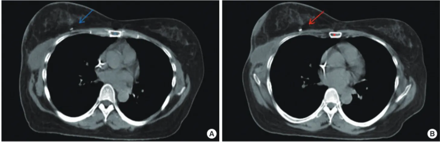
Univariate analysis revealed significant associations of outer quadrant lesion (p=0.040) and seroma ≥15 mL (p=0.004) with displacement in the lateromedial direction. Seroma (p=0.005) and the time interval between surgery and radio- therapy (p=0.040) were significantly associated with displace- Figure 1. Estimation of a surgical clip displacement. (A) A surgical clip (blue arrow) near breast seroma in a right breast cancer patient who received breast-conserving surgery was seen in initial simulation computed tomography (CT). (B) Re-simulation CT for a boost radiotherapy was undertaken 5 weeks after whole breast irradiation. The surgical clip (red arrow) was shifted 22 mm medially, 14 mm posteriorly, 10 mm inferiorly, and 27.9 mm three-dimensionally. Blue dot is a reference point in the initial simulation CT, and red dot is a reference point in the re-simulation CT.
A B
Table 1. Patients characteristics (n=178)
Characteristic No. (%)
Age (yr)* 50 (22–79)
BMI (kg/m2)* 24.1 (15.0–37.3)
Breast size
Small 113 (63.5)
Medium 58 (32.6)
Large 7 (3.9)
Location
Right 86 (48.3)
Left 92 (51.7)
Quadrant
Upper-outer 102 (57.3)
Upper-inner 39 (21.9)
Lower-outer 20 (11.2)
Lower-inner 17 (9.6)
Axillary surgery
No/SLNB 34 (19.1)
ALND 144 (80.9)
pT stage
pTis 19 (10.7)
pT1 114 (64.0)
≥pT2 45 (25.3)
pN stage
pN0 79 (44.4)
pN1 67 (37.6)
pN2–3 32 (18.0)
Seroma (mL)
<15 164 (92.1)
≥15 14 (7.9)
Postoperative chemotherapy
No 42 (23.6)
Yes 136 (76.4)
BMI=body mass index; SLNB=sentinel lymph node biopsy; ALND=axillary lymph node dissection.
*Median (range).
ment in the superoinferior direction. Multivariate analyses re- vealed that seroma ≥15 mL was the only significant factor as- sociated with displacement in the lateromedial (p=0.002) and superoinferior (p=0.003) directions. Outer quadrant lesion and time interval between surgery and radiotherapy showed a trend associated with displacement in the lateromedial (p=
0.063) and superoinferior (p=0.057) directions (Supplemen- tary Table 1).
Subgroup analyses of patients with seroma ≥15 mL were performed. The 90th percentile of displacement of the surgi- cal clips was 15.1 mm in the lateromedial direction, 12.7 mm in the anteroposterior direction, 10.0 mm in the superoinferior direction, and 21.75 mm in the 3D distance (Table 4).
DISCUSSION
BCS has become a standard treatment for early stage breast cancer, allowing breast preservation in patients with breast
cancer. Radiotherapy is an essential treatment after BCS to achieve an oncological outcome equivalent to mastectomy [1- 3,12]. Because tumor recurrences reportedly appear adjacent to the primary tumor, the surgical bed should be covered with a high radiation dose. Radiation delivery to exact surgical bed on CT scan is a major concern for radiation oncologists. Ra- diotherapy to patients with breast cancer takes 5 to 6 weeks using a conventional fractionation schedule. Radiation should be delivered to the intended target site accurately from the be- ginning to the end of treatment. Geometric uncertainties exist during radiotherapy because of setup error, intrafractional motion, and interfractional target change. An adequate mar- gin to the radiation target can ensure that the target is not missed. The proper margin differs according to the treatment sites and modalities.
The lumpectomy cavity may change after breast cancer sur- gery. Patients who do not receive adjuvant chemotherapy start postoperative radiotherapy within 6 weeks after surgery. Post- Table 2. Displacement of surgical clips in early breast cancer
Direction
Lateromedial direction Anteroposterior direction Superoinferior direction 3D distance Lat
(n=75) Med
(n=103) Total
(n=178) Ant
(n=94) Post
(n=84) Total
(n=178) Sup
(n=89) Inf
(n=89) Total
(n=178) Total (n=178) Distance (mm)* 2.0
(0.0–12.2) 2.7
(0.0–22.2) 2.6
(0.0–22.2) 2.6
(0.0–14.2) 2.3
(0.0–9.8) 1.8
(0.0–14.2) 3.0
(0.0–10.0) 3.0
(0.0–10.0) 2.0
(0.0–10.0) 5.3 (0.0–28.2)
90th percentile 5.3 5.4 5.3 7.5 7.0 7.1 6.0 6.0 6.0 9.8
3D=three-dimensional; Lat=lateral; Med=medial; Ant=anterior; Post=posterior; Inf=inferior; Sup =superior.
*Median (range).
Table 4. Displacement of clips in patients with seroma ≥15 mL Direction
Lateromedial direction Anteroposterior direction Superoinferior direction 3D distance Lat
(n=5)
Med (n=9)
Total (n=14)
Ant (n=6)
Post (n=8)
Total (n=14)
Sup (n=7)
Inf (n=7)
Total (n=14)
Total (n=14) Distance (mm)* 4.5
(0.0–7.8) 4.4 (0.8–22.2)
4.5 (0.0–22.2)
4.5 (1.8–14.2)
3.1 (0.9–5.4)
3.1 (0.9–14.2)
5.0 (2.0–7.0)
4.0 (0.0–10.0)
4.5 (0.0–10.0)
7.7 (3.4–28.2)
90th percentile - - 15.1 - - 12.7 - - 10.0 21.8
3D=three-dimensional; Lat=lateral; Med=medial; Ant=anterior; Post=posterior; Inf=inferior; Sup =superior.
*Median (range).
Table 3. Association between displacement of surgical clips and clinicopathologic factors in early breast cancer Factor
Lateromedial direction (mm)
No. (%) Anteroposterior direction (mm)
No. (%) Superoinferior direction (mm) No. (%)
≤3.5 >3.5 p-value ≤4.5 >4.5 p-value ≤5.0 >5.0 p-value
Horizontal 0.040 0.294 0.464
Outer 82 (64.1) 40 (80.0) 94 (66.7) 28 (75.7) 104 (67.5) 18 (75.0)
Inner 46 (35.9) 10 (20.0) 47 (33.3) 9 (24.3) 50 (32.5) 6 (25.0)
Seroma (mL) 0.004 0.455 0.005
<15 123 (96.1) 41 (82.0) 131 (92.9) 33 (89.2) 146 (94.8) 18 (75.0)
≥15 5 (3.9) 9 (18.0) 10 (7.1) 4 (10.8) 8 (5.2) 6 (25.0)
Surgery-radiation interval (wk) 0.162 0.880 0.040
≤23 92 (71.9) 41 (82.0) 105 (74.5) 28 (75.7) 111 (72.1) 22 (91.7)
>23 36 (28.1) 9 (18.0) 36 (25.5) 9 (24.3) 43 (27.9) 2 (8.3)
operative healing may continue during radiotherapy. Fibrosis, contraction, and absorption of fluid can influence the size and shape of the lumpectomy cavity. Disturbance of the lymphatic flow may induce an edematous change in breast tissue of pa- tients who undergo lymph node dissection. The breast may be influenced by postoperative and radiation-induced change more than other solid organs, because the breast is not a fixed organ; it consists of fat and lacks an external wall, so is easily movable. With this background, we investigated the possible factors influencing the surgical cavity in patients with breast cancer including breast size, quadrant, lymph node dissection, adjuvant chemotherapy, the time interval between surgery and radiotherapy, and seroma.
The 90th percentile of displacement of the surgical clips be- tween initial simulation CT before the start of radiotherapy and re-simulation CT after 50.4 Gy of radiotherapy was with- in 10 mm in all patients, but more than 10 mm in patients with seroma. Conventional radiotherapy consists of whole breast irradiation and surgical bed boost. Simulation CT scan for surgical bed boost after whole breast irradiation allows adaptive planning for change of surgical cavity. However, this takes time and involves extra cost and extra radiation expo- sure. Presently, displacements of surgical clips were within 10 mm in all directions. Given a 10 mm margin to the surgical cavity, a plan based on initial simulation CT scan would be sufficient to cover the primary tumor bed. Recently, intensity- modulated radiotherapy (IMRT) has been adopted to treat patients with breast cancer. The heart, coronary arteries, and lungs can be saved from a high dose radiation using IMRT [13,14]. External beam-partial breast irradiation using IMRT also can be considered for selected patients. IMRT preserves the normal organs but has a higher risk of missing the target.
Interfractional change in the target volume can cause unin- tended dose delivery [15]. Our results suggest that a 10 mm margin may be sufficient for IMRT planning to compensate for the alteration of the surgical cavity during radiotherapy.
Presently, seroma ≥15 mL on initial simulation CT was as- sociated with >10 mm displacement of the surgical clips. Se- roma has been reported as a significant factor associated with tumor bed volumetric change of 5% or greater during radio- therapy [16]. Tumor volume was observed to increase in 30%
of patients and to decrease in 70% of patients. In another study, the presence of seroma was significantly associated with a change in the lumpectomy cavity volume of more than 40%
(p=0.021). A significant seroma volume change was also re- ported elsewhere [17]. Mean seroma volume was 65.7 mL be- fore whole breast irradiation and 35.6 mL at boost planning.
Two of 24 patients showed an increase in the volume of sero- ma by 9.7% and 10.7%, respectively. Our data suggest that for
patients who have seroma ≥15 mL, re-simulation CT scan should be considered before boost planning. Boost margin of 20 mm from the clinical target volume may be sufficient to cover the displacement of lumpectomy cavity in patients with seroma, but could unnecessarily irradiate normal tissue. Ac- cording to previous reports, the lumpectomy cavity undergoes volume reduction rather than volume expansion during ra- diotherapy in most cases. CT scans for boost plan and adjust- ment of target volume may be a suitable option to cover the lumpectomy cavity adequately and to minimize the irradia- tion of normal breast tissue.
Patients who received radiotherapy within 23 weeks after surgery showed a trend to have a larger displacement of the surgical clips in the superoinferior direction. When the time interval between surgery and radiotherapy is shorter, there is a greater possibility that postoperative healing changes the lumpectomy cavity. An analysis of the changes in the breast during whole breast irradiation reported that the mean vol- ume reduction in the excision cavity was 22.5% (p<0.0001) and was inversely correlated with the time between surgery and radiotherapy (p<0.01) [18]. Another study compared the volume of the lumpectomy cavity from postoperative CT scan to planning CT scan. Change in the tumor bed volume be- tween the two CT scans was 2.1% per day in patients who re- ceived immediate postoperative radiotherapy after surgery and 0.4% per day in patients who had a delay for adjuvant chemotherapy between surgery and radiotherapy [19]. These findings suggested that the volumetric change is inversely pro- portional to the interval between surgery and radiotherapy.
However, the study was limited by its retrospective nature and relatively small sample size [20]. The results from the retro- spective data should be interpreted with caution. We showed the displacement of selected single clip for each patient and analyzed it using the 90th percentile as a reference value [21].
Variation between all clips in the same patient was not ana- lyzed in this study.
In conclusion, displacements of the surgical clips during whole breast irradiation typically range under 10 mm. Given a margin of 10 mm, a target volume based on initial simula- tion CT scan would sufficiently cover the lumpectomy cavity.
However, in patients who have seroma ≥15 mL after BCS, re- simulation before a boost treatment may be necessary to make exact planning adaptive to volumetric changes in the lumpectomy cavity.
CONFLICT OF INTEREST
The authors declare that they have no conflict of interests.
REFERENCES
1. Veronesi U, Cascinelli N, Mariani L, Greco M, Saccozzi R, Luini A, et al.
Twenty-year follow-up of a randomized study comparing breast-con- serving surgery with radical mastectomy for early breast cancer. N Engl J Med 2002;347:1227-32.
2. Blichert-Toft M, Nielsen M, Düring M, Møller S, Rank F, Overgaard M, et al. Long-term results of breast conserving surgery vs. mastectomy for early stage invasive breast cancer: 20-year follow-up of the Danish ran- domized DBCG-82TM protocol. Acta Oncol 2008;47:672-81.
3. Litière S, Werutsky G, Fentiman IS, Rutgers E, Christiaens MR, Van Limbergen E, et al. Breast conserving therapy versus mastectomy for stage I-II breast cancer: 20 year follow-up of the EORTC 10801 phase 3 randomised trial. Lancet Oncol 2012;13:412-9.
4. Bartelink H, Horiot JC, Poortmans P, Struikmans H, Van den Bogaert W, Barillot I, et al. Recurrence rates after treatment of breast cancer with standard radiotherapy with or without additional radiation. N Engl J Med 2001;345:1378-87.
5. Vrieling C, Collette L, Fourquet A, Hoogenraad WJ, Horiot JH, Jager JJ, et al. The influence of patient, tumor and treatment factors on the cos- metic results after breast-conserving therapy in the EORTC ‘boost vs.
no boost’ trial. EORTC Radiotherapy and Breast Cancer Cooperative Groups. Radiother Oncol 2000;55:219-32.
6. Bartelink H, Horiot JC, Poortmans PM, Struikmans H, Van den Bogaert W, Fourquet A, et al. Impact of a higher radiation dose on local control and survival in breast-conserving therapy of early breast cancer: 10-year results of the randomized boost versus no boost EORTC 22881-10882 trial. J Clin Oncol 2007;25:3259-65.
7. Romestaing P, Lehingue Y, Carrie C, Coquard R, Montbarbon X, Ardiet JM, et al. Role of a 10-Gy boost in the conservative treatment of early breast cancer: results of a randomized clinical trial in Lyon, France. J Clin Oncol 1997;15:963-8.
8. Hunter MA, McFall TA, Hehr KA. Breast-conserving surgery for pri- mary breast cancer: necessity for surgical clips to define the tumor bed for radiation planning. Radiology 1996;200:281-2.
9. Harrington KJ, Harrison M, Bayle P, Evans K, Dunn PA, Lambert HE, et al. Surgical clips in planning the electron boost in breast cancer: a qualitative and quantitative evaluation. Int J Radiat Oncol Biol Phys 1996;34:579-84.
10. Bra size. Wikipedia. https://en.wikipedia.org/wiki/Bra_size. Accessed
June 10th, 2016.
11. Chung MJ, Lee GJ, Suh YJ, Lee HC, Lee SW, Jeong S, et al. Setup error and effectiveness of weekly image-guided radiation therapy of Tomo- Direct for early breast cancer. Cancer Res Treat 2015;47:774-80.
12. Fisher B, Anderson S, Bryant J, Margolese RG, Deutsch M, Fisher ER, et al. Twenty-year follow-up of a randomized trial comparing total mas- tectomy, lumpectomy, and lumpectomy plus irradiation for the treat- ment of invasive breast cancer. N Engl J Med 2002;347:1233-41.
13. Jin GH, Chen LX, Deng XW, Liu XW, Huang Y, Huang XB. A compara- tive dosimetric study for treating left-sided breast cancer for small breast size using five different radiotherapy techniques: conventional tangen- tial field, filed-in-filed, tangential-IMRT, multi-beam IMRT and VMAT. Radiat Oncol 2013;8:89.
14. Mansouri S, Naim A, Glaria L, Marsiglia H. Dosimetric evaluation of 3-D conformal and intensity-modulated radiotherapy for breast cancer after conservative surgery. Asian Pac J Cancer Prev 2014;15:4727-32.
15. Kim KS, Shin KH, Choi N, Lee SW. Hypofractionated whole breast ir- radiation: new standard in early breast cancer after breast-conserving surgery. Radiat Oncol J 2016;34:81-7.
16. Chung MJ, Suh YJ, Lee HC, Kang DG, Kim EJ, Kim SH, et al. Tumor bed volumetric changes during breast irradiation for the patients with breast cancer. Radiat Oncol J 2013;31:228-33.
17. Sharma R, Spierer M, Mutyala S, Thawani N, Cohen HW, Hong L, et al.
Change in seroma volume during whole-breast radiation therapy. Int J Radiat Oncol Biol Phys 2009;75:89-93.
18. Oh KS, Kong FM, Griffith KA, Yanke B, Pierce LJ. Planning the breast tumor bed boost: changes in the excision cavity volume and surgical scar location after breast-conserving surgery and whole-breast irradia- tion. Int J Radiat Oncol Biol Phys 2006;66:680-6.
19. Petersen RP, Truong PT, Kader HA, Berthelet E, Lee JC, Hilts ML, et al.
Target volume delineation for partial breast radiotherapy planning:
clinical characteristics associated with low interobserver concordance.
Int J Radiat Oncol Biol Phys 2007;69:41-8.
20. Lee SW, Hwang TK, Hong SH, Lee JY, Chung MJ, Jeong SM, et al. Out- come of postoperative radiotherapy following radical prostatectomy: a single institutional experience. Radiat Oncol J 2014;32:138-46.
21. Sung KC, Chang Y, Ryu S, Chung HK. High levels of serum vitamin D are associated with a decreased risk of metabolic diseases in both men and women, but an increased risk for coronary artery calcification in Korean men. Cardiovasc Diabetol 2016;15:112.