Received: May 18, 2018 Revised: July 31, 2018 Accepted: November 18, 2018 AnnAls of
CliniCAl
neurophysiology
Case RepoRt
Ann Clin Neurophysiol 2019;21(1):44-47 https://doi.org/10.14253/acn.2019.21.1.44
Correspondence to Sang-Won Ha
Department of Neurology, Veterans Health Service Medical Center, 53 Jinhwangdo-ro 61-gil, Gangdong-gu, Seoul 05368, Korea Tel: +82-2-2225-4601
Fax: +82-2-2225-1327 E-mail: [email protected]
ORCID Inha Hwang
http://orcid.org/0000-0002-4964-3010 UnKyu Yun
http://orcid.org/0000-0001-6469-5874 Heewon Bae
http://orcid.org/0000-0002-2266-9288 Jeong Ho Han
http://orcid.org/0000-0003-3197-4898 Sang-Won Ha
http://orcid.org/0000-0001-8881-5519 Doo-eung Kim
http://orcid.org/0000-0003-4343-4709
http://www.e-acn.org pISSN 2508-691X eISSN 2508-6960
Copyright © 2019 the Korean society of Clinical Neurophysiology
This is an Open Access article distributed under the terms of the Creative Commons Attribution Non-Commercial License (http://
creativecommons.org/licenses/by-nc/4.0) which permits unrestricted non-commercial use, distribution, and reproduction in any medium, provided the original work is properly cited.
Zoster-associated limb paresis present- ing as femoral neuropathy
Inha Hwang, UnKyu Yun, Heewon Bae, Jeong Ho Han, Sang-Won Ha, and Doo-eung Kim
Department of Neurology, Veterans Healthcare Service Medical Center, Seoul, Korea
Zoster-associated limb paresis is a relatively uncommon complication of herpes zoster that is characterized by focal motor weakness. Awareness of this disorder is important to avoid un- necessary invasive investigations and to ensure appropriate treatment. We report a case of a herpes zoster involving the femoral nerve.
Key words: Femoral neuropathy; Herpes zoster; Varicella zoster virus
Shingles (herpes zoster) may cause a variety of neurological complications in the central and peripheral nervous systems, including postherpetic neuralgia, varicella zoster virus myelitis, segmental weakness, and delayed ischemic cerebral infarction due to zoster virus-associated granulomatous vasculitis.1 Zoster-associated limb paresis (ZALP) is a rela- tively uncommon complication of herpes zoster characterized by focal motor weakness, which may occur in limbs affected by herpes zoster.2 Zoster-associated mononeuropathy (ZAM) as a cause of herpes zoster-associated paresis is even rarer.3 Here we report a case of a herpes zoster involving the femoral nerve.
CASE
A 79-year-old man was admitted to the dermatology department of our hospital with pain and skin eruptions over the left medial thigh. He was diagnosed as having herpes zoster and was treated with 750 mg of famciclovir daily for 7 days. The patient reported that he could not walk due to pain but did not have weakness in the leg. He was not evaluated for gait difficulty at that time. Two weeks after the treatment, the patient revisited the hospital due to weakness in the left leg and was admitted to our neurology department.
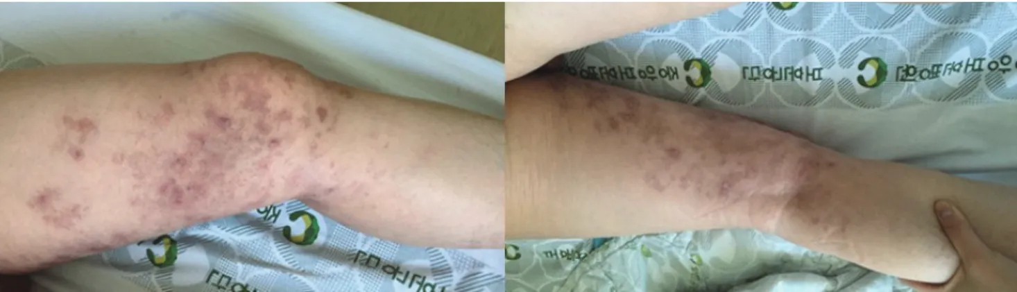
Upon admission he showed scars of herpetic eruption and pigmentation over the left medial thigh and medial calf, but not over the lateral thigh (Fig. 1). Muscle weakness in the left lower limb was mostly noted for hip flexion (MRC grade 2) and knee extension (MRC
45
http://www.e-acn.org https://doi.org/10.14253/acn.2019.21.1.44
Inha Hwang, et al. Zoster associated femoral neuropathy
Zoster-associated limb paresis present- ing as femoral neuropathy
Inha Hwang, UnKyu Yun, Heewon Bae, Jeong Ho Han, Sang-Won Ha, and Doo-eung Kim
Department of Neurology, Veterans Healthcare Service Medical Center, Seoul, Korea
Zoster-associated limb paresis is a relatively uncommon complication of herpes zoster that is characterized by focal motor weakness. Awareness of this disorder is important to avoid un- necessary invasive investigations and to ensure appropriate treatment. We report a case of a herpes zoster involving the femoral nerve.
Key words: Femoral neuropathy; Herpes zoster; Varicella zoster virus
grade 2). The left tibialis anterior, gastrosoleus group, hip ad- ductors, and abductors were all 5/5. The patient had no left patellar muscle stretch reflex and a normal left Achilles re- flex. Sensation was impaired over the left anteromedial thigh and medial calf, but not over the lateral thigh. His right lower
limb was entirely normal with respect to strength, sensation, and muscle stretch reflexes.
The findings of routine laboratory studies were normal.
Magnetic resonance imaging (MRI) of the lumbar spine showed only small herniations of the intervertebral discs Fig. 1. The patient showed scars of herpetic eruption and pigmentation over the left medial anterior thigh and medial calf, but not over the lateral thigh.
Fig. 2. (A) Compound muscle action potential in femoral nerve conduction study and (B) sensory nerve action potential in medial femoral cutaneous nerve conduction study were unobtainable on the left, while the amplitude responses of those were normal on the right.
A
B
46 https://doi.org/10.14253/acn.2019.21.1.44 http://www.e-acn.org
Annals of Clinical Neurophysiology Volume 21, Number 1, January 2019
from levels L1/2 to L4/5, mild spinal stenosis at level L4/5, and degenerative minute spondylolisthesis at level L4/5. MRI did not reveal any nerve or spinal cord abnormalities such as prolonged T2-weighted signals, nerve enlargement, or post- gadolinium enhancement. An electrodiagnostic evaluation was performed. Compound muscle action potential in fem- oral nerve conduction study and sensory nerve action po- tential in medial femoral cutaneous nerve conduction study were unobtainable on the left, while the amplitude respons- es of those were normal on the right (Fig. 2). The amplitude of the left peroneal motor nerve response was slightly de- creased, while the left posterior tibial motor nerve response was normal. Needle electromyography (EMG) revealed pos- itive sharp waves in the left L2-S1 paraspinal, vastus lateralis, and iliacus muscles. The EMG findings suggested left femoral neuropathy with coexistent lumbosacral radiculopathy. We assumed that the lumbosacral radiculopathy was attribut- able to the preexisting herniations of the intervertebral discs at levels L1 to L5, because a diffuse denervation process was not observed in the corresponding peripheral limb muscles.
We thought that herpes zoster accounted for the left femo- ral neuropathy but not for the radiculopathy.
Combining the history with the findings of the physical examination, imaging studies, and electrodiagnostic evalua- tion led to a diagnosis of herpes zoster femoral neuropathy with coexisting polyradiculopathy. We treated the patient with intravenous acyclovir at 10 mg/kg/day for 7 days and physical therapy. The patient’s muscle strength improved to MRC grade 3 at the time of discharge (2 weeks after starting acyclovir). He could not walk at the time of discharge, but his left leg strength had improved to MRC grade 4 when he visited the outpatient department approximately 2 months after starting acyclovir. However, he still complained of pos- therpetic neuralgia. The patient refused an electrodiagnostic follow-up examination due to his improving symptoms.
DiSCuSSion
Segmental zoster paresis is a rare occurrence. The incidence of segmental limb paresis with cutaneous zoster has been reported at 3% to 5%.3 ZAM as a cause of zoster-associated paresis is even rarer. In one series of 49 patients with ZALP, it was caused by radiculopathy (37%), plexopathy (41%),
mononeuropathy (14%), or radiculoplexus neuropathy (8%).3 In another series, ZAM was associated with prolonged symp- toms, significant weakness, and a high rate of postherpetic neuralgia.4 Our patient also complained of postherpetic neu- ralgia at 2 months of follow-up.
ZALP may localize to the root, plexus, or more-peripheral nerve, and is difficult to localize clinically in part because the involved myotomes often do not correspond to the dermatomes affected by the rash.1 For example, mononeu- ropathies may be difficult to distinguish clinically from more-proximal focal plexopathies or radiculopathies. Elec- trophysiological studies are useful for correctly diagnosing and evaluating the extent of lesions. Nerve conduction stud- ies usually disclose reduced sensory nerve action potentials and compound muscle action potentials in the affected segments.5 Needle EMG generally reveals abnormal spon- taneous activities, such as fibrillations and positive sharp waves in clinically weak muscles. Imaging studies such as magnetic resonance neurography can be invaluable in the identification and localization of affected areas. One recent retrospective study found MRI imaging abnormalities in seven of 10 patients (70%) with zoster-associated plexopa- thy.1 Jones et al. reviewed the nerve imaging features of 26 patients with zoster paresis: nine of the 14 patients (64%) with postganglionic electrodiagnostic localization (at the plexus or nerve level) demonstrated imaging abnormalities in the affected plexus or nerve, and one patient showed enhancement with gadolinium contrast.3 More commonly, nerve enlargement or increased T2-weighted signals within the affected nerve were observed. None (0%) of the 12 pa- tients with preganglionic localization (at the anterior horn or root level) demonstrated abnormal findings in nerve or spinal cord imaging, but abnormalities have been described in other cases of zoster paresis.3,6 We did not perform an imaging study to explore the affected femoral nerve in the present patient.
The precise mechanism underlying zoster-associated paresis is poorly understood.4 Virally mediated injury at the level of the anterior horn cell or ventral root has been sug- gested as the cause of herpes zoster paresis.4 A postmortem observation showed the degeneration of anterior spinal roots with lymphocytic infiltration of the posterior and an- terior horns.7 An MRI study revealed contrast enhancement of the anterior roots of affected segments, suggesting the
47
http://www.e-acn.org https://doi.org/10.14253/acn.2019.21.1.44
Inha Hwang, et al. Zoster associated femoral neuropathy
presence of hypervascularity or disruption of the blood- nerve barrier caused by viral-induced inflammation in pa- tients with zoster paresis.8 Anterior spinal roots seem to be common sites of inflammation and degeneration, but ante- rior horn cells, the brachial or lumbar plexus, and peripheral nerves can also be involved. Individual reports of ZAMs sug- gest that a distally mediated neuropathic mechanism could underlie herpes zoster paresis in some patients.4
The prognosis of zoster-associated limb paresis is general- ly good. Patients usually recover most functions, and more than half of them recover completely.2 It remains unclear whether a diagnosis failure affects the prognosis. Mondelli et al.9 reported that antiviral therapy at the appropriate dose and duration was associated with a reduced incidence of segmental zoster paresis. It is unclear whether additional intravenous antiviral or steroid treatment is beneficial once paresis occurs. Further studies of the optimal treatment for promoting functional recovery are necessary. The protective effect provided by the zoster vaccine against zoster-associ- ated paresis would also be of particular interest.
Conflicts of Interest
The authors have no financial conflicts of interest.
REFEREnCES
1. Zubair AS, Hunt C, Watson J, Nelson A, Jones LK Jr. Imaging find- ings in patients with zoster-associated plexopathy. AJNR Am J Neuroradiol 2017;38:1248-1251.
2. Kawajiri S, Tani M, Noda K, Fujishima K, Hattori N, Okuma Y. Seg- mental zoster paresis of limbs: report of three cases and review of literature. Neurologist 2007;13:313-317.
3. Jones LK Jr, Reda H, Watson JC. Clinical, electrophysiologic, and imaging features of zoster-associated limb paresis. Muscle Nerve 2014;50:177-185.
4. Reda H, Watson JC, Jones LK Jr. Zoster-associated mononeuropa- thies (ZAMs): a retrospective series. Muscle Nerve 2012;45:734-739.
5. Haanpää M, Häkkinen V, Nurmikko T. Motor involvement in acute herpes zoster. Muscle Nerve 1997;20:1433-1438.
6. Haanpää M, Dastidar P, Weinberg A, Levin M, Miettinen A, Lapin- lampi A, et al. CSF and MRI findings in patients with acute herpes zoster. Neurology 1998;51:1405-1411.
7. Murakami T, Shibazaki K, Kurokawa K, Ichikawa Y, Ohsawa Y, Sun- ada Y. Conduction block of varicella zoster virus neuropathy. Neu- rology 2003;61:1153-1154.
8. Hanakawa T, Hashimoto S, Kawamura J, Nakamura M, Suenaga T, Matsuo M. Magnetic resonance imaging in a patient with seg- mental zoster paresis. Neurology 1997;49:631-632.
9. Mondelli M, Romano C, Rossi S, Cioni R. Herpes zoster of the head and limbs: electroneuromyographic and clinical findings in 158 consecutive cases. Arch Phys Med Rehabil 2002;83:1215-1221.