서 론
어깨 관절 운동 범위(range of motion)는 견관절의 기능 평가 및 질환을 이해하는 데 있어 중요한 임상적 매개 변수이다.1,2) 관절 운동 범위를 측정하기 위해 일반적으로는 재래식 측정 방법인 각 도기가 이용되어 왔다. 여러 연구를 통해 각도기를 통한 측정은
Copyright © 2021 by The Korean Orthopaedic Association
“This is an Open Access article distributed under the terms of the Creative Commons Attribution Non-Commercial License (http://creativecommons.org/licenses/by-nc/4.0/) which permits unrestricted non-commercial use, distribution, and reproduction in any medium, provided the original work is properly cited.”
The Journal of the Korean Orthopaedic Association Volume 56 Number 1 2021 Received February 7, 2020 Revised February 19, 2020 Accepted April 10, 2020 Correspondence to: Eugene Kim, M.D., Ph.D.
Department of Orthopaedic Surgery, Kangbuk Samsung Hospital, Sungkyunkwan University School of Medicine, 29 Saemunan-ro, Jongno-gu, Seoul 03181, Korea TEL: +82-2-2001-1820 FAX: +82-2-2001-2176 E-mail: eugene0809.kim@samsung.com ORCID: https://orcid.org/000-0002-5979-3389
깊이 센서 기반 모션 분석 시스템을 사용한 어깨 운동학 조사
이인규 • 박재형 • 손동욱 • 조용운* • 하상훈 • 김유진
성균관대학교 의과대학 강북삼성병원 정형외과학교실, *건국대학교 의과대학 건국대학교 충주병원 정형외과학교실
Investigation for Shoulder Kinematics Using Depth Sensor-Based Motion Analysis System
Ingyu Lee, M.D., Jai Hyung Park, M.D., Ph.D., Dong-Wook Son, M.D., Ph.D., Yongun Cho, M.D. *, Sang Hoon Ha, M.D., and Eugene Kim, M.D., Ph.D.
Department of Orthopaedic Surgery, Kangbuk Samsung Hospital, Sungkyunkwan University School of Medicine, Seoul, *Department of Orthopaedic Surgery, Konkuk University Chungju Hospital, College of Medicine, Konkuk University, Chungju, Korea
Purpose: The purpose of this study was to analyze the motion of the shoulder joint dynamically through a depth sensor-based motion
analysis system for the normal group and patients group with shoulder disease and to report the results along with a review of the relevant literature.Materials and Methods: Seventy subjects participated in the study and were categorized as follows: 30 subjects in the normal group
and 40 subjects in the group of patients with shoulder disease. The patients with shoulder disease were subdivided into the following four disease groups: adhesive capsulitis, impingement syndrome, rotator cuff tear, and cuff tear arthropathy. Repeating abduction and adduction three times, the angle over time was measured using a depth sensor-based motion analysis system. The maximum abduction angle (θmax), the maximum abduction angular velocity (ωmax), the maximum adduction angular velocity (ωmin) , and the abduction/adduction time ratio (tabd/ tadd) were calculated. The above parameters in the 30 subjects in the normal group and 40 subjects in the patients group were compared. In addition, the 30 subjects in the normal group and each subgroup (10 patients each) according to the four disease groups, giving a total of five groups, were compared.Results: Compared to the normal group, the maximum abduction angle (θ
max), the maximum abduction angular velocity (ωmax), and the maximum adduction angular velocity (ωmin) were lower, and abduction/adduction time ratio (tabd/tadd) was higher in the patients with shoulder disease. A comparison of the subdivided disease groups revealed a lower maximum abduction angle (θmax) and the maximum abduction angular velocity (ωmax) in the adhesive capsulitis and cuff tear arthropathy groups than the normal group. In addition, the abduction/adduction time ratio (tabd/tadd) was higher in the adhesive capsulitis group, rotator cuff tear group, and cuff tear arthropathy group than in the normal group.Conclusion: Through an evaluation of the shoulder joint using the depth sensor-based motion analysis system, it was possible to measure
the range of motion, and the dynamic motion parameter, such as angular velocity. These results show that accurate evaluations of the function of the shoulder joint and an in-depth understanding of shoulder diseases are possible.Key words: shoulder, range of motion, kinect, dynamic, angular velocity
신뢰할 수 있는 것으로 밝혀졌으며, 견관절의 운동 범위를 측정 하기 위해 널리 사용되었다.3-5)
그러나 각도기는 관찰자의 경험 및 회전 중심을 측정하는 방법 등과 같은 여러 요소들의 영향을 받을 수 있어6-10) 관찰자 간 측정 각도가 달라져 신뢰성과 재현성이 낮아질 수 있는 것이 제한점이 다.11) 이에 반해 깊이 센서 기반 동작 분석 시스템을 통한 전자식 측정 방법은 사용자의 동작을 인식하여 어깨 관절의 운동 범위를 측정할 수 있기 때문에 관찰자 간의 측정 각도의 오류를 교정할 수 있다.12-19)
또한 현재까지 견관절 운동에 대한 평가는 관절 운동 범위 등 의 정적 운동에 중점을 두고 있었다. 건측 수준의 양호한 관절 운 동 범위를 보이나, 느리거나 떨리는 등 이상 운동 양상을 보이 는 환자가 많다는 것을 고려할 때 정적 운동 평가를 통한 양적 평 가뿐 아니라 동적 운동 평가를 통한 질적 평가도 이루어져야 한
다.20,21) 이러한 견관절의 운동 기능 평가를 위해서는 동적 관절
상태를 수치화할 수 있는 측정 방식이 필요하다. 깊이 센서 기반 동작 시스템은 견관절의 위치를 시간에 따라 측정하여 속도 및 가속도와 같은 부가적인 운동 상태를 평가할 수 있다.22,23) 본 연구에서는 깊이 센서 기반 동작 분석 시스템을 이용하여 정적 운동 지표인 관절 운동 범위뿐 아니라 각속도 등의 동적 운 동을 측정하고자 한다. 정상 견관절과 이환된 견관절의 운동을 측정하고 비교하여 운동 기능 평가 도구로서 깊이 센서 기반 동 작 분석 시스템의 유용성을 확인하고, 더 나아가 여러 견관절 질 환의 생역학적 분석에 도움이 될 수 있도록 문헌 고찰과 함께 보 고하는 바이다.
대상 및 방법
1. 깊이 센서 기반 동작 분석 시스템 기반 측정 방법
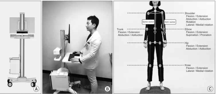
인체 움직임을 파악하기 위해 출시된 Kinect-V2 (Microsoft, Seattle, WA, USA; 2014)를 통해 피험자의 견관절 위치 및 움 직임을 인식하였다. Kinect-V2는 적외선을 발사한 후 되돌아오 는 시간(time of flight)을 측정하는 RGB-D sensor를 통해 환 자의 표면 영상을 획득할 수 있다. 딥러닝(deep-learning) 기술 및 칼만 필터(kalman filter)를 적용한 프로그램 POM checker (Team Elysium, Seoul, Korea; 2018)를 통해 영상 및 관절의 위 치를 수치화하여 데이터를 얻었다(Fig. 1A). 측정의 객관성을 유 지하고 깊이 센서 기반 동작 분석 시스템을 활용한 신체 인식 시 최적의 측정 환경을 구성하기 위해 깊이 센서 기반 동작 분석 시 스템의 위치는 지면과 수직으로 1.5 m 떨어진 곳에 설치하고, 측 정 대상과의 거리는 2 m로 고정하였다(Fig. 1B).
깊이 센서를 통해 입력 받은 깊이 영상으로부터 총 25곳의 주 요 관절의 위치를 인식할 수 있으며, 인식한 관절 두 곳을 이은 벡 터(vector) 간의 관절 운동 각도를 측정할 수 있었다. 본 연구에서 는 견관절의 각도를 측정하기 위해 경추 관절과 고관절의 중심을 이은 척추 벡터(spine vector)와 팔 벡터(arm vector) 간의 각도 를 측정하였다(Fig. 1C).
2. 어깨 관절 운동 범위 측정
적외선을 통해 피험자의 표면 영상과 관절 위치가 측정되기 때문 에 적외선을 반사하는 폴리에스테르 소재로 이루어진 옷이나 검
A B C
Trunk
Flexion / Extension Abduction / Adduction
Spine vector Arm vector
Shoulder Flexion / Extension Abduction / Adduction Rotation
Lateral / Medial rotation Elbow
Flexion / Extension Supination / Pronation
Hip
Flexion / Extension Abduction / Adduction
Knee
Flexion / Extension Lateral / Medial rotation
Figure 1. (A) Kinect-V2 (Microsoft, Seattle, WA, USA; 2014). (B) The location of the depth sensor-based motion analysis system was installed at a distance of 1.5 m to the ground, and the distance to the patient was fixed at 2 m. (C) In order to measure the angle of the shoulder joint, a spinal vector connecting the center of the cervical joint and the hip joint was used, and the angle between the spine vector and the arm vector was measured.
은색 옷은 측정 시 방해요소로 작용할 수 있다. 따라서 측정 전 적 외선의 반사 확률이 적은 면 소재의 옷으로 환복이 필요하다.
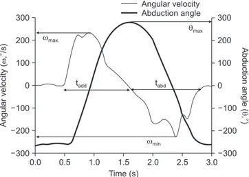
피험자는 기계에서 일정 거리 떨어진 위치에서 센서를 정면으 로 바라보고 정자세로 선다. 손목 관절은 중립위(neutral posi- tion), 팔꿈치 관절은 최대 신전(full extension) 상태에서 실험자 의 지시에 따라 견관절의 외전 및 내전을 반복한다. 인식된 신체 부위는 실시간으로 움직임이 기록되었으며, 측정 간 휴지기간 없 이 총 3회 반복하여 시행한 결과를 분석하였다. 속도의 곡선의 상 향 기울기는 0.1°/s 및 -0.1°/s를 초과해야 하며 각 이벤트는 적절 한 시작 및 종결을 보장하기 위해 수작업으로 검사되었다. 각 반 복마다 시간에 따른 견관절의 외전 각도를 측정하였다. 이를 통 해 최대 외전 각도(θmax)를 측정하였다. 시간에 따른 외전 각도의 변화를 통해 각속도를 도출하였으며, 최대 내전 각속도(ωmin)와 최대 외전 각속도(ωmax)를 도출하였다. 견관절이 최대 외전 각도 (θmax)로 도달하는 데까지 걸리는 시간을 외전 시간(tabd)으로 구하 였으며, 이후 내전 상태로 돌아가는 데 걸리는 시간을 내전 시간 (tadd)으로 정하고, 외전/내전 시간 비(tabd/tadd)를 측정하였다(Fig.
2).
3. 실험 대상
70명의 피험자가 자원하여 연구에 참여하였다. 먼저 정상군 (normal group) 30명과 견관절 질환을 가진 환자군(disease group) 40명으로 분류하였으며 정상군에서는 (1) 견관절 질환 (회전근개 파열, 유착성 활액막염, 충돌 증후군, 회전근개 관절병 증)으로 진단된 적이 있는 자, (2) 통증을 포함한 혼합된 임상 증
상으로 인하여 불확실하게 진단된 적이 있는 자, (3) 심각한 어 깨 부상(골절, 탈구 또는 수술)의 병력이 있는 자, (4) 선천성 기형 이 있는 자를 제외하였다. 관절의 운동 양상을 측정, 분석하는 본 연구의 목적에 따라 견관절 질환은 근육 병변이 있거나 관절 운 동 범위에 제한이 있는 4가지 질환(회전근개 파열[rotator cuff tear], 유착성 활액막염[adhesive capsulitis], 충돌 증후군[im- pingement syndrome], 회전근개 관절병증[cuff tear arthrop- athy])을 선택하였고 각 질병군당 10명의 환자, 총 40명의 환자 로 구성되었다. 회전근개 파열 환자는 자기공명영상(magnetic resonance imaging, MRI)상 극상근(supraspinatus muscle) 의 전층 파열이 있는 환자 중 수술(관절경하 회전근개 봉합술) 예 정인 환자 10명을 대상으로 하였다. 회전근개 관절병증의 환자의 경우 MRI상 Tangent 징후 양성이며, Hamada 분류 2단계 이상 의 회전근개 관절병증이 확인되는 환자 중 수술(역행성 인공 견 관절 전치환술) 예정인 환자 10명을 대상으로 하였다. 충돌 증후 군 및 유착성 활액막염 환자는 신체 진찰상 질환에 해당하는 임 상 징후를 보이고, 초음파 또는 MRI상 다른 질환이 배제되는 각 10명의 환자를 대상으로 하였다. 각 질환군의 진단 및 배정은 1 명의 정형외과 전문의에 의해 시행되었다.
본 연구는 강북삼성병원 Institutional Review Board (KB- SMC 2019-07-012)로부터 연구 승인을 받고 진행되었다.
4. 통계적 분석
모든 값은 매개 변수의 특성에 따라 평균±표준편차로 표시되었 다. 30명의 정상군과 40명의 질병군, 두 군 간의 비교에는 Stu- dent t-test가 이용되었다.
매개변수의 분포의 정규성 여부를 검정하기 위해 Shapiro–
Wilk test 및 그래픽 기법(graphical approach, histogram)을 이용하였고, 정규분포에 위배되는 매개변수들(최대 외전 각도 [θmax], 외전/내전 시간 비[tabd/tadd])에 대해서는 자연로그 변환 후 분석에 이용하였다.
Table 1. Comparison of the Range of Motion and Angular Velocity between Patients with Shoulder Disease and a Normal Group
Characteristic Normal group (n=30)
Patient group
(n=40) p-value
θmax (°) 165.4±7.7 122.6±38.0 0.012 ωmax (°/s) 342.9±90.0 191.6±93.6 <0.001 ωmin (°/s) -301.9±85.6 -226.7±72.3 0.023 tabd/tadd 0.9±0.3 2.1±1.2 <0.001 Values are presented as mean±standard deviation. θmax, maximum abduction angle; ωmax, maximum abduction angular velocity; ωmin, maximum adduction angular velocity; tabd/tadd, time ratio between adduction and abduction.
0.0 0.5 1.0 1.5 2.0 2.5
300
200
100
0
100
200
300
3.0
Angularvelocity(,/s)
Time (s)
Angular velocity Abduction angle
Abductionangle(,)
300
200
100
0
100
200
300
max
max
t
abdt
addmin
Figure 2. Kinetic measurements in the normal groups: abduction angle (thick line) and velocity (thin line) for one repeated shoulder abduction of a typical subject collected using Kinect-V2. The variables include the maximum abduction angle (θmax), maximum angular velocity during abduction (ωmax), angular velocity during adduction (ωmin), time during abduction (tabd) and time during adduction (tadd).
30명의 정상군, 각 10명의 질환군 4군, 총 5군 간의 비교에는 일원 분산 분석(ANOVA, analysis of variance) 통계가 이용되었 다. 모든 p<0.05는 통계적으로 유의한 것으로 간주되었고, 통계 프로그램 IBM SPSS ver. 25.0 (IBM Corp., Armonk, NY, USA) 이 사용되었다.
결 과
견관절 질환을 가진 환자군에서는 정상군에 비하여 팔의 최 대 외전 각도(θmax [정상군 vs. 환자군]: 165.4°±7.7° vs. 122.6°
±38.0°; p=0.012), 최대 외전 각속도(ωmax: 342.9±90.0°/s vs.
191.6±93.6°/s; p<0.001), 최대 내전 각속도(ωmin: -301.9±
Table 2. Comparison of the Range of Motion and Angular Velocity between Patients with Shoulder Disease and the Normal Group with One-Way ANOVA
Characteristics Normal group (n=30) AC group (n=10) IS group (n=10) RC group (n=10) CT group (n=10)
θmax (°) 165.4±7.7ab 83.9±34.6acd 161.0±10.6de 140.3±24.9c 107.0±21.3be ωmax (°/s) 342.9±90.0f 110.9±37.5fgh 263.8±80.0hj 254.0±73.6gk 115.4±43.0fjk ωmix (°/s) -301.9±85.6 -182.4±64.4 -239.4±68.5 -275.3±71.0 -183.0±15.8 tabd/tadd 0.9±0.3lmn 2.7±2.0l 1.5±0.4 1.9±0.6m 2.1±0.6n Values are presented as mean±standard deviation. a–nPairs of data marked with the same alphabet mean a statistically significant difference. AC, adhesive capsulitis; IS, impingement syndrome; RC, rotator cuff tear; CT, cuff tear arthropathy; θmax, maximum abduction angle; ωmax, maximum abduction angular velocity; ωmin, maximum adduction angular velocity; tabd/tadd, time ratio between adduction and abduction.0 1 2 3 4 5
100
50
0
100
150
6
Angularvelocity(,/s)
Time (s)
Abduction(,) angle
160 140 120 100 80 60 40 20 0 Angular velocity Abduction angle
0 0.5 1 1.5 2 2.5
100
50
0
100
150
4
Angularvelocity(,/s)
Time (s)
Angular velocity Abduction angle
Abduction(,) angle
160 140 120 100 80 60 40 20 0 3 3.5
1 3 5 7 9 11 13 15 17 19 21 23 25 27 29 31 100
50 0
100 150 200 250 300
Angularvelocity(,/s)
Time (s)
Angular velocity Abduction angle
Abduction(,) angle
160 140 120 100 80 60 40 20 0
0 0.5 1 1.5 2 2.5
300
200
100
0
200
300
4
Angularvelocity(,/s)
Time (s)
Angular velocity Abduction angle
Abduction(,) angle
160 140 120 100 80 60 40 20 0 3 3.5
A B
C D
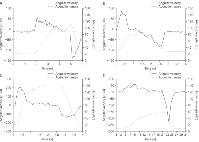
Figure 3. Kinetic measurements in the patients with shoulder diseases. (A) Rotator cuff tear group. (B) Adhesive capsulitis group. (C) Impingement syndrome group. (D) Cuff tear arthropathy group.
85.6°/s vs. -226.7±72.3°/s; p=0.023)가 유의하게 감소되었다.
외전/내전 시간 비(tabd/tadd: 0.9±0.3 vs. 2.1±1.2; p<0.001)는 정 상군에 비해 환자군에서 증가하여 유의한 차이가 있었다(Table 1).
세분화된 질환군별 비교에서는 최대 외전 각도(θmax)가 정상군 에 비해 유착성 활액막염 환자군 및 회전근개 관절병증 환자군에 서 감소되었고, 최대 외전 각속도(ωmax)가 정상군에 비해 유착성 활액막염 환자군과 회전근개 관절병증 환자군에서 감소되었으 며, 외전/내전 시간 비(tabd/tadd)가 정상군에 비해 유착성 활액막 염 환자군, 회전근개 파열 환자군 및 회전근개 관절병증 관절병 증 환자군에서 증가되었다(Table 2, Fig. 3).
고 찰
어깨 관절 운동 범위는 견관절의 기능 평가 및 질환을 이해하는 데 있어 중요한 임상적 매개 변수이다. 하지만 관절 운동 범위만 이 어깨 관절 기능의 유일한 평가 요소는 아니다. Arzi 등24)과 Uri 등25)이 보고했듯이, 수술 후 정상적인 운동 범위의 회복만이 견관 절의 치료 효과를 나타내는 것은 아니기 때문이다. 이에 동적 운 동 평가의 필요성이 대두되었으며 실용적이며 신뢰 가능한 동적 운동 평가 도구의 개발이 요구되었다.
Kniect-V2의 출시 이후 적외선 센서를 통한 동작 분석에 대한 관심이 높아졌으며, 깊이 센서 기반 동작 분석 시스템과 기존의 관절 운동 측정 방식의 정확도를 비교한 여러 연구가 보고되었 다. Matsen 등26)은 사진 촬영을 통해 측정한 관절 운동 범위 값과 깊이 센서 기반 동작 분석 시스템을 이용한 측정값이 일치한다고 보고하였다. 또한 Huber 등27)과 Lee 등28)은 깊이 센서 기반 동작 분석 시스템을 이용한 관절 운동 범위 측정이 높은 급내상관계수 (intraclass correlation coefficient)를 보였으며, 각도기를 이용 한 측정치와 비교할 때 높은 일치를 보인다고 보고하였다.
하지만 깊이 센서 기반 동작 분석 시스템을 이용하여 어깨 관 절 운동 범위를 측정한 기존 연구들은 지면을 활용한 측정 방식 을 사용하였다.21,25,28,29) 이는 팔꿈치와 팔을 이은 벡터와 지면과 수직인 벡터 간 각도를 측정하여 관절 운동 범위를 평가하는 방 식이었다. 이러한 측정방식의 경우 한 쪽 방향으로 기울어진 측 정 대상에 대한 관절 운동 범위 측정 시 부정확한 측정값을 얻을 수 있는 가능성이 존재한다. 본 논문에서는 기존 방법보다 정확 한 측정값을 얻기 위해 어깨 관절 운동 범위 측정 시 지면과 수직 인 벡터를 사용하지 않고 척추의 중심과 목을 이어서 구성한 척 추 벡터를 사용하여 관절 운동 범위를 측정하였다.
깊이 센서 기반 동작 분석 시스템의 또 다른 문제 중 하나는 측 정 대상이 빠른 움직임을 보였을 때 신체 부위 오 인식으로 인한 노이즈 값이 측정될 수 있다는 것이다. 이러한 문제점을 해결하 기 위해 노이즈에 강인하고 연속적인 움직임을 보이는 물체의 일 정 시간 후 위치를 예측할 수 있는 칼만 필터 알고리즘을 응용한
측정 시스템을 설계하여 측정 시 활용하였다.
본 연구에서 정상군의 최대 외전 각도는 165.4°±7.7°로 측정 되었으며, 이는 이전에 보고된 정상 견절의 관절 운동 범위와 일 치한다.4) 이는 환자군의 관절 운동 범위 평균인 122.6°±38.0°
와 유의한 차이가 있었으며, 해당 질환군에서 나타날 수 있는 관 절 운동 범위의 제한(limitation of range of motion)이 깊이 센서 기반 동작 분석 시스템에서도 측정되었음을 확인할 수 있
었다.30,31) 또한 환자군에서는 정상군에 비해 최대 외전 각속도
(ωmax) 및 최대 내전 각속도(ωmin)가 유의하게 감소된 것을 확인할 수 있다. 이는 통증 및 근력 약화로 인해 최대 외전 각도에 도달하 고 중립위로 복귀하는 동적 운동 과정 또한 병적인 양상을 보였 다는 것을 의미한다. 외전/내전 시간비(tabd/tadd)는 정상군에 비해 환자군에서 유의하게 길어졌다. 환자군에서 삼각근, 회전근개 등 의 근육 운동을 요하는 외전 운동이 중력에 의해 수동적으로 일 어날 수 있는 내전 운동에 비해 더 많은 시간을 요구하였고, 수동 적인 관절 운동에 비해 능동적인 관절 운동에 더 어려움이 있었 다고 추측할 수 있다(Table 1).
각 질환에 대한 비교에서는 유착성 활액막염군과 회전근개 파 열 관절병증군의 운동 이상이 두드러졌다. 두 질병군은 정상군 뿐 아니라 다른 질병군에 비해서도 낮은 최대 외전 각도 및 최 대 외전 각속도를 보였다. 유착성 활액막염군에서는 관절의 유착 으로 인한 통증이 관절 운동 범위 및 능동 운동 속도를 감소시켰 을 것으로 생각되며, 회전근개 파열 관절병증군에서는 근력 약화 가 두드러져 이러한 결과가 나타났을 것으로 추정된다.32,33) 특히 회전근개 파열군과 회전근개 파열 관절병증군의 비교에서 유의 한 차이가 보인 것을 통해 자연 경과를 공유하는 두 질환이 질병 의 심각도에 따라 다른 관절 운동 이상을 나타내며 깊이 센서 기 반 동작 분석 시스템을 통해 이 차이를 측정할 수 있다는 것을 확 인하였다(Table 2). 각 질환군의 대표적인 운동 곡선에서는 질병 의 생역학적 특징이 두드려졌다(Fig. 3).34) 충돌증후군 환자의 경 우(Fig. 3C) 정상과 유사한 모양으로 사인함수 형태의 각도 및 각 속도 변화를 보였다. 충돌 증후군의 임상 증상이 관절 운동 범위 의 제한이나 근력 약화보다 동통으로 나타나는 것을 고려할 때, 깊이 센서 기반 동작 분석 시스템을 통한 측정에서도 이에 부합 하는 결과가 나타났다. 유착성 관절막염 환자의 경우(Fig. 3B) 정 상군과 비슷한 사인함수 형태의 곡선을 보이나 외전 시 움직임이 많이 떨리는 것과 관절 운동 범위의 제한을 확인할 수 있었다.35) 회전근개 파열 환자(Fig. 3A) 및 회전근개 파열 관절병증 환자의 경우(Fig. 3D) 외전 시 떨림, 관절 운동 범위의 제한과 더불어 내 전 시 짧은 시간 동안 급격히 각속도의 감소가 일어나는 상완 낙 하 징후(drop arm sign)36,37)가 나타나는 것을 확인할 수 있었다.
깊이 센서 기반 동작 분석 시스템은 정확성과 재현성이 우수하 며 임상적으로 사용하기 쉬운 동적 움직임 평가 방법이다. 상기 시스템을 이용하여 정상군과 질환군의 견관절 운동을 효과적으
로 측정하고 감별할 수 있었다. 각 질병군에 따른 관절 운동의 차 이를 관찰하였으며 같은 경과를 공유하는 질병 간에도 진행 정도 에 따라 유의한 차이가 나타남을 확인하였다. 깊이 센서 기반 동 작 분석 시스템은 견관절의 정적, 동적 운동의 표준을 제시하고, 임상적으로 견관절 질환의 진행 및 치료 효과의 판정에 도움이 될 것으로 생각된다.
본 연구의 한계는, 우선 증례 수가 많지 않다는 점이다. 따라서 유의한 차이를 보일 것으로 예상되었던 변수들에 대해서도 통계 적인 차이를 확인하기 어려웠다. 두 번째로, 견관절의 외전 및 내 전 운동만을 평가하였다. 깊이 센서 기반 동작 시스템을 통해 모 든 방향의 견관절 운동이 측정 가능하였으나 내/외회전 등의 다 른 방향 운동에 대해서는 내재적 오차가 발생하였다. 이러한 오 차는 관절 운동 범위가 심하게 감소된 환자군에서 두드러지게 나 타났으며, 이는 측정 센서 또는 프로그램의 개선을 통해 보완해 야 할 부분으로 생각된다. 세 번째로, 피험자에 대한 견관절 운동 지시가 모호했을 수 있다. 운동을 지시할 때 시간 및 속도에 대한 언급이 포함되지 못했으며, 따라서 각속도(ωmax, ωmin) 및 외전/
내전 시간 비(tabd/tadd)가 부정확하게 측정되었을 수 있다. 네 번 째로, 측정 최소 시간 간격이 0.1초로 제한되어 진동수가 낮은 떨 림 측정은 하지 못하였다. 다섯 번째로, 환자군에서 현저히 감소 된 각속도의 원인을 정확히 감별하기 어려웠다. 유착성 활액막염 의 경우 통증으로 인해, 회전근개 파열 관절병증의 경우 근력 약 화로 인해 각속도의 저하가 발생했을 것이라고 추정했으나 본 연 구의 도구를 통해 이를 감별할 방법이 없었다.
결 론
깊이 센서 기반 동작 분석 시스템을 통해 정상 및 질병에 이환된 견관절의 관절 운동 범위뿐 아니라 각속도 등의 동적 운동 변수 를 측정할 수 있었다. 운동 변수의 분석을 통해 정상 견관절과 이 환된 견관절 운동의 차이를 확인할 수 있었으며, 견관절 질환별 이상 운동 양상을 확인할 수 있었다. 깊이 센서 기반 동작 분석 시 스템을 통해 더 정확한 견관절의 운동 기능 평가 및 심도 있는 견 관절 질환의 이해가 가능할 것이다.
CONFLICTS OF INTEREST
The authors have nothing to disclose.
ORCID
Ingyu Lee, https://orcid.org/0000-0001-9804-3612 Jai Hyung Park, https://orcid.org/0000-0001-9735-4291 Dong-Wook Son, https://orcid.org/0000-0002-5037-0304
Yongun Cho, https://orcid.org/0000-0002-9213-6989 Sang Hoon Ha, https://orcid.org/0000-0002-7806-2469 Eugene Kim, https://orcid.org/0000-0002-5979-3389
REFERENCES
1. Humphries A, Cirovic S, Bull AM, Hearnden A, Shaheen AF.
Assessment of the glenohumeral joint's active and passive ax- ial rotational range. J Shoulder Elbow Surg. 2015;24:1974-81.
2. Muir SW, Corea CL, Beaupre L. Evaluating change in clinical status: reliability and measures of agreement for the assess- ment of glenohumeral range of motion. N Am J Sports Phys Ther. 2010;5:98-110.
3. Cools AM, De Wilde L, Van Tongel A, Ceyssens C, Rycke- waert R, Cambier DC. Measuring shoulder external and in- ternal rotation strength and range of motion: comprehensive intra-rater and inter-rater reliability study of several testing protocols. J Shoulder Elbow Surg. 2014;23:1454-61.
4. Mullaney MJ, McHugh MP, Johnson CP, Tyler TF. Reliability of shoulder range of motion comparing a goniometer to a digital level. Physiother Theory Pract. 2010;26:327-33.
5. Elveru RA, Rothstein JM, Lamb RL. Goniometric reliability in a clinical setting. Subtalar and ankle joint measurements.
Phys Ther. 1988;68:672-7.
6. Riddle DL, Rothstein JM, Lamb RL. Goniometric reliability in a clinical setting. Shoulder measurements. Phys Ther.
1987;67:668-73.
7. Bovens AM, van Baak MA, Vrencken JG, Wijnen JA, Ver- stappen FT. Variability and reliability of joint measurements.
Am J Sports Med. 1990;18:58-63.
8. Giphart JE, Brunkhorst JP, Horn NH, Shelburne KB, Torry MR, Millett PJ. Effect of plane of arm elevation on glenohu- meral kinematics: a normative biplane fluoroscopy study. J Bone Joint Surg Am. 2013;95:238-45.
9. Hayes K, Walton JR, Szomor ZR, Murrell GA. Reliability of five methods for assessing shoulder range of motion. Aust J Physiother. 2001;47:289-94.
10. van de Pol RJ, van Trijffel E, Lucas C. Inter-rater reliability for measurement of passive physiological range of motion of upper extremity joints is better if instruments are used: a sys- tematic review. J Physiother. 2010;56:7-17.
11. Terwee CB, de Winter AF, Scholten RJ, et al. Interobserver reproducibility of the visual estimation of range of motion of the shoulder. Arch Phys Med Rehabil. 2005;86:1356-61.
12. Aizawa J, Masuda T, Hyodo K, et al. Ranges of active joint motion for the shoulder, elbow, and wrist in healthy adults.
Disabil Rehabil. 2013;35:1342-9.
13. Alta TD, de Toledo JM, Veeger HE, Janssen TW, Willems WJ.
The active and passive kinematic difference between primary reverse and total shoulder prostheses. J Shoulder Elbow Surg.
2014;23:1395-402.
14. Bonnechère B, Jansen B, Salvia P, et al. Validity and reliability of the Kinect within functional assessment activities: com- parison with standard stereophotogrammetry. Gait Posture.
2014;39:593-8.
15. Bonnechère B, Sholukha V, Jansen B, Omelina L, Rooze M, Van Sint Jan S. Determination of repeatability of Kinect sen- sor. Telemed J E Health. 2014;20:451-3.
16. Cippitelli E, Gasparrini S, Spinsante S, Gambi E. Kinect as a tool for gait analysis: validation of a real-time joint ex- traction algorithm working in side view. Sensors (Basel).
2015;15:1417-34.
17. Fernandez-Baena A, Susín A, Lligadas X. Biomechanical validation of upper-body and lower-body joint movements of Kinect motion capture data for rehabilitation treatments.
Paper presented at: 4th International Conference on Intelli- gent Networking and Collaborative Systems; 2012 Sep 19-21;
Bucharest, Romania. p.656-61.
18. Hawi N, Liodakis E, Musolli D, et al. Range of motion assess- ment of the shoulder and elbow joints using a motion sensing input device: a pilot study. Technol Health Care. 2014;22:289- 95.
19. Mousavi Hondori H, Khademi M. A review on technical and clinical impact of Microsoft Kinect on physical therapy and rehabilitation. J Med Eng. 2014;2014:846514.
20. Weiss PL, Kizony R, Elion O, et al. Development and valida- tion of tele-health system for stroke rehabilitation. Int J Dis- abil Hum Dev. 2014;13:361-8.
21. Kurillo G, Han JJ, Obdržálek S, et al. Upper extremity reach- able workspace evaluation with Kinect. Stud Health Technol Inform. 2013;184:247-53.
22. Games for Health Europe. 3rd Games for Health Europe Conference; 2013 Oct 28-29; Amstelveen, Netherlands. Wi- esbaden: Springer Vieweg, 2013.
23. Werner BC, Holzgrefe RE, Griffin JW, et al. Validation of an innovative method of shoulder range-of-motion measure- ment using a smartphone clinometer application. J Shoulder Elbow Surg. 2014;23:e275-82.
24. Arzi H, Krasovsky T, Pritsch M, Liebermann DG. Movement control in patients with shoulder instability: a comparison between patients after open surgery and nonoperated pa- tients. J Shoulder Elbow Surg. 2014;23:982-92.
25. Uri O, Pritsch M, Oran A, Liebermann DG. Upper limb ki- nematics after arthroscopic and open shoulder stabilization. J Shoulder Elbow Surg. 2015;24:399-406.
26. Matsen FA 3rd, Lauder A, Rector K, Keeling P, Cherones AL.
Measurement of active shoulder motion using the Kinect, a commercially available infrared position detection system. J Shoulder Elbow Surg. 2016;25:216-23.
27. Huber ME, Seitz AL, Leeser M, Sternad D. Validity and reli- ability of Kinect skeleton for measuring shoulder joint angles:
a feasibility study. Physiotherapy. 2015;101:389-93.
28. Lee SH, Yoon C, Chung SG, et al. Measurement of shoulder range of motion in patients with adhesive capsulitis using a Kinect. PLoS One. 2015;10:e0129398.
29. Otte K, Kayser B, Mansow-Model S, et al. Accuracy and re- liability of the Kinect version 2 for clinical measurement of motor function. PLoS One. 2016;11:e0166532.
30. Collin P, Matsumura N, Lädermann A, Denard PJ, Walch G.
Relationship between massive chronic rotator cuff tear pat- tern and loss of active shoulder range of motion. J Shoulder Elbow Surg. 2014;23:1195-202.
31. Balci N, Balci MK, Tüzüner S. Shoulder adhesive capsulitis and shoulder range of motion in type II diabetes mellitus:
association with diabetic complications. J Diabetes Compli- cations. 1999;13:135-40.
32. James-Belin E, Lasbleiz S, Haddad A, et al. Shoulder adhesive capsulitis: diagnostic value of active and passive range of motion with volume of gleno-humeral capsule as a reference.
Eur J Phys Rehabil Med. Published online November 18, 2019; doi:10.23736/S1973-9087.19.05890-8.
33. Ecklund KJ, Lee TQ, Tibone J, Gupta R. Rotator cuff tear ar- thropathy. J Am Acad Orthop Surg. 2007;15:340-9.
34. Goldstein B. Shoulder anatomy and biomechanics. Phys Med Rehabil Clin N Am. 2004;15:313-49.
35. Fu FH, Harner CD, Klein AH. Shoulder impingement syndrome. A critical review. Clin Orthop Relat Res.
1991;269:162-73.
36. Post M, Silver R, Singh M. Rotator cuff tear. Diagnosis and treatment. Clin Orthop Relat Res. 1983;173:78-91.
37. Murrell GA, Walton JR. Diagnosis of rotator cuff tears. Lan- cet. 2001;357:769-70.
깊이 센서 기반 모션 분석 시스템을 사용한 어깨 운동학 조사
이인규 • 박재형 • 손동욱 • 조용운* • 하상훈 • 김유진
성균관대학교 의과대학 강북삼성병원 정형외과학교실, *건국대학교 의과대학 건국대학교 충주병원 정형외과학교실
목적:
견관절의 운동 기능에 대한 평가는 견관절 질환의 진단 및 경과를 파악하는 데 있어 중요하다. 본 연구는 정상군 및 견관절 질환을 가진 환자군에 대해 깊이 센서 기반 동작 분석 시스템을 통한 관절 운동을 동적으로 분석하여 문헌 고찰과 함께 보고하는 바이다.대상 및 방법:
70명의 피험자가 연구에 참여하였으며 정상군 30명과 견관절 질환을 가진 환자 40명으로 분류하였다. 견관절 질환 을 가진 환자 40명은 4가지 질환(회전근개 파열, 유착성 활액막염, 충돌 증후군, 회전근개 관절병증)으로 세분화하였다. 총 3회 반 복된 외전 및 내전 운동 시 시간에 따른 각도를 깊이 센서 기반 동작 분석 시스템으로 측정하였으며, 최대 외전 각도, 최대 외전 각 속도, 최대 내전 각속도, 외전/내전 시간 비를 계산하였다. 상기 매개 변수들에 대해 정상군 30명과 환자군 40명을 비교하는 한편, 정상군 30명과 4가지 질환군별 10명, 총 5개 군을 비교하였다.결과:
견관절 질환을 가진 환자군에서는 정상군에 비해 감소된 최대 외전 각도(θmax), 최대 외전 각속도(ωmax), 최대 내전 각속도 (ωmin)를 보였으며, 증가된 외전/내전 시간 비(tabd/tadd)를 보였다. 세분화된 질환군별 비교에서는 최대 외전 각도(θmax)와 최대 외전 각속도(ωmax)가 정상군에 비해 유착성 활액막염 환자군 및 회전근개 관절병증 환자군에서 감소되었고, 외전/내전 시간 비(tabd/tadd) 가 정상군에 비해 유착성 활액막염 환자군, 회전근개 파열 환자군 및 회전근개 관절병증 환자군에서 증가되었다.결론:
깊이 센서 기반 동작 분석 시스템을 사용한 견관절의 운동 분석을 통해 관절 운동 범위뿐 아니라 각속도 등의 동적 운동 변수 를 측정할 수 있었으며, 이를 통해 견관절의 더 정확한 기능 평가 및 심도 있는 질환의 이해가 가능할 것이다.색인단어: 견관절, 운동 범위, 키넥트, 동적, 각속도
접수일 2020년 2월 7일 수정일 2020년 2월 19일 게재확정일 2020년 4월 10일 책임저자 김유진
03181, 서울시 종로구 새문안로 29, 성균관대학교 의과대학 강북삼성병원 정형외과학교실
TEL 02-2001-1820, FAX 02-2001-2176, E-mail eugene0809.kim@samsung.com, ORCID https://orcid.org/0000-0002-5979-3389
Copyright © 2021 by The Korean Orthopaedic Association
“This is an Open Access article distributed under the terms of the Creative Commons Attribution Non-Commercial License (http://creativecommons.org/licenses/by-nc/4.0/) which permits unrestricted non-commercial use, distribution, and reproduction in any medium, provided the original work is properly cited.”