154 Copyright © 2013 Korean Neurotraumatology Society
Introduction
Temporal fossa hollowing is a noted sequela of surgical dissection in this region for obtaining access to the tempo- ral area.10) In the field of neurosurgery, this finding is ob- served after oncological resection or post-traumatic surgery for providing sufficient decompression.1) Several methods are generally used to augment temporal fossa hollowing using materials such as bone grafts, hydroxyapatite, meth- yl methacrylate, autogenous tissue, and high-density poly- ethylene (HDPE) implants. However, such methods are as- sociated with problems such as infection, resorption, hard to mold, and uncommon neurosurgical techniques.2-4,6,8,11,12,15,16)
We describe an augmentation method for temporal fos- sa hollowing using titanium mesh in addition to previous- ly performed cranioplasty.
Case Report
A 22-year-old man presented with a history of left mid- dle cerebral infarction due to moyamoya disease and right- sided hemiplegia. He had undergone decompressive crani- ectomy at another hospital. At that time, his temporalis muscle had been resected for achieving sufficient decom- pression.
After that, he underwent 3 additional operations for post- operative epidural hematoma, intracerebral hemorrhage, and hydrocephalus. After tracheostomy and ventriculoperi- toneal shunt, his condition was stable and brain swelling de- creased.
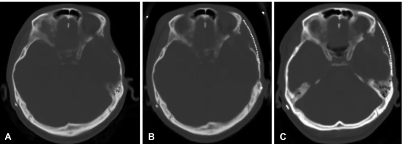
Cranioplasty was performed 43 days after decompressive craniectomy. He started rehabilitation treatment, and his condition steadily improved. However, temporal fossa hol- lowing became more prominent. The patient and his parents requested augmentation surgery for a favorable cosmetic appearance. On computerized tomography (CT) scan (Fig- ure 1A), the left temporal area showed a defect when com- pared to the right temporal area, because of previous tem- poral muscle resection. Surgery was performed under general anesthesia at 6 months after cranioplasty. Temporal fossa augmentation was performed using titanium mesh (74×50
Temporal Hollowing Augmentation with Titanium Mesh after Autologous Cranioplasty in Temporal Muscle Resected Case: A Case Report
Tae-Yong Park, MD, Ji-Hee Kim, MD, Won Seok Chang, MD, Jin Woo Chang, MD, Yong Gou Park, MD and Hyun Ho Jung, MD
Division of Stereotactic and Functional Neurosurgery, Brain Research Institute, Department of Neurosurgery, Yonsei University College of Medicine, Seoul, Korea
Temporal fossa hollowing can represent a serious cosmetic concern to patients after post-traumatic surgery, oncological re- section, or surgical dissection for obtaining access to the temporal area. Various methods have been described to augment temporal fossa hollowing, such as use of autogenous bone and cartilage implants, high-density polyethylene implants, and dermal fat grafts. We report a case of 22-year-old man with temporal fossa hollowing after post-traumatic surgery, includ- ing temporal muscle resection, whose defect was augmented by using titanium mesh even though long after cranioplasty.
(Korean J Neurotrauma 2013;9:154-156) KEY WORDS: Temporal fossa hollowing ㆍAugmentation ㆍTitanium mesh.
Received: August 18, 2013 / Revised: September 21, 2013 Accepted: September 21, 2013
Address for correspondence: Hyun Ho Jung, MD
Division of Stereotactic and Functional Neurosurgery, Brain Re- search Institute, Department of Neurosurgery, Yonsei University College of Medicine, 50 Yonsei-ro, Seodaemun-gu, Seoul 120- 752, Korea
Tel: +82-2-2228-2150, Fax: +82-2-393-9979 E-mail: [email protected]
CASE REPORT
Korean J Neurotrauma 2013;9:154-156
pISSN 2234-8999 / eISSN 2288-2243 http://dx.doi.org/10.13004/kjnt.2013.9.2.154
Tae-Yong Park, et al.
http://www.kjnt.org 155 mm, Jaeil Medical Corp., Seoul, Korea), which was fas-
tened at the zygomatic arch, lateral orbital rim, and frontal and temporal bone with screws (Figure 1B, 2). After 6 months, comparative CT showed that the cosmetic im- provement persisted throughout the outpatient follow-up period (Figure 1C).
Discussion
In the field of neurosurgery, temporal fossa hollowing can be observed after oncological resection, post-traumat- ic resection for providing sufficient decompression, or sur- gical dissection for obtaining access to the temporal area.1) Temporal fossa hollowing that occurs after surgical dissec- tion for the pterional approach, bicoronal, or Dandy incision is theorized to be associated with atrophy of the superficial temporal fat pad secondary to ischemia, displacement, or denervation of the fat pad.8) Temporal fossa hollowing may also occur because of temporalis muscle resection for achiev- ing further decompression as same with in our case.
Various methods have been described to augment the defect, including the use of HDPE implant, polymaleinate ionomeric prosthesis (IONOS®) bone cement, methyl meth- yl methacrolate, lipotransfer, autogenous bone and carti- lage.2-4,6,8,9,11,12,15,16)
Maas et al.9) suggested that autogenous bone and carti- lage were proper materials for covering temporal fossa hollowing. However, autogenous bone and cartilage im- plants are associated with considerable donor site morbidi- ty, increased surgical time and complexity, the possibility of graft resorption, as well as difficulties in controlling appro- priate graft volume.14)
More recently, many synthetic alloplastic materials have been developed and used. The ideal alloplastic materials have been described as inert, noncarcinogenic, noninflam- matory, and nonallergenic. The material should resist me- chanical strain and be easily malleable.1,6)
Porous HDPE implants were developed in the early 1970s.6) HDPE is flexible at room temperature, and when heated in hot water, it becomes malleable. These characteristics of
A B C
FIGURE 1. Sequential brain computerized tomography (CT) scan. A: Before the surgery for temporal hollowing, the left temporal area shows a defect compared to the right temporal area, because of previous temporal muscle resection. B: Two months after op- eration, the scan shows temporal fossa hollowing disappeared by augmentation using titanium mesh. C: Six months after opera- tion, the cosmetic improvement persisted throughout the outpatient follow-up period.
FIGURE 2. Immediate postoperative skull anteroposterior and lateral X-ray shows temporal fossa augmentation us- ing titanium mesh. The mesh was fas- tened with screw on frontal and parietal bone and lateral orbital limb, and zygo-
matic arch. A B
156 Korean J Neurotrauma 2013;9:154-156
Temporal Hollowing Augmentation with Titanium Mesh after Autologous Cranioplasty in Temporal Muscle Resected Case
HDPE allow surgeons to maneuver the material with ease.
Several authors have demonstrated rapid ingrowth of fi- brous tissue through the characteristic porous structure of these materials, resulting in firm attachment and integra- tion of the implant to the surrounding tissue. Furthermore, the rapidity of vascularized tissue ingrowth makes these materials more resistant to infection. However, although they are produced in a number of sizes, which can be shaped, their rigid nature can make it rather difficult to contour to complex surfaces and high cost problem.6,11,15) As in our case, we could not get firm fixation with HDPE because the hol- lowing is not due to bony defect but is partial resection of temporalis muscle.
The use of dermal fat grafts for restoring facial contour was first reported in 1931 by Figi,5) and its success has been well documented. In general, grafts are harvested from any preexisting abdominal scar in which the tissue is com- posed of a deep dermis layer and a subdermal fat layer. The graft is sutured securely below with its dermis facing super- ficially. In this way, tissue perfusion in this region is suffi- cient to allow graft acceptance. McNichols et al.10) reported no case of graft loss or fat necrosis. However, most neuro- surgeons are not familiar with this technique, which is more recommended only when the patients have a scar in abdo- men skin.
The use of Mersilene mesh for craniofacial reconstruc- tion has been reported since 1976, when Weidenbecher et al.13) described its use in skull sinus reconstruction. This mesh is composed of polyester with interlocked fiber junc- tions. This characteristic allows it to be cut without fold- ing the edges, while maintaining its easy maneuverability.
It is smooth and pliable, and its elastic properties allow it to adapt to mechanical stresses and contour well to under- lying defect, producing a soft reconstruction.1) There were several reports of temporal hollowing augmentation using titanium mesh, but the procedures were usually done by plastic surgeons with complication of zygomaticotemproal fractures or its repair.7) In our case, the temporal hollowing was iatrogenic cause for the decompression, resecting tem- poralis muscle, during severe brain swelling in traumatic brain injury management. In such case, because of loss of temporal muscle, rather rigid titanium mesh would be much easier to make contour using zygomatic arch than using Mersilene mesh. Though there was time interval, about 6 months after cranioplasty, augmentation could be selected for the cosmetic cause. Also in case of temporalis muscle resection for decompression, temporal hollowing augmen- tation should be done at the time of cranioplasty to avoid an- other operation.
Conclusion
We have described the augmentation of temporal hollow- ing using titanium mesh after temporal muscle resected case. Augmentating with titanium mesh could be done long after the cranioplasty, but in case of temporal muscle resec- tion, augmentation should be considered in one stage with cranioplasty.
■ The authors have no financial conflicts of interest.
REFERENCES
1) Atherton DD, Joshi N, Kirkpatrick N. Augmentation of temporal fossa hollowing with Mersilene mesh. J Plast Reconstr Aesthet Surg 63:1629-1634, 2010
2) Badie B. Cosmetic reconstruction of temporal defect following pterional [corrected] craniotomy. Surg Neurol 45:383-384, 1996 3) Cheung LK, Samman N, Tideman H. The use of mouldable acryl-
ic for restoration of the temporalis flap donor site. J Craniomaxil- lofac Surg 22:335-341, 1994
4) Ducic Y, Pontius AT, Smith JE. Lipotransfer as an adjunct in head and neck reconstruction. Laryngoscope 113:1600-1604, 2003 5) Figi FA. Chronic stenosis of the larynx with special consideration
of skin grafting. 1940. Ann Otol Rhinol Laryngol 103(4 Pt 1):
249-264, 1994
6) Frodel JL, Lee S. The use of high-density polyethylene implants in facial deformities. Arch Otolaryngol Head Neck Surg 124:
1219-1223, 1998
7) Guo J, Tian W, Long J, Gong H, Duan S, Tang W. A retrospective study of traumatic temporal hollowing and treatment with titani- um mesh. Ann Plast Surg 68:279-285, 2012
8) Kim S, Matic DB. The anatomy of temporal hollowing: the super- ficial temporal fat pad. J Craniofac Surg 16:760-763, 2005 9) Maas CS, Merwin GE, Wilson J, Frey MD, Maves MD. Compari-
son of biomaterials for facial bone augmentation. Arch Otolar- yngol Head Neck Surg 116:551-556, 1990
10) McNichols CH, Hatef DA, Cole P, Hollier LH, Thornton JF. Con- temporary techniques for the correction of temporal hollowing:
augmentation temporoplasty with the classic dermal fat graft. J Craniofac Surg 23:e234-e238, 2012
11) Rapidis AD, Day TA. The use of temporal polyethylene implant after temporalis myofascial flap transposition: clinical and radio- graphic results from its use in 21 patients. J Oral Maxillofac Surg 64:12-22, 2006
12) Schick B, Draf W, Schauss F. [Augmentation of the temporal fos- sa with Ionos bone cement]. HNO 44:467-470, 1996
13) Weidenbecher M, Waller G, Lehmann W. [Mersilene mesh in re- construction of the osseous walls of the pneumatised skull (au- thor’s transl)]. HNO 24:351-353, 1976
14) Welling DB, Maves MD, Schuller DE, Bardach J. Irradiated ho- mologous cartilage grafts. Long-term results. Arch Otolaryngol Head Neck Surg 114:291-295, 1988
15) Worley CM, Strauss RA. Augmentation of the anterior temporal fossa after temporalis muscle transfer. Oral Surg Oral Med Oral Pathol 78:146-150, 1994
16) Wright S, Bekiroglu F, Whear NM, Grew NR. Use of Palacos R-40 with gentamicin to reconstruct temporal defects after maxillofa- cial reconstructions with temporalis flaps. Br J Oral Maxillofac Surg 44:531-533, 2006