Abstract
Purpose: The aim of the present in vitro study was to compare bacterial adhesion on four different implant abutment materials with diverse surface roughness.
Materials and Methods: The specimens of gold and zirconia were prepared and modified from the commercially available titanium rods (Ti) and TiN coating on machined titanium (TiN). Fusobacterium nucleatum, Porphyromonas gingivalis, and Treponema denticola in the saliva-coated specimens were analyzed by real-time polymerase chain reaction using the extracted DNA.
Results: F. nucleatum adhered well with an exception of gold. P. gingivalis adhered well to zirconia and T. denticola adhered more to zirconia, Ti than gold and TiN. In conclusion, bacterial adhesion in the saliva-coated specimens had less effect on surface roughness.
Conclusion: In stages of early adhesion, zirconia exhibited higher adhesion rate, but Ti coated TiN and gold materials show a low adhesion tendency.
Key Words: adhesion, implant abutment, oral bacteria
임플란트 지대주 재료에 따른 구강세균부착
이성훈1, 신현승2, 박정철2, 조인우2, 송영균3
단국대학교 치과대학 1구강미생물학교실, 2치주과학교실, 3치과보철학교실
The Adhesion of Oral Bacteria on Saliva-Coated Abutments
Sung-Hoon Lee1, Hyun-Seung Shin2, Jung-Chul Park2, In-Woo Cho2, Young-Gyun Song3
Departments of
1Oral Microbiology and Immunology,
2Periodontology, and
3Prosthodontics, College of Dentistry, Dankook University, Cheonan, Korea
ISSN 1229-5418 Implantology 2016; 20(3): 124~131
Reprint requests: Young-Gyun Song
Department of Prostodontics, College of Dentistry, Dankook University, 119 Dandae-ro, Dongnam-gu, Cheonan 31116, Korea
Tel: 82-41-550-1932, Fax: 82-41-550-1859 E-mail: [email protected]
Received for publication: August 16, 2016 Revised for publication: August 22, 2016 Accepted for publication: August 23, 2016
교신저자: 송영균
(31116) 천안시 동남구 단대로 119 단국대학교 치과대학 치과보철학교실 Tel: 82-41-550-1932, Fax: 82-41-550-1859 E-mail: [email protected]
원고접수일: 2016년 8월 16일 원고수정일: 2016년 8월 22일 게재확정일: 2016년 8월 23일
Copyright © 2016. The Korean Academy of Oral & Maxillofacial Implantology
This is an Open Access article distributed under the terms of the Creative Commons Attribution Non-Commercial License (http://creativecommons.org/licenses/by-nc/4.0/) which permits
대주에 이미 널리 사용되고 있는 실정이다.
표면 경도의 증가는 임플란트의 유지관리 시, 초음파 기구나 금속으로 된 큐렛에 의한 표면의 손상을 감소시 킬 수 있기 때문에 유리하다9,10. 지대주 표면의 손상은 표 면 거칠기를 증가시키고 세균의 부착을 용이하게 하기 때문에11, 표면의 견고함은 장기적인 예후에 유리할 것으 로 예상된다.
TiN 코팅은 치은을 통해서 투과되어 보일 때, 기존의 티타늄 지대주에 비하여 치은의 색을 보다 정상적으로 보이게 하므로 심미적으로도 유리하다. 최근 많은 임플 란트 제조사들이 지대주를 TiN으로 코팅하고 있다. 비슷 한 이유로 ZrN 코팅도 지대주의 기계적 성질을 개선시키 는 방법으로 연구되고 있다.
임플란트 표면에 대한 세균 부착에 관해서는 연구가 많지 않지만, 세균의 부착은 세균의 종류, 임플란트 표면 의 물리, 화학적 특징, 그리고 구강 분비액의 도말 여부 등에 좌우되는 것으로 알려지고 있다12-15. 상이한 임플란 트 재료에 대한 구강 세균 부착에 대한 연구에 따르면, 재료의 표면 거칠기는 표면의 자유에너지보다 더 중요하 며, 이는 재료에 따라 세균의 부착 정도가 차이가 있다는 것을 의미하는 것이다13.
본 연구에서는 이러한 여러 가지 지대주 재료에 치주 세균이 부착하는 정도를 비교분석해 보고자 하였다.
치과 임플란트 술식의 보편화와 더불어 구강 내 임플란트 주위 감염과 연관된 세균총에 대한 연구는 비교적 활발히 진행되어 왔다. 임플란 트와 관련한 치주조직의 미생물에 대한 기존의 연구에 따르면 건강한 상태의 치주조직 및 치유단계에 있는 치 주조직에서는 치주질환에 이환된 조직과 비교하여 적은 숫자의 세균이 치은 열구 내에 존재하며 분포하는 세균 군에 있어서 다른 양상을 보인다1,2.
치주염은 치은 주변에서 기인하는 경우가 대부분이며, 임플란트의 경우 치은과 접하고 있는 임플란트의 지대주 부근에서 임플란트 주위염이 기인한다. 기존의 임플란트 지대주는 금주조합금이 대부분을 차지하였으나, 최근에 는 재료 및 표면처리도 매우 다양해지고 있다. 초기에는 임플란트 치료가 완전무치악자를 위한 치료에서 시작되 었기 때문에 임플란트 매식체와 마찬가지로 기계적으로 가공된 순수 티타늄이 임플란트 지대주의 주된 재료였 다. 그러나 티타늄 지대주는 다양한 임상적 상황에 유연 하게 적용되는 데에는 한계가 있었다. UCLA 지대주는 금합금을 주조하여 각각의 임상적 상황에 맞도록 제작할 수 있도록 고안된 지대주이다3,4. 이 지대주의 형태나 각 도는 임의로 조절될 수 있기 때문에 식립된 매식체의 각 도나 위치가 원래 치아의 위치에서 많이 벗어난 경우, 또 는 전치부에서 임플란트 보철물의 emergence profile을 자연치와 유사하게 모방할 수 있기 때문에 심미적인 보 철물의 제작을 위해서도 이용되었다.
최근 기성 지대주에 사용되는 titanium nitride (TiN)와 zirconium nitride (ZrN)와 같은 질화물 코팅 기술을 적용 하고 있다. 질화물 코팅 기술은 우선 금속 구조물을 금빛
Original Article
II
연구재료 및 방법1. 시편의 준비
Gold, Ti, Ti-TiN, zirconia 4가지 시편을 직경 4 mm, 높이 1 mm의 디스크 형태로 제작하였다.
1) Ti: 임플란트 가공을 위해 상업적으로 유통되는 직 경 4 mm의 원통형 티타늄 봉을 절삭 가공하여 제작하였 다.
2) Gold: 직경 4.2 mm, 높이 1.2 mm의 왁스패턴을 이 용한 금주조를 시행하여 연마 후 직경 4 mm, 높이 1 mm 에 가장 근접한 시편을 제작하였다.
3) Ti-TiN: 위의 Ti 시편 위에 TiN을 증착시켰다.
4) Zirconia: 치과용 computer aided design/computer aided manufacturing (CAD/CAM)을 이용하여 직경 4 mm 높이 1 mm의 시편을 제작하였다.
2. 세균의 준비
Fusobacterium nucleatum
ATCC 25586과Porphyromonas gingivalis
W50은 5 mg/ml의 hemin과 0.1 μg/μl의 vitamin K가 첨가된 brain heart infusion 배지를 이용하여 배양하였다.
Treponema denticola
ATCC 35321은 OMIZ-Pat 배지를 이용하였다. 모든 세균은 37oC의 혐기성 환경(5% H2, 10% CO2 및 85% N2)에서 배 양하여 준비하였다.3. 프라이머의 설계
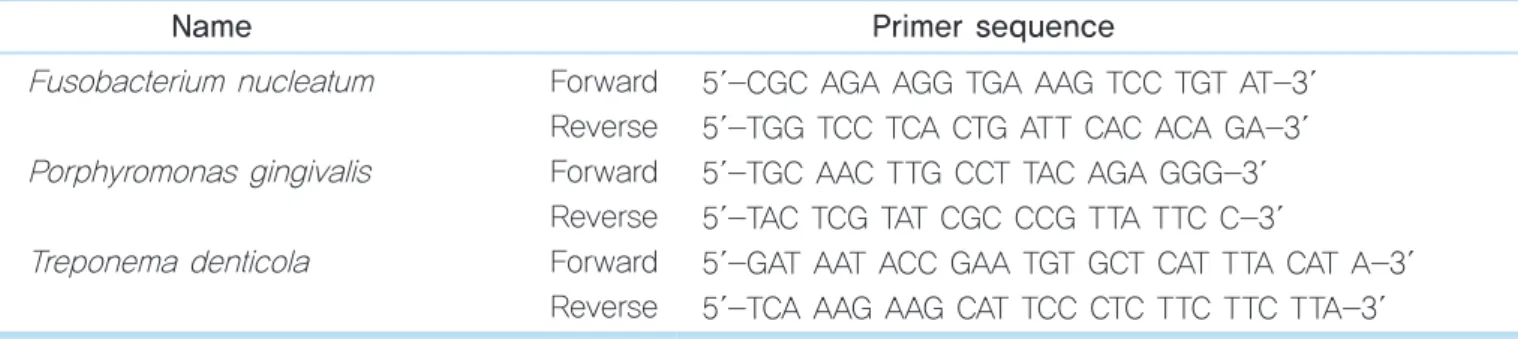
각 세균의 16S ribosomal DNA 염기서열을 이용하여 프 라이머(primer)를 설계하였다. Melting temperature는 60oC로 설정하였고 중합연쇄반응의 product size는 200~300 bp로 설계하였다(Table 1).
4. 정량 분석을 위한 표준 DNA 농도의 생성
세균 양에 대한 염색체의 표준곡선(standard curve)을 얻기 위하여 배양된 각 세균의 수를 Petroff-Hausser bacterial counting chamber (Hausser Scientific, Horsham, PA, USA)를 이용하여 109에서 103 범위로 10 배 단위로 측정하고, G-spinTM Genomic DNA extraction kit (iNtRON Biotech, Seongnam, Korea)를 사용하여 genomic DNA를 추출하였다.
5. 시편에의 세균 부착
건강한 성인의 비자극성 타액을 냉장 보관후 6,500
Table 1.
The primer sequences for real-time PCRName Primer sequence
Fusobacterium nucleatum
Porphyromonas gingivalis
Treponema denticola
Forward Reverse Forward Reverse Forward Reverse
5´-CGC AGA AGG TGA AAG TCC TGT AT-3´
5´-TGG TCC TCA CTG ATT CAC ACA GA-3´
5´-TGC AAC TTG CCT TAC AGA GGG-3´
5´-TAC TCG TAT CGC CCG TTA TTC C-3´
5´-GAT AAT ACC GAA TGT GCT CAT TTA CAT A-3´
5´-TCA AAG AAG CAT TCC CTC TTC TTC TTA-3´
PCR: polymerase chain reaction.
Sung-Hoon Lee et al. : The Adhesion of Oral Bacteria on Saliva-Coated Abutments. Implantology 2016
한 각 세균 100 μl (파장 660 nm에서 OD=0.6)를 식균하 였다. 혐기상태에서
T. denticola
는 48시간, 그 외의 세균 은 24시간 배양하였다. 시편을 인산완충액으로 세척하고 세균을 시편에서 분리하기 위해서 파쇄 완충액(lysis buffer; 0.5 M Tris pH 9.0, 10 mM NaCl, 20 mM ethyl- ene diamine tetraacetic acid, 1% sodium dodecyl sul- fate)을 넣고 vortexing하였다.6. 정량 실시간 중합효소연쇄반응
추출된 구강 내 미생물 DNA의 측정을 위해 정량 실시 간 중합효소연쇄반응이 실시되었다. 표준곡선의 형성을 위해 제작되었던 표준용액과 추출된 구강 내 미생물 DNA를 각각 SYBR Premix Ex Taq (Takara, Kyoto, Japan), forward primer, reverse primer, ROX Reference Dye II, 증류수와 혼합하였다. 혼합된 용액은
이 단일가닥으로 될 때의 온도(형광 소멸 온도)를 측정하 여 단일증폭생성물임을 확인하는 dissociation curve 단 계를 프로그램상으로 추가하였다. 표준곡선을 이용하여 얻어진 중합연쇄반응 생산물에서 세균 양을 측정하였다.
각 primer에 대하여 3회씩 실시하였으며 평균값을 통계 분석에 이용하였다.
7. 통계적 분석
통계적 분석을 위해 IBM SPSS Statistics version 23 (IBM Co., Armonk, NY, USA) 프로그램을 사용하였다.
통계적인 유의성은 one-way ANOVA와 multiple com- parison Scheffé test를 시행하여 유의수준 5%에서 검정 하였다.
2 10
1
0.1
0.01
0.001
0.0001
0.00001
0.000001
Rn
40 Cycle
4 6 8 10 12 14 16 18 20 22 24 26 28 30 32 34 36 38 0 1E+10 1E+09 100,000,000 10,000,000 1,000,000 100,000 10,000 1000 100 10 1
Bacterial count
40 Amplification plot
10 20 30
Log concentration of sample
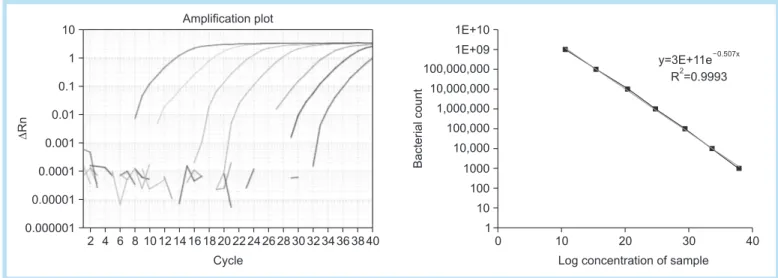
Fig. 1.
Standard curve of Fusobacterium nucleatum.Sung-Hoon Lee et al. : The Adhesion of Oral Bacteria on Saliva-Coated Abutments. Implantology 2016
Original Article
III
연구결과구강 내 미생물의 정량분석을 위해 실시간 중합효소연
쇄반응을 이용하였다. 특정 primer를 이용하여 증폭시킬 때 효과적인 증폭이 이루어지는지 알아보고 세균 수와 임계값 주기의 상관관계가 있는지 알아보기 위하여 표준 곡선을 확인하였다. 그 결과 세균 수의 log 값과 임계값 수치가 함수관계를 가짐을 확인할 수 있었다(Fig. 1).
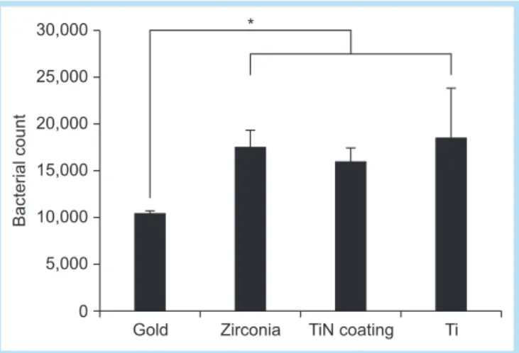
F. nucleatum
은 gold, TiN, zirconia, Ti 순으로 부착이 증가하였다. Gold는 다른 시편에 비해 세균부착수가 적 게 나타났다(p<0.05; Fig. 2).P. gingivalis
는 gold, TiN, Ti, zirconia 순으로 부착이 증가하였다. Zirconia 시편은 다른 시편에 비해 통계적으 로 유의하게 많은 부착을 보였다(p<0.05; Fig. 3).T. denticola
는 gold, TiN, zirconia, Ti 순으로 부착이 증가하였다. Gold와 TiN 코팅은 zirconia와 Ti에 비해 적 은 부착을 보였다(p<0.05; Fig. 4).Gold 30,000
25,000
20,000
15,000
10,000
5,000
0
Bacterial count
*
Zirconia TiN coating Ti
Fig. 2.
Mean bacterial count of Fusobacterium nucleatum on specimens asterisks indicate significant difference (p<0.05). TiN: titanium nitride, Ti: titanium.Sung-Hoon Lee et al. : The Adhesion of Oral Bacteria on Saliva-Coated Abutments.
Implantology 2016
Gold 35,000
25,000
20,000
15,000
10,000
5,000
0
Bacterial count
*
Zirconia TiN coating Ti 30,000
Fig. 3.
Mean bacterial count of Porphyromonas gingivalis on specimens asterisks indicate significant difference (p<0.05). TiN: titanium nitride, Ti: titanium.Sung-Hoon Lee et al. : The Adhesion of Oral Bacteria on Saliva-Coated Abutments.
Implantology 2016
Gold 140,000
100,000
80,000
60,000
40,000
20,000
0
Bacterial count
Zirconia TiN coating Ti 120,000
a
b
b
a
Fig. 4.
Mean bacterial count of Treponema denticola on specimens. Means with different letters are significantly different (p<0.05). TiN: titanium nitride, Ti: titanium.Sung-Hoon Lee et al. : The Adhesion of Oral Bacteria on Saliva-Coated Abutments.
Implantology 2016
본 연구의 실험에서 추출된 구강 내 미생물 DNA 측정 을 위해 실시된 정량 실시간 중합효소연쇄반응은 fluo- rescence signal을 사용하여 미생물의 종류와 양을 확인 하는 정확성과 민감성이 높은 방법으로 효과적이고 신속 한 장점이 있어 치주 병원성 미생물의 검출에 적절히 활 용될 수 있다16,17.
임플란트 지대주는 점막을 관통하는 특성상, 이곳에 치태가 형성되면 결과적으로 임플란트 주위염을 유발하 여, 하부의 임플란트 매식체의 건강한 골유착에 위해한 영향을 줄 수 있다. 매식체의 표면이 너무 거칠면 세균의 부착이 쉽게 일어나고, 염증이 발생하는 경우, 일상적인 구강 위생 행위에 의해 염증이 조절되지 않기 때문에 예 후가 불리해진다18,19. 임플란트 지대주 주변의 세균 조성 에 대한 연구에서, 표면 거칠기가 증가함에 따라 치태 형 성이 증가하는 것이 관찰되었으나, 지대주의 표면 거칠 기가 0.2 μm 이하인 경우에는, 치은연하 또는 치은연상 의 세균 조성에는 거칠기에 따른 차이가 거의 없는 것으 로 보고되었다15,20. 그러나 타액의 여부에 따른 결과는 일 반 실험실 논문의 결과와는 다른 결과를 보였다. 타액 여 부에 따른 구강세균 부착실험에서 타액 도말하지 않은 재료들에 대한
Streptococcus mutans
의 부착은 재료의 표면 자유 에너지의 변화와 높은 상관성이 있었다. 그러 나 사용한 재료들을 타액 또는 혈청으로 도말한 경우에 는S. mutans
의 부착과 표면 자유 에너지의 변화와는 상 관성이 없었다12.이번 연구에서는 기존에 알려진 것과 다르게 zirconia 에서 세균의 부착이 높게 나타났다. 이것은 단시간에 이 루어진 실험 때문으로 생각된다. 재료에 따라 타액이 개
때문에 지르코니아의 세균 부착이 높게 나타났을 것으로 생각된다. 장기간의 실험에서는 지르코니아의 세균부착 수가 티타늄보다 월등히 적은 것으로 보고되고 있다22. Kohavi 등23은 구강에 장착된 티타늄 지대주에 타액의 아 밀라제와 혈청 알부민이 주로 흡착함을 보고하였다. 여 러 연구 보고에 따르면, 세균은 피막의 일정한 단백질에 선택적으로 부착한다. Okte 등24은 전자현미경에 의한 관 찰을 통하여, 0.14~1.00 μm의 표면 거칠기와 약 35 mN/
m의 표면 에너지를 보이는 티타늄 시편들에 대한 세균의 부착을 비교하였다. 타액 비도말 시편으로 행한 실험에 서는, 연마된 티타늄 시편에 비하여 Ti-ZrN, Ti-TiN 또 는 열산화 처리(thermal oxidation)한 티타늄(TiO2)에 세 균이 유의하게 적게 부착하였다. 한편 이 시편들을 타액 으로 도말하여 실험하면,
S. mutans
는 TiO2, Ti pol- ished, Ti-ZrN, Ti-TiN, 그리고 레이저 방사선으로 변모 시킨 티타늄(Ti-laser)의 순으로 많이 부착하였다.T.
denticola
는 가장 거친 표면의 시료인 Ti에 더 많이 부착 하고, 여타의 시료에도 그람 음성 세균 가운데서 제일 많 이 부착하였다.구강에서는 치아와 임플란트 지대주 재료들이 타액 및 치은 열구액과 같은 체액으로 젖어 있고, 이런 체액에는 여러 물질들이 들어 있어 세균의 부착에 영향을 준다23. In vivo에서 치태형성은 피막으로 덮힌 치아와 티타늄 표 면에 연쇄 구균과 여타의 조기 정착자들이 군락하면서
시작된다24,25. 치태가 성숙함에 따라 치주낭에 혐기성 환
경이 형성 되면서 영양 요구가 까다로운 그람 음성 혐기 성 세균들이 자라게 된다26. 그리고 이 혐기성 세균들이 치은 연하 치태에서 탈락될 때 어떤 세균은 표피에 부착
Original Article
하거나 또는 치은 조직을 침입할 수도 있고, 또는 치과 기구를 사용하여 청결이 한 티타늄 표면에 재정착할 수 도 있는 것이다. 본 실험의 결과는 혐기성 세균들이 피막 으로 덮힌 티타늄 표면에 직접 부착할 수 있음을 시사한 다. 또한 gold와 TiN 코팅 재료에서 세균의 낮은 부착률 을 보여줌을 알 수 있다. 이것은 표면거칠기 외에 타액이 개재된 상태에서는 타액성분 등 다른 요소가 작용함을 알 수 있었다. 일반적으로 임플란트 지대주 재료를 일정 한 수준까지 부드럽게 하면 세균의 부착과 정착을 감소 시킬 수 있다19,27. 그러나 세균의 in vitro에서 행동은 in vivo에서의 행동과 상이할 수 있다는 것을 주지해야 할 것이다. 앞으로 단순 타액이 아닌 타액의 성분에 따른 세 균부착 경향에 대한 연구가 추가적으로 이루어져야 할 것이다.
V
결론타액에 도말된 지대주 재료는 구강 내 세균부착양상이 표면거칠기와는 다른 양상으로 나타난다. 초기부착의 경 우 zirconia가 다른 재료들에 비해 더 높은 경향을 보이 며, 기성지대주의 재료인 TiN과 gold의 경우 전반적으로 낮은 세균 부착률을 보인다.
References