In patients with rheumatoid arthritis (RA), life expectancy is reduced by 3 to 10 years compared to that of the normal population, and this in- creased mortality is largely due to cardiovascular disease (CVD) caused by accelerated athero- sclerosis found in RA.1 A recent meta-analysis in- dicated that the risk of CVD-associated death and cerebrovascular disease could be as much as 50%
higher in patients with RA compared with controls.2 Enhanced vascular risk is not limited to individuals with diagnosed RA, because in- creased mortality in non-RA patients who have early inflammatory polyarthritis or elevated levels of rheumatoid factor (RF) has been reported.3
RA activity has been associated with CVD, and it is not clear whether the increased car-
Original Article
Effect of Rheumatoid Factor on Vascular Stiffness in General Population without Joint Symptoms
Ji Hyun Lee1, Hee Sang Tag2, Geun Tae Kim2, Min Jeong Kim3, Seung Geun Lee4, Eun Kyung Park4, Dong Wan Koo4
1Division of Rheumatology, Department of Internal Medicine, Maryknoll Medical Center, Busan, Korea
2Division of Rheumatology, Department of Internal Medicine, College of Medicine, Kosin University, Busan, Korea
3Department of Neurology, College of Medicine, Kosin University, Busan, Korea
4Division of Rheumatology, Department of Internal Medicine, Pusan National University Hospital, Busan, Korea
Objectives: The role of rheumatoid factor (RF) in vascular stiffness and cardiovascular risk in subjects without joint symptoms remains unclear. We investigated vascular stiffness in subjects without joint symptoms using pulse wave velocity (PWV), calculated Framingham risk scores (FRS), an estimator of cardiovascular risk, and analyzed whether vascular stiffness and FRS were affected by RF.
Methods: Two hundred forty-two subjects were included in this population-based study. RF was quantified with turbid immunometry using a cut-off of RF > 15 IU/ml to denote RF positivity. Information was then obtained on joint symptoms. Brachial-ankle PWV (baPWV) was measured using an automated device.
Results: Of the 242 subjects, 15 were RF-positive. RF-positive subjects without joint symptoms had a higher baPWV and FRS than RF-negative subjects without joint symptoms, but the difference did not reach statistical significance. However, when we stratified the subjects into two groups (group A – high RF: RF ≥ 40 IU/ml;
group B – low RF: RF < 40 IU/ml), group A showed significantly higher baPWV (1640.7 ± 179.6 ㎝/s vs. 1405.7
± 225.7 ㎝/s, P = 0.008) and FRS (25.7 ± 4.87 vs. 11.8 ± 9.6, P < 0.001). Multiple regression analysis was used to examine potential confounders, and RF exhibited significant but modest effects on baPWV (adjusted R-squared = 0.038, P = 0.030).
Conclusions: In a sample of the general population without joint symptoms, higher levels of RF were associated with increased vascular stiffness, suggesting a pathophysiologic link between RF and endothelial dysfunction.
Key Words: Pulse wave analysis, Rheumatoid factor, Vascular stiffness,
Corresponding Author: Geun Tae Kim, Division of Rheumatology, Department of Internal Medicine, College of Medicine, Kosin University, 262, Gamcheon-ro, Seo-gu, Busan 49267, Korea Tel: +82-51-990-6415 Fax: +82-51-990-3010 E-mail: gtah@hanmail.net
Received:
Revised:
Accepted:
Jul. 09, 2015 Sep. 10, 2015 Sep. 21, 2015
diovascular risk seen in RA patients is dependent on traditional cardiovascular risk factors. Del Rincon ID et al. suggested that the incidence of cardiovascular events in RA patients might ac- tually be independent of traditional car- diovascular risk factors.4 In his study, the in- cidence rate ratio of cardiovascular events in RA patients was meaningful after adjusting for tradi- tional cardiovascular risk factors. Furthermore, a study in people without chronic arthritis de- scribed an association between high RF titers and increased cardiovascular and all-cause mortality after adjusting for traditional risk factors.5 Also, RA patients who are positive for anti-cyclic cit- rullinated peptide antibodies (anti-CCP Abs) have greater subclinical atherosclerosis than those who are not.6 A recent study revealed that ischemic heart disease in RA is independently associated with positive anti-CCP Abs.7
Although RF has been shown to be associated with increased cardiovascular mortality in RA, whether RF directly influences CVD remains unclear. RF is associated with smoking, which is a known cardiovascular risk factor.8 RF is present in up to 15% of elderly subjects. RF may arise through polyclonal B cell activation caused by an infection or antigen-driven proliferation of B cells associated with autoimmune diseases, suggesting that immunological factors may have a role.9 There is also the possibility that the effect of RF is mediated by inflammation, which has been found to predict CVD and mortality.
In this study, we investigated whether the pres-
ence of RF is associated with endothelial dysfunc- tion in subjects without joint symptoms or inflammation. We evaluated the effect of RF on endothelial function using pulse wave velocity (PWV), which assesses arterial stiffness by meas- uring the status of large and small arteries in the lower extremities. We also calculated the Framingham Risk Score (FRS) to estimate car- diovascular risk.10 To isolate the effect of RF, we restricted the analysis to RF-positive subjects without joint symptoms and with C-reactive pro- tein (CRP) levels within normal range.
MATERIALS AND METHODS
1. Study Population
We performed cross-sectional analysis using 242 consecutive subjects without joint symptoms who underwent brachial-ankle (ba) PWV assess- ment from January 2010 to December 2012 in our hospital for health screening (Fig. 1). At enroll- ment, musculoskeletal symptoms during the pre- ceding 12 months, including joint pain, joint swel- ling, and morning stiffness, were probed. Those who answered ‘no’ to all three questions were in- cluded in this study. We excluded subjects who were being treated for arthritis or had a history of rheumatoid arthritis. Subjects with CRP levels outside of the normal range were also excluded from the study. Subjects with autoimmune rheu- matic diseases, ANA titers above 1:160, history of thyroid diseases, palpable goiter, or abnormal
free T4 (FT4) or thyroid stimulating hormone (TSH) levels were additionally excluded. Subjects younger than 30 years or older than 74 years were excluded because of the limits on the age range in the FRS.11 Informed consent from the patients was not required because we examined the data retrospectively from medical records and de-identified it after collection to ensure patient confidentiality. This study protocol was approved by the ethics review boards (MMC/2013/09/
24-1[174]).
2. Baseline Data
Information on smoking (never, former, or cur- rent), alcohol, and medication use was obtained
from a questionnaire. Body mass index (BMI) was calculated by dividing weight in kilograms by the square of height in meters. Blood pressure (BP) was measured with a standard mercury manometer.
Hypertension was denoted by persistent blood pressures at or above 140/90 mmHg, as recom- mended by the Joint National Committee VII, or if the subject was being treated for hypertension.
3. Laboratory Evaluation
All subjects fasted for at least 12 hours at the beginning of the study before blood tests. Plasma glucose, total cholesterol, triglycerides, low-den- sity lipoprotein (LDL), and high-density lip- oprotein (HDL) were determined by standard lab- Fig. 1. Study population flowchart.
oratory procedures. The American Diabetes Association criteria were used to define Diabetes Mellitus (DM) and we considered a subject to have DM when fasting plasma glucose levels were ≥ 126 ㎎/dL on two consecutive assessments or if the subject was being treated for DM. Rheumatoid factor was quantified with turbid immunometry (Advia 1800, Siemens) using a cut-off of RF > 15 IU/ml to designate RF-positivity. The plasma con- centration of high-sensitivity CRP (hsCRP) was measured by performing fully automated turbid immunometry (Advia 1800, Siemens). The study participants were also subdivided into two groups based on RF (group A: RF ≥ 40 IU/ml; group B:
RF < 40 IU/ml).
4. FRS Measurement
The FRS was determined by summing the Framingham points assigned to each risk factor, such as age, LDL, HDL, BP (regardless of using antihypertensive medications), cigarette smok- ing, and DM.
5. Measurement of Arterial Stiffness
Arterial stiffness was assessed by measuring the baPWV using an automatic waveform analyzer (VP-1000; Colin Co., Komaki, Japan). The VP-1000 simultaneously records pulse waves, blood pressure (BP; both arms and ankles), an- kle-brachial pressure index (ABI), ECG, and heart sounds, as described elsewhere.12 ABI was calcu- lated as the ratio of ankle systolic BP to arm sys- tolic BP, with the lowest measured values of the
ankle systolic BP used for the calculation. For the measurement of baPWV, pulse waves obtained from the brachial and tibial arteries were re- corded simultaneously and the transmission time was defined by the time interval between the ini- tial rise in brachial and tibial waveforms. The transmission distance from the arm to each ankle was calculated using body height. The baPWV was automatically computed as the transmission dis- tance divided by the transmission time. All partic- ipants included in the present study had a normal ABI ( > 0.9). High baPWV values were defined as those in the gender-specific highest quartile among the study subjects [baPWV (the mean of the right and left values) ≥ 1,490 ㎝/s in females].
6. Statistical Analysis
Statistical analysis was performed using SPSS for Windows version 21.0 (Chicago, IL). Results are presented as the mean ± standard deviation (SD) or percentage. Because the number of RF-positive subjects and subjects with RF ≥ 40 IU/ml was rela- tively small and the assumption of the gen- eral-linear models was not met, comparisons be- tween those groups were performed using the Mann-Whitney U test. Correlation coefficients were calculated using the Spearman correlation tests. Statistical significance was defined as a P-value < 0.05.
RESULTS
1. Clinical Characteristics of Subjects
Two hundred forty-two subjects (164 men, 78 women, 51.2 ± 8.9 years) were included in this population-based study. Of the 242 subjects, 15 were RF-positive (11 men and 4 women).
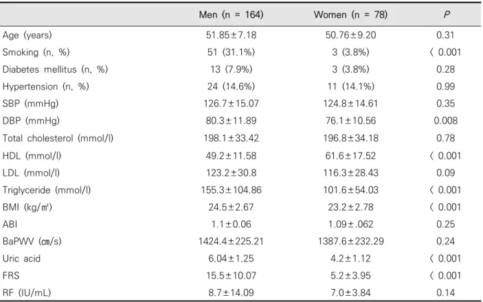
Traditional risk factors, including smoking, ele- vated diastolic BP, TG, and BMI, were significantly more frequent in men than in women.
Furthermore, HDL was significantly lower and FRS was significantly higher in men than in women.
The baseline characteristics of participants are shown in (Table 1).
2. Association between RF and Arterial Stiffness Systolic and diastolic BP, BMI, HDL, LDL, total cholesterol, and triglycerides showed no correla- tion with the presence of elevated RF levels.
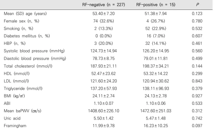
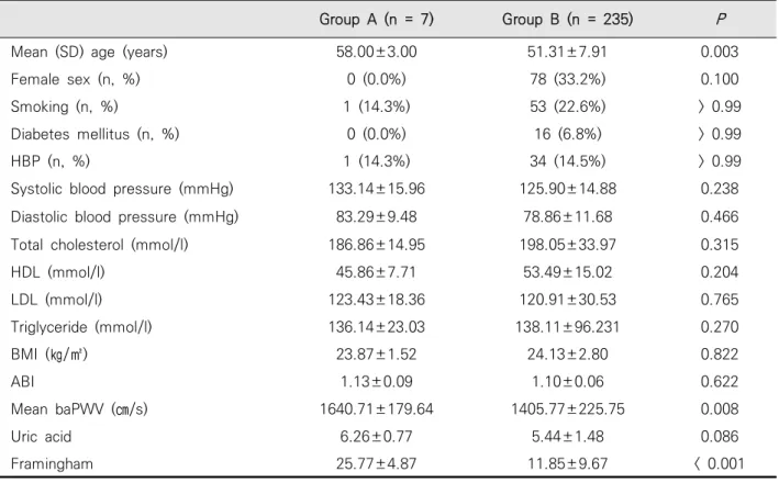
RF-positive subjects without joint symptoms had a higher baPWV and FRS than RF-negative sub- jects, but neither comparison reached statistical significance (Table 2). The study participants were further subdivided into two groups based on the RF value (group A, RF ≥ 40 IU/ml; group B, RF
< 40 IU/ml). Group A was significantly older. There were no significant differences in SBP, DBP, total cholesterol, HDL, LDL, TG, uric acid, BMI, or ABI
Men (n = 164) Women (n = 78) P
Age (years) 51.85±7.18 50.76±9.20 0.31
Smoking (n, %) 51 (31.1%) 3 (3.8%) < 0.001
Diabetes mellitus (n, %) 13 (7.9%) 3 (3.8%) 0.28
Hypertension (n, %) 24 (14.6%) 11 (14.1%) 0.99
SBP (mmHg) 126.7±15.07 124.8±14.61 0.35
DBP (mmHg) 80.3±11.89 76.1±10.56 0.008
Total cholesterol (mmol/l) 198.1±33.42 196.8±34.18 0.78
HDL (mmol/l) 49.2±11.58 61.6±17.52 < 0.001
LDL (mmol/l) 123.2±30.8 116.3±28.43 0.09
Triglyceride (mmol/l) 155.3±104.86 101.6±54.03 < 0.001
BMI (kg/㎡) 24.5±2.67 23.2±2.78 < 0.001
ABI 1.1±0.06 1.09±.062 0.25
BaPWV (㎝/s) 1424.4±225.21 1387.6±232.29 0.24
Uric acid 6.04±1.25 4.2±1.12 < 0.001
FRS 15.5±10.07 5.2±3.95 < 0.001
RF (IU/mL) 8.7±14.09 7.0±3.84 0.14
Data are presented as number (%) or mean SD unless otherwise indicated. P < 0.05 was considered statistically significant. SBP, Systolic blood pressure; DBP, Diastolic blood pressure; HDL, high density lipoprotein; LDL, low density lipoprotein; BMI, body mass index; ABI,ankle brachial index; baPWV, brachial ankle pulse wave velocity; FRS, Framingham risk score; RF, rheumatoid factor
Table 1. Subjects’ characteristics
between the two groups. However, significantly higher baPWV and FRS values were noted in group A when compared with those of group B (Table 3).
3. Correlation of Brachial Artery Pulse Wave Velocity Values with Clinical Features of Participants
Age (Spearman coefficient 0.355, P < 0.001), SBP (coefficient 0.631, P < 0.001), DBP (coefficient 0.455, P < 0.001), RF (coefficient 0.135, P = 0.035), and TG (coefficient 0.133, P = 0.038) showed sig-
nificant correlations with baPWV. However, baPWV did not correlate with total cholesterol, HDL, LDL, uric acid, or BMI. In multiple regression analysis, age, SBP, and RF were significant con- tributors to increased baPWV (Table 4).
DISCUSSION
In this study, we analyzed the association of RF with arterial stiffness and endothelial function in subjects without joint symptoms or inflammation RF-negative (n = 227) RF-positive (n = 15) P
Mean (SD) age (years) 53.40±7.20 51.38±7.94 0.123
Female sex (n, %) 74 (32.6%) 4 (26.7%) 0.780
Smoking (n, %) 2 (13.3%) 52 (22.9%) 0.532
Diabetes mellitus (n, %) 0 (0.0%) 16 (7.0%) 0.607
HBP (n, %) 3 (20.0%) 32 (14.1%) 0.461
Systolic blood pressure (mmHg) 124.73±14.94 126.20±14.95 0.560
Diastolic blood pressure (mmHg) 78.73±8.75 79.01±11.81 0.499
Total cholesterol (mmol/l) 187.93±21.11 198.37±34.21 0.144
HDL (mmol/l) 52.47±23.62 53.32±14.22 0.299
LDL (mmol/l) 121.60±24.20 120.94±30.62 0.843
Triglyceride (mmol/l) 137.20±57.93 138.11±96.93 0.379
BMI (㎏/㎡) 24.11±2.74 24.13±2.78 0.927
ABI 1.10±0.07 1.10±0.06 0.533
Mean baPWV (㎝/s) 1408.60±226.10 1472.60±251.03 0.312
Uric acid 5.50±1.42 5.47±1.48 0.742
Framingham 11.99±9.78 16.23±10.25 0.097
Data are presented as number (%) or mean SD unless otherwise indicated. P < 0.05 was considered statistically significant. SBP, Systolic blood pressure; DBP, Diastolic blood pressure; HDL, high density lipoprotein; LDL, low density lipoprotein; BMI, body mass index; ABI, ankle brachial index; baPWV, brachial ankle pulse wave velocity; FRS, Framingham risk score
Table 2. Baseline characteristics of RF-negative and RF-positive subjects. RF-positive subjects without joint symptoms had higher baPWV and FRS than RF-negative subjects, but neither comparison reached statistical significance
using baPWV. The main findings are as follows:
1) subjects with high levels of RF had significantly higher baPWV and FRS, reflecting 10-year car- diovascular risk, and 2) RF was identified using multiple regression analysis as a contributing fac- tor to increased baPWV.
RF is a family of autoantibodies that recognize epitopes on the Fc portion of IgG. The Fc portion of IgG is essential for complement fixation and interaction with the Fc receptor, and thus for up- take of immune complexes. A transient increase of RF is part of the normal immunoregulatory process that occurs during bacterial and viral in-
fections, probably in response to immune com- plexes containing microbial antigens. A low titer of RF can be found in 10 to 15% of healthy in- dividuals, whereas chronic persistence of high-affinity IgM-type RF at elevated titers and the presence of IgG and IgA subtypes are charac- teristic features of RA. RF can be found in lower titers in many other rheumatic autoimmune dis- eases as well.13 At the commonly used cut-off val- ue of 15 to 20 IU/mL, RF shows only moderate specificity for RA; specificity is considerably in- creased at higher titers, and several studies have found RF levels above 40 to 50 IU/mL to be quite Group A (n = 7) Group B (n = 235) P
Mean (SD) age (years) 58.00±3.00 51.31±7.91 0.003
Female sex (n, %) 0 (0.0%) 78 (33.2%) 0.100
Smoking (n, %) 1 (14.3%) 53 (22.6%) > 0.99
Diabetes mellitus (n, %) 0 (0.0%) 16 (6.8%) > 0.99
HBP (n, %) 1 (14.3%) 34 (14.5%) > 0.99
Systolic blood pressure (mmHg) 133.14±15.96 125.90±14.88 0.238
Diastolic blood pressure (mmHg) 83.29±9.48 78.86±11.68 0.466
Total cholesterol (mmol/l) 186.86±14.95 198.05±33.97 0.315
HDL (mmol/l) 45.86±7.71 53.49±15.02 0.204
LDL (mmol/l) 123.43±18.36 120.91±30.53 0.765
Triglyceride (mmol/l) 136.14±23.03 138.11±96.231 0.270
BMI (㎏/㎡) 23.87±1.52 24.13±2.80 0.822
ABI 1.13±0.09 1.10±0.06 0.622
Mean baPWV (㎝/s) 1640.71±179.64 1405.77±225.75 0.008
Uric acid 6.26±0.77 5.44±1.48 0.086
Framingham 25.77±4.87 11.85±9.67 < 0.001
Data are presented as number (%) or mean SD unless otherwise indicated. P < 0.05 was considered statistically significant. SBP, Systolic blood pressure; DBP, Diastolic blood pressure; HDL, high density lipoprotein; LDL, low density lipoprotein; BMI, body mass index; ABI, ankle brachial index; baPWV, brachial ankle pulse wave velocity; FRS, Framingham risk score.
Table 3. Comparison between high (≥ 40 IU/mL) and low (< 40 IU/mL) RF group. Significantly higher baPWV and FRS values were noted in group A when compared with those of group B
specific for RA.14,15 High titers of RF have consid- erable prognostic value because they are asso- ciated with severe RA, more rapid disease pro- gression, worse outcome, extra-articular manifes- tations, and an increased likelihood of developing CVD. We found that RF-positive subjects had a higher baPWV than RF-negative subjects, but the association was not statistically significant.
However, subjects with higher levels of RF showed significantly higher baPWV and FRS, suggesting that higher levels of RF are associated with arterial stiffness and cardiovascular mortality.
We previously reported that endothelial dys- function, as measured by decreased elastic prop- erties of the carotid artery wall, was more preva- lent in the patients with RA.16 Several studies sug-
gested that RF has been associated with increased cardiovascular mortality in subjects with RA.3,17,18 In addition, studies in people without arthritis showed an association between RF and car- diovascular mortality.5,19 Our findings add to the previous findings on effects of RF on arterial stiffness. We used baPWV to quantify the severity of arterial stiffness; baPWV is defined as the time delay between the rapid upstroke of the feet and the simultaneously recorded pulse waves in the brachial artery and tibial artery, and has been re- ported to be a good marker for arterial stiffness.20 Though the value of baPWV in predicting car- diovascular events has been suggested to be lim- ited, there are some studies showing baPWV as an independent predictor of cardiovascular death Univariate analysis Multivariate analysis
Coefficient
(β) 95% CI P R2 Coefficient
(β) P
Age 11.897 8.555-15.238 < 0.001 0.170 8.367 < 0.001
SBP (mmHg) 9.516 8.000-11.031 < 0.001 0.389 7.720 < 0.001
DBP (mmHg) 8.066 5.797-10.335 < 0.001 0.170 1.771 0.122
Total cholesterol (mmol/L) 0.442 -0.417-1.302 0.312 0.004 -0.593 0.421
HDL (mmol/L) -1.061 -2.999-0.877 0.282 0.005 -0.859 0.338
LDL (mmol/L) 0.287 -0.670-1.245 0.555 0.001 1.106 0.148
Triglyceride (mmol/L) 0.262 -0.042-0.565 0.091 0.012 -0.096 0.518
BMI (㎏/㎡) 4.100 -6.322-14.522 0.775 0.002 -7.128 0.081
RF (IU/mL) 3.743 1.341-6.145 0.002 0.038 1.194 0.030
baPWV(R2 = 0.497, adjusted R2 = 0.477 in multivariate analysis)
Data are presented as number (%) or mean SD unless otherwise indicated. P < 0.05 was considered statistically significant. SBP, Systolic blood pressure; DBP, Diastolic blood pressure; HDL, high density lipoprotein; LDL, low density lipoprotein; BMI, body mass index; RF, rheumatoid factor.
Table 4. Multiple regression analysis between baPWV and clinical parameters. Age, SBP, and RF were identified as significant contributors to increased baPWV in multiple regression analysis
and cardiac events in elderly persons in the com- munity and in patients with CVD.21,22 BaPWV measurement is noninvasive and cost effective when conducted using simple techniques, and thus has significant potential for screening applications.
Exactly how RF leads to arterial stiffness re- mains unclear. However, there are several hy- potheses concerning the vascular effect of RF.
First, RF titer levels are positively associated with age (a known cardiovascular risk factor).23 This association is found in this study, and similarly reported in other population cohorts and patients with RA. However, adjusting for age did not re- move the effect of RF on baPWV, so the effect of RF cannot be explained by age alone. There were some studies suggesting that RF is associated with other cardiovascular risk factors, including smoking, DM, and serum cholesterol levels,5,24 but we could not find any association between RF and these other cardiovascular risk factors. Second, inflammation has been reported to predict car- diovascular events. Previous studies in patients without RA pointed out the importance of in- flammation in the atherosclerotic process.25-27 The effect of RF might be explained by in- flammation, so we recruited subjects with normal levels of C-reactive protein (CRP) and without joint symptoms to reduce the possible confound- ing influence of inflammation. Finally, it is possi- ble that either immunologic factors play a role in atherosclerosis or RF has direct pathological effects on the endothelium. One study that im-
munohistochemically evaluated the role of RF in rheumatoid vascular injury suggested that vas- cular injury involves the production of RF on the endothelial cell surface in rheumatoid nodules.28
There are several limitations to this study. The first is the small number of subjects. Future studies with larger sample sizes should be undertaken to overcome this limitation. Another limitation is the cross-sectional study design, which cannot dem- onstrate causal associations. Also, it is possible that some subjects included in this study devel- oped RA after the study was completed, so some results might be driven by RA itself. We evaluated hsCRP levels only once, which did not reflect the effects of changes in the inflammatory marker over time. Finally, we did not account for all of the medications that may have some effect on en- dothelial function.
In conclusion, higher levels of RF were asso- ciated with increased arterial stiffness in a sample of the general population without joint symptoms, suggesting a pathophysiologic link between RF and endothelial dysfunction. Longitudinal studies employing larger samples are needed to de- termine the prognostic implication of increased arterial stiffness in RF-positive subjects.
Higher levels of RF were associated with in- creased vascular stiffness in the general pop- ulation, suggesting a pathophysiologic link be- tween RF and endothelial dysfunction.
REFERENCES
1. Kaplan MJ. Cardiovascular complications of rheumatoid arthritis: assessment, prevention, and treatment. Rheum Dis Clin North Am 2010;36:405-26.
2. Aviña-Zubieta JA, Choi HK, Sadatsafavi M, Etminan M, Esdaile JM, Lacaille D. Risk of car- diovascular mortality in patients with rheuma- toid arthritis: a meta-analysis of observational studies. Arthritis Rheum 2008;59:1690-7.
3. Goodson NJ, Wiles NJ, Lunt M, Barrett EM, Silman AJ, Symmons DP. Mortality in early inflammatory polyarthritis: cardiovascular mortality is in- creased in seropositive patients. Arthritis Rheum 2002;46:2010-9.
4. del Rincon ID, Williams K, Stern MP, Freeman GL, Escalante A. High incidence of car- diovascular events in a rheumatoid arthritis co- hort not explained by traditional cardiac risk factors. Arthritis Rheum 2001;44:2737-45.
5. Tomasson G, Aspelund T, Jonsson T, Valdimarsson H, Felson DT, Gudnason V. Effect of rheumatoid factor on mortality and coronary heart disease.
Ann Rheum Dis 2010;69:1649-54.
6. Gerli R, Bartoloni Bocci E, Sherer Y, Vaudo G, Moscatelli S, Shoenfeld T. Association of an- ti-cyclic citrullinated peptide antibodies with subclinical atherosclerosis in patients with rheumatoid arthritis. Ann Rheum Dis 2008;67:
724-5,
7. Lopez-Longo FJ, Oliver-Miňarro D, de la Torre I, González-Diáz de Rabago E, Sánchez-Ramón
S, Rodriquez-Mahou M, et al. Association between anti-cyclic citrullinated peptide antibodies and ischemic heart disease in patients with rheuma- toid arthritis. Arthritis Rheum 2009;61:419-24.
8. Wolfe F. The effect of smoking on clinical, labo- ratory, and radiographic status in rheumatoid arthritis. J Rheumatol 2000;27:630-7.
9. Danesh J, Wheeler JG, Hirschfield GM, Eda S, Eiriksdottir G, Rumley A, et al. C-reactive protein and other circulating markers of inflammation in the prediction of coronary heart disease. N Engl J Med 2004;350:1387-97.
10. Expert Panel on Detection, Evaluation, and Treatment of High Blood Cholesterol In Adults.
Executive Summary of The Third Report of The National Cholesterol Education Program (NCEP) Expert Panel on Detection, Evaluation and Treatment of High Blood Cholesterol In Adults (Adult Treatment Panel Ⅲ). JAMA 2001;
285:2486-97.
11. Wilson PW, D’Agostino RB, Levy D, Belanger AM, Silbershatz H, Kannel WB. Prediction of cor- onary heart disease using risk factor categories.
Circulation 1998;97:1837-47.
12. Marc CH, Alan JS, Josef SS, Michal EW, Michael HW. Rheumatology. 5th ed. p.887-95, London, Mosby, 2011.
13. Jansen AL, van der Horst-Bruinsma I, van Schaardenburg D, Van der Stadt RJ, de Koning MH, Dijkmans BA. Rheumatoid factor and anti- bodies to cyclic citrullinated peptide differ- entiate rheumatoid arthritis from undifferentiated polyarthritis in patients with early arthritis. J
Rheum 2002;29:2074-6.
14. Nell VP, Machold KP, Stamm TA, Eberl G, Heinzl H, Uffmann M, et al. Autoantibody profiling as early diagnostic and prognostic tool for rheuma- toid arthritis. Ann Rheum Dis 2005;64:1731-6.
15. Tomiyama H, Yamashina A, Arai T, Hirose K, Koji Y, Chikamori T, et al. Influences of age and gender on results of noninvasive bra- chial-ankle pulse wave velocity measurement -- a survey of 12517 subjects. Atherosclerosis 2003;166:303-9.
16. Lee JH, Cho KI, Kim SM. Carotid arterial stiffness in patients with rheumatoid arthritis assessed by speckle tracking strain imaging: its associa- tion with carotid atherosclerosis. Clin Exp Rheumatol 2012;30:720-8.
17. Gonzalez A, Icen M, Kremers HM, Crowson CS, Davis JM 3rd, Therneau TM, et al. Mortality trends in rheumatoid arthritis: the role of rheu- matoid factor. J rheumatol 2008;35:1009-14.
18. van Schaardenburg D, Hazes JM, de Boer A, Zwinderman AH, Meijers KA, Breedveld FC.
Outcome of rheumatoid arthritis in relation to age and rheumatoid factor at diagnosis. J Rheumatol 1993;20:45-52.
19. Heliövaara M, Aho K, Knekt P, Aromaa A, Maatela J, Reunanen A. Rheumatoid factor, chronic arthri- tis and mortality. Ann Rheum Dis 1995;54:811-4.
20. Boutouyrie P, Tropeano AI, Asmar R, Gautier I, Benetos A, Lacolley P, et al. Aortic stiffness
is an independent predictor of primary coronary events in hypertensive patients: a longitudinal study. Hypertension 2002;39:10-5.
21. Tomiyama H, Koji Y, Yambe M, Shiina K, Motobe K, Yamada J, et al. Brachial--ankle pulse wave velocity is a simple and independent predictor of prognosis in patients with acute coronary syndrome. Circ J 2005;69:815-22.
22. Rhee MY, Lee HY, Park JB. Measurements of arterial stiffness: methodological aspects.
Korean Circ J 2008;38:343-50.
23. Marc CH, Alan JS, Josef SS, Michal EW, Michael HW. Rheumatology. 5th ed. p.887-95, London, Mosby, 2011.
24. Wolfe F. The effect of smoking on clinical, labo- ratory, and radiographic status in rheumatoid arthritis. J Rheumatol 2000;27:630-7.
25. Libby P. Inflammation in atherosclerosis. Nature 2002;420:868-74.
26. Wick G, Knoflach M, Xu Q. Autoimmune and inflammatory mechanisms in atherosclerosis.
Annu Rev Immunol 2004;22:361-403.
27. Paoletti R, Gotto AM Jr, Hajjar DP. Inflammation in atherosclerosis and implications for therapy.
Circulation 2004;109:Ⅲ20-6.
28. Kato H, Yamakawa M, Ogino T. Complement medi- ated vascular endothelial injury in rheumatoid nod- ules: a histopathological and immunohistochemical study. J Rheumatol 2000;27:1839-47.