122 PISSN 0304-128X, EISSN 2233-9558
폴리디메틸실록산 기반 마이크로패턴 채널 시스템을 이용한 단일 세포의 극성 신호에 관한 연구
서정수* · 이찬빈* · 판이자*** · 왕잉샤이오*** · 정영미*,** · 김태진*,**,†
*부산대학교 생명시스템학과 46241 부산광역시 금정구 부산대학로63번길 2
**부산대학교 생명과학과
46241 부산광역시 금정구 부산대학로63번길 2
***캘리포니아 샌디에고 대학교 공과대학 생명공학과 92093 USA 캘리포니아 라 호야 9500길만길
(2019년 11월 26일 접수, 2019년 12월 16일 수정본 접수, 2019년 12월 17일 채택)
A Study on Single Cell Polarized Signals Using Polydimethylsiloxane-based Micropatterned Channel System
Jung-Soo Suh*, Chanbin Lee*, Yijia Pan***, Yingxiao Wang***, Youngmi Jung*,** and Tae-Jin Kim*,**,†
*Department of Integrated Biological Science, Pusan National University, 2, Busandaehak-ro 63beon-gil, Geumjeong-gu, Busan 46241, Korea
**Department of Biological Sciences, Pusan Nationl University, 2, Busandaehak-ro 63beon-gil, Geumjeong-gu, Busan 46241, Korea
***Department of Bioengineering, University of California at San Diego, La Jolla, CA 92093, USA (Received 26 November 2019; Received in revised form 16 December 2019; accepted 17 December 2019)
요 약
본 연구에서는 폴리디메틸실록산(PDMS)과 모세관-미세몰딩(MIMIC) 기술을 활용하여 마이크로패턴 채널 시스템을 제작하고, 단일 세포 수준에서 극성화 패턴으로 형성되는 분자 신호를 고해상도 세포 이미징을 통해 분석하였다. 이 과 정에서 혈소판유래성장인자(PDGF)가 처리된 세포에서는 세포 이동에 중요한 세 종류의 신호인 포스포이노시티드 3- 인산화효소(PI3K), Rac 및 액틴(Actin) 신호가 선두(front)영역에서 후미(rear)영역에 비해 강하게 활성화 하는 데 반해, 마이오신 경쇄(MLC) 신호는 비특이적 경향성을 보여주었다. 본 연구 결과는 향후 마이크로패턴의 미세환경에서 세포 극성화 신호와 세포 이동과의 상관 관계를 연구하는 데 중요한 도움이 될 것으로 사료된다.
Abstract − In this study, we produced the micropatterned channel system using polydimethylsiloxane (PDMS) and micromolding in capillaries (MIMIC) technology and evaluated cellular polarity signals through high-resolved imaging at the single-cell level. In cells treated with platelet-derived growth factor (PDGF), three types of key signals in cell migration; phosphoinositide 3-kinase (PI3 K), Rac, and Actin, were strongly activated in the front area compared to the rear region, whereas myosin light chain (MLC) showed no notable activity in the front and rear areas. Our results will, therefore, provide important information and methodology for studying the correlation between cell polarity signals and cell migration under the newly defined microenvironment.
Key words: Polydimethylsiloxane, Micromolding in capillaries, Micropattern, Cell polarity, Cell imaging
†To whom correspondence should be addressed.
E-mail: [email protected]
This is an Open-Access article distributed under the terms of the Creative Com- mons Attribution Non-Commercial License (http://creativecommons.org/licenses/by- nc/3.0) which permits unrestricted non-commercial use, distribution, and reproduc- tion in any medium, provided the original work is properly cited.
1. 서 론
최근 생체 재료와 화학 공학적 기술들을 접목한 융합 연구는 조 직 공학과 재생 의학 연구에 큰 활력을 불어넣고 있다. 이러한 융합
기술들은 다양한 세포미세환경을 적절하게 모사하도록 활용되어 세포 신호 및 기능 연구에서 많은 진전을 가져다 주었다[1-5]. 특히, 미세환경에 민감한 조직(tissue)의 발생 및 생성과정에서 세포의 방향성과 이동성에 필요한 분자적 의사 결정 기전은 매우 중요한 요소로 판단되고 있으나 아직 비밀에 싸여있다[6,7]. 뿐만 아니라, 암세포의 전이과정에서 흔히들 나타나는 형태적 극성화(polarity) 전략과 이동에 관한 분자적 신호 체계도 규명되어야 할 요소들이 매우 많다[8,9]. 그러나, 세포 행동 및 생리과정에서 동반되는 세포
의 극성 신호 연구는 일반적으로 사용되는 세포배양시스템 환경에 서는 통제가 어려워 실험적으로 연구하는데 많은 제약이 따르고 있어, 패턴화된 미세유체 환경 시스템의 적용이 필요할 것으로 사 료된다.
폴리디메틸실록산을 활용한 소프트 리소그래피(soft lithography) 기술은 패턴화가 필요한 다양한 몰드 제작에 매우 유용하게 활용되 고 있으며[10], 입체적으로 통제된 2D, 3D미세환경을 제작하거나, 세포외기질의 미세지형적인(micro-topographical) 특징을 모사하는 등 세포-세포외기질(extracellular matrx, ECM) 및 세포-세포 상호 작용 연구에 폭넓게 활용되고 있다[11,12]. 특히 최근에는 3D 프린 팅 기술과 나노리소그래피(Nanolithography) 등과 함께 세포의 마 이크로/나노 구조 환경을 정밀하게 모사 구현하는 시스템으로 크게 발전하고 있다[13,14]. 본 연구에서는 이러한 소프트 리소그래피 중 MIMIC (Micromolding in Capillaries) 기술을 활용하여 마이크로 패턴 채널시스템을 제작하고 최적화함으로써, 단일 세포(Single cell)의 극성 신호 연구에 활용하였다. 모사된 마이크로패턴 환경에 서 극성화 된 단일 세포의 단백질 분자 신호를 실시간 고해상도 이 미징을 통해 측정함으로써 단일 세포 수준에서 극성화 신호를 비교 분석하고자 하였다.
2. 실 험 2-1. 폴리디메틸실록산 몰드의 제조
폴리디메틸실록산(Polydimethylsiloxane, PDMS) 몰드의 제조는 실리콘 마스터(Silicon master)와 포토리소그래피(Photolithography) 방법을 통한 PDMS pre-폴리머의 주물 과정을 통해 제작되었다. 실 리콘 와퍼(Silicon wafer)는 piranha wet etch (H2SO4와 H2O2의 7:3 비율) 처리와 함께 3차 증류수에 의한 세척작업 후, 잔여 수분을 완 전히 제거하기 위해 섭씨 200도에서 5분간 처리과정을 거쳤다. 뒤 이어 와퍼는 포토레지스트 SU8 2015 처리 후 3000 rpm 으로 30초 간 스핀 코팅을 진행하였고 이후 섭씨 95도에서 3분간 열처리를 수 행하였다. 포토레지스트가 코팅된 와퍼는 22초간 UV 처리 (140 mJ/cm2)를 진행하였고, 반복해서 섭씨 95도에서 3분간 열처리를 진행하였다. 이후 실리콘 와퍼는 SU8 디벨로퍼 시약을 3분간 처리 후 2% SDS (sodium dodecyl sulfate) 용액을 통한 세척 및 건조 과정을 거 쳤다. 이후 PDMS pre-폴리머는 실리콘 와퍼에 부어 섭씨 60도에서 15시간 처리한 후 탄성중합체의 PDMS 몰드를 제작하였다.
2-2. MIMIC을 통한 마이크로패턴채널 제작
Glass coverslip을 piranha wet etch (H2SO4와 H2O2의 7:3 비율)로 20분 동안 처리한 후, 3차 증류수를 통해 세척 및 건조과정을 거쳤 다. 세척된 glass coverslip은 2% dimethyldichlorosilane이 포함된 chlorobenzene에서 10초간 담근 후, 아세톤, 에탄올 및 3차 증류수 로 세척 후 건조 시킨 후 UV-생성 오존에서 1분간 산화 과정을 거 쳤다. PDMS 몰드도 아세톤, 에탄올 및 3차 증류수를 통해 세척 과 정을 거친 후 silanized glass coverslip 에 놓았다. 주입구(Inlet)과 배출구(outlet)의 입구를 2 mm 직경으로 뚫은 후, Rhodamine- labeled 피브로넥틴(Fibronectin) 용액(40 μg/ml)을 20 μl 가량 주입 구에 도포하였다. 이후 시료는 진공상태를 적용하여 용액을 입구로 부터 채널로 유도한 후, 배출구로 다시 유도하는 채널의 충진 및 출 력기법을 사용하였다. 미세유체(microfluidic) 시료는 PDMS 몰드
를 제거하기 전, 피브로넥틴이 표면적에 퇴적하도록 밤새 상온에 보관되었다. 이후 유리 덮개를 petri dish로 조립하여 0.5% (w/v) Pluronic F127을 1시간 동안 도포한 후 PBS (phosphate-buffered saline)로 세척함으로써 세포 배양에 적합한 마이크로패턴 채널을 제작되었다.
2-3. 세포배양 및 DNA 플라스미드 삽입
본 연구에서는 사용된 HeLa 세포와 CHO (Chinese hamster ovary) 세포는 10% FBS (fetal bovine serum), 2 mM L-glutamine, 100 unit/
ml penicillin, 100 ug/ml streptomycin과 1 mM sodium pyruvate를 첨가한 Dulbecco’s modified Eagle’s medium (DMEM)에서 배양 되었다. 세포는 95% air와 5% CO2가 혼합된 가스가 주입되고 섭 씨 37도의 환경조건을 가진 세포배양기에서 배양되었다. DNA 플 라스미드의 삽입은 제조사의 프로토콜에 따라 Lipofectamine 2000 (Invitrogen)을 통해 수행되었다. 사용된 DNA 플라스미드는 PH- AKT-GFP, PAK-PBD-YFP, MLC-GFP, Actin-GFP이며 Addgene으 로부터 구입하였다.
2-4. 세포이미징 및 분석
DNA 플라스미드가 주입된 세포는 0.5% FBS가 함유된 CO2-비 의존성 배양액(Gibco BRL)을 채운 후, Zeiss Axiovert 도립형광현 미경(40x objective, CCD-Cascade 512B; Photometrics) 하에서 MetaFluor 6.2 software를 활용하여 이미징 및 분석 실험이 수행되었다. 성장 인자에 대한 단일 세포 극성 신호 연구를 위해 세포는 재조합 rat platelet-derived growth factor (PDGF, 50ng/ml) (Sigma Aldrich)가 1시간 처리되었다.
2-5. 통계분석
모든 통계 데이터는 평균 및 표준오차로 표현되었다. 그룹 간 통 계적 차이를 검증하기 위해 Student’s t-test을 마이크로소프트 엑셀 소프트웨어를 사용하여 분석하였다. 통계상 유의적 차이는 P-값 (*p<0.05, **p<0.01)에 의해 판정하였다.
3. 결과 및 고찰 3-1. 마이크로패턴 채널에서의 극성화 된 세포
실레인화 유리(silanized glass)의 상부에 마이크로패턴 형태를 갖춘 미세환경을 제작하기 위해, 본 연구자는 Fig. 1에서 보인 바와 같이 MIMIC(Micromolding in Capillaries)기법과 채널아웃가스기 술(channel-outgas techniques)을 응용하였다. 채널아웃가스 기술은 피브로넥틴 용액을 채널의 주입구에 가한 후 마이크로채널의 통로 로 스며들게 하고, 기압의 변화를 통해 피브로넥틴 용액을 배출구 까지 흐르도록 하였다. 피브로넥틴은 가장 대표적인 세포외기질 (ECM) 단백질로 세포가 표면에 접착할 때 필요한 단백질이다. 초 기 접착 단계에서 피브로넥틴과 같은 ECM 단백질을 코팅해주지 않게 되면 세포는 잘 접착하지 않게 된다. 마이크로채널 내에 피브 로넥틴 층이 형성되고 건조 시킨 후 폴리디메틸실록산 몰드를 제거 한 후 Pluronic 127 용액을 가하게 되면 10 μm의 strip을 가진 피브 로넥틴 코팅 마이크로패턴을 제작하였다. Pluronic 127은 무독성 중합체(triblock polymer, polyethylene glycolpolypropylene glycol- polyethylene glycol)로써 피브로넥틴 흡수와 세포접착으로부터 유
리 표면을 보호하게 된다[15]. 따라서 폴리디메틸실록산 기반의 소 프트 리소그래피 기법에 의해 제작된 마이크로패턴 채널에서만 세 포는 접착하고 배양된 세포는 극성화(polarization)된 형태적 특징
을 보였다. Fig. 2에서 보여진 바와 같이 피브로넥틴 단백질은 채널 내부로 효과적으로 주입되었고 세포외기질의 구성 단백질로 유리 표면에 코팅이 되었다. 형광현미경 상에서 적색형광시료인 rhodamine 에 의해 표지된 피브로넥틴은 균일하게 잘 나타났으며(Fig. 2B), 피 브로넥틴이 코팅된 채널 지역에서만 세포는 통제되어 위치하였다 (Fig. 2C).
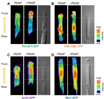
3-2. 세포의 극성 신호 측정
세포의 극성 신호를 비교 분석하기 위해 배양된 세포에서 극성화를 가지고 이동하는 세포의 front 영역과 rear 영역간의 신호 활성을 분석하였다. 혈소판유래성장인자(PDGF, platelet-derived growth factor)를 처리하게 되었을 때, 이는 PDGF 수용체와 결합하고 하위 그룹으로 신호를 전달하게 된다. Fig. 3에서 보여준 바와 같이, 세 포에 PDGF를 처리한 후, phosphoinositide 3-kinase(PI3K)의 주요 신호체계인 PH-AKT-GFP의 신호는 front 영역에서 강하게 활성화 된 데 반해, rear 영역에서는 거의 활성화 되지 않았다. AKT는 단 백질 인산화효소 B(PKB)로도 알려져 있으며 세린/트레오닌(serin/
threonine)에 특이적인 단백질 키나아제로 Pleckstrin Homology (PH) 도메인을 가지고 있어 PI3K에 의해 활성화된다. PDGF 처리 전 PH-AKT-GFP가 발현된 세포의 FIR (Fluorescence intensity ratio, front/rear) 값이 1.2129±0.079 인데 반해 PDGF 처리 후 FIR값은 1.766±0.101로 상승하였다(n=5, **p<0.01, mean±s.e.m.) (Fig. 4A).
이는 극성화 된 세포가 이동 방향을 결정할 때 PDGFR-PI3K-AKT 를 선택적으로 활성화시키는 것을 의미한다고 사료된다[16,17]. 이와 유사하게도, PAK-PBD-YFP와 Actin-GFP신호도 PDGF 처리 후 rear 영역에 비해 front 영역이 특이적으로 활성화되었다. PAK은 Fig. 1. Schematic drawing of the procedure for micromolding in capillaries and micropatterning on the silanized glass.
Fig. 2. Imaging of micropattern. (A) Fluorescence image of a micropat- tern of NHS-rhodamine-conjugated fibronectin on silanized glass. Scale bar, 20 µm. (B) Profiling analysis of the micropat- tern that matches with the line shown in (A). (C) Phase-contrast image (40x) of HeLa cells cultured on the micropatterned glass. Scale bar, 10 µm.
p21-activated protein kinase (PAK)로 p21-binding domain (PBD)를 가지고 있어서 Rac에 의해 특이적으로 결합된다. 따라서 Rac이 활 성화되면 황색형광단백질(YFP)이 표지된 PAK-PBD의 형광신호가 증가하게 된다. PAK-PBD-YFP와 Actin-GFP의 PDGF 처리 전과 후의 세포의 FIR값은 각각 1.293±0.073과 1.576±0.087 (n=6, *p<0.05, mean±s.e.m.), 1.099±0.048과 1.332±0.084(n=5, *p<0.05, mean±s.e.m.) 로 측정되었다(Fig. 4B와 C). PAK-PBD는 Rac 신호의 표지자이기에 Rac에 의해 매개된 PAK-PBD의 활성은 세포가 이동할 때 형성되는 라멜리포디아(Lamellipodia)의 확장에 중요하게 관여하는 신호임 이 분명하다[18,19]. Actin도 미세섬유를 형성함으로써 세포의 극 성화와 이동에 영향을 끼치며 라멜리포디아 조직에 중요한 매개자 역할을 한다[20,21]. 극성화 된 세포의 front 영역에서 라멜리포디 아의 형성이 높음에 따라 이들 형성과정에 필요한 PAK-PBD와 Actin 신호의 활성이 높은 것으로 여겨진다.
이와는 대조적으로 MLC (Myosin light chain)-GFP의 신호는 front 영역이 rear 영역과 비교하여 뚜렷한 활성을 보이지 않았다.
MLC-GFP는 PDGF 처리 전과 후에서 FIR 값이 각각 0.990±0.096과 0.963±0.055 (n=6, mean±s.e.m.)로 측정되었으며 통계 분석결과 유 의하지 않았다(Fig. 4D). MLC는 Actin의 구성과 세포의 수축 이완 운동에 중요한 기능을 하는 인자이지만[22], 극성화 된 세포에서의 front 영역에서는 특이적으로 활성화되지 않은 것으로 보아, 세포의 극성화 선택과 이동에는 비의존성 신호로 판단된다. 비록 선행연구 에서는 PDGFR이 활성화될 때 RhoA-ROCK 신호경로를 거쳐 MLC 가 활성화되는 것으로 알려져 있지만, 대부분의 연구가 일반적인 세포배양용기에서 수득 된 결과들이다[23]. 따라서 본 연구에서는 이전 연구에서는 관찰 할 수 없었던 MLC에 대한 새로운 분자적 행 동(molecular behavior)이 탐지되었고, 이러한 특성은 MLC가 스트 레스 섬유(stress fiber) 형성과 극성화가 이미 진행된 세포에서는 활성 민감도가 높지 않다고 사료된다.
4. 결 론
본 연구에서는 폴리디메틸실록산 몰드와 소프트 리소그래피 기 술을 사용하여 마이크로패턴을 제작하였다. 마이크로패턴은 세포 의 극성화 미세환경을 조성하여, 일반적인 세포배양환경에서 측정 하기 어려운 극성화 된 세포의 단백질 분자 신호의 역동성을 측정 하는 데 활용되었다. 그 결과, 네 종류의 단백질 신호인 PH-AKT, PAK-PBD, MLC, Actin의 활성을 세포의 front 영역과 rear 영역을 비교 분석하였고, PDGF 처리 후 PH-AKT, PAK-PBD 와 Actin의 신호가 front 영역에서 강하게 활성화 되는 현상을 확인하였다. 이는 세포가 극성화 전략에 따라 세포 이동 시, 신호 분자를 특이적으로 활성화 시키는 것을 의미한다고 사료된다.
감 사
이 논문은 부산대학교 국립대학육성사업(2018-2019)의 지원을 받아 연구되었음.
References
1. Lutolf, M. P. and Hubbell, J. A, “Synthetic Biomaterials as Fig. 4. The fluorescent intensity ratios of the four key signals in cell
polarization. (A-D) The bar graphs represent the polarity, or intensity ratios between the front and the rear areas as described in methods (mean±s.e.m., n=5 for AKT and Actin, n=6 for PAK and MLC, *represents P<0.05 and **P<0.01, ns: not significant). –PDGF: before PDGF; +PDGF: after PDGF stimulation.
Fig. 3. The polarization of molecular signals. CHO cells transfected with various fluorescent indicators were cultured on fibronec- tin-coated micropatterned surface (10 µm in width), with one opened end (front) and the other closed end (rear) contacting a neighboring cell. The fluorescent intensity distributions of (A) PH-AKT-GFP, (B) PAK-PBD-YFP, (C) Actin-GFP or (D) MLC-GFP before and after PDGF stimulation.
Instructive Extracellular Microenvironments for Morphogenesis in Tissue Engineering,” Nat. Biotechnol., 23(1), 47-55(2005).
2. Rice, J. J., Martino, M. M., De Laporte, L., Tortelli, F., Briquez, P. S. and Hubbell, J. A., “Engineering the Regenerative Micro- environment with Biomaterials,” Adv. Healthc. Mater., 2(1), 57- 71(2013).
3. Liu, W. F. and Chen, C. S., “Engineering Biomaterials to Control Cell Function,” Materials Today, 8(12), 28-35(2005).
4. Shoichet, M. S., “Polymer Scaffolds for Biomaterials Applica- tions,” Macromolecules, 43(2), 581-591(2009).
5. Little, L., Healy, K. E. and Schaffer, D., “Engineering Biomaterials for Synthetic Neural Stem Cell Microenvironments,” Chemical Reviews, 108(5), 1787-1796(2008).
6. Stroka, K. M., Gu, Z., Sun, S. X. and Konstantopoulos, K., “Bioen- gineering Paradigms for Cell Migration in Confined Microenvi- ronments,” Current Opinion in Cell Biology, 30, 41-50(2014).
7. McLennan, R., Dyson, L., Prather, K. W., Morrison, J. A., Baker, R. E., Maini, P. K. and Kulesa, P. M., “Multiscale Mechanisms of Cell Migration During Development: Theory and Experiment,”
Development, 139(16), 2935-2944(2012).
8. Polacheck, W. J., Zervantonakis, I. K. and Kamm, R. D., “Tumor Cell Migration in Complex Microenvironments,” Cellular and Molecular Life Sciences, 70(8), 1335-1356(2013).
9. Clark, A. G. and Vignjevic, D. M., “Modes of Cancer Cell Inva- sion and the Role of the Microenvironment,” Current Opinion in Cell Biology, 36, 13-22(2015).
10. Qin, D., Xia, Y. and Whitesides, G. M., “Soft Lithography for Micro-and Nanoscale Patterning,” Nature Protocols, 5(3), 491(2010).
11. Ghibaudo, M., Trichet, L., Le Digabel, J., Richert, A., Hersen, P.
and Ladoux, B., “Substrate Topography Induces a Crossover from 2D to 3D Behavior in Fibroblast Migration,” Biophysical Journal, 97(1), 357-368(2009).
12. Kim, D. H., Provenzano, P. P., Smith, C. L. and Levchenko, A.,
“Matrix Nanotopography as a Regulator of Cell Function,” The Journal of Cell Biology, 197(3), 351-360(2012).
13. Vozzi, G., Flaim, C., Ahluwalia, A., and Bhatia, S., “Fabrication of PLGA Scaffolds Using Soft Lithography and Microsyringe Deposition,” Biomaterials, 24(14), 2533-2540(2013).
14. Kamei, K. I., Mashimo, Y., Koyama, Y., Fockenberg, C., Nakashima, M., Nakajima, M. and Chen, Y., “3D Printing of Soft Lithogra- phy Mold for Rapid Production of Polydimethylsiloxane-based Microfluidic Devices for Cell Stimulation with Concentration Gradients,” Biomedical Microdevices, 17(2), 36(2015).
15. Dewez, J. L., Lhoest, J. B., Detrait, E., Berger, V., Dupont-Gil- lain, C. C., Vincent, L. M. and Rouxhet, P. G., “Adhesion of Mammalian Cells to Polymer Surfaces: from Physical Chemis- try of Surfaces to Selective Adhesion on Defined Patterns,” Bio- materials, 19(16), 1441-1445(1998).
16. Veevers-Lowe, J., Ball, S. G., Shuttleworth, A. and Kielty, C. M.,
“Mesenchymal Stem Cell Migration is Regulated by Fibronectin Through α5β1-integrin-mediated Activation of PDGFR-β and Potentiation of Growth Factor Signals,” J. Cell. Sci., 124(8), 1288- 1300(2011).
17. Tallquist, M. and Kazlauskas, A., “PDGF Signaling in Cells and Mice,” Cytokine & Growth Factor Reviews, 15(4), 205-213(2014).
18. Pertz, O., Hodgson, L., Klemke, R. L. and Hahn, K. M., “Spa- tiotemporal Dynamics of RhoA Activity in Migrating Cells,”
Nature, 440(7087), 1069-1072(2006).
19. Duchek, P., Somogyi, K., Jékely, G., Beccari, S. and Rørth, P.,
“Guidance of Cell Migration by the Drosophila PDGF/VEGF Receptor,” Cell, 107(1), 17-26(2001).
20. Yamaguchi, H. and Condeelis, J., “Regulation of the Actin Cyto- skeleton in Cancer Cell Migration and Invasion,” Biochimica et Biophysica Acta (BBA)-Molecular Cell Research, 1773(5), 642- 652(2007).
21. Gardel, M. L., Schneider, I. C., Aratyn-Schaus, Y. and Waterman, C. M., “Mechanical Integration of Actin and Adhesion Dynamics in Cell Migration,” Annual Review of Cell and Developmental Biology, 26, 315-333(2010).
22. Plotnikov, S. V., Pasapera, A. M., Sabass, B. and Waterman, C.
M., “Force Fluctuations Within Focal Adhesions Mediate ECM- Rigidity Sensing to Guide Directed Cell Migration,” Cell, 151(7), 1513-1527(2012).
23. Ridley, A. J., “Rho GTPases and Cell Migration,” Journal of Cell Science, 114(15), 2713-2722(2001).