PGHN
Original Article
Received:June 14, 2015, Revised:September 6, 2015, Accepted:September 17, 2015
Corresponding author: Yvan Vandenplas, Department of Pediatrics, Universitair Ziekenhuis Brussel, Laarbeeklaan 101, 1090 Brussels, Belgium.
Tel: +32-24775780, Fax: +32-24775784, E-mail: [email protected] Copyright ⓒ 2015 by The Korean Society of Pediatric Gastroenterology, Hepatology and Nutrition
This is an openaccess article distributed under the terms of the Creative Commons Attribution NonCommercial License (http://creativecommons.org/licenses/by-nc/4.0/) which permits unrestricted noncommercial use, distribution, and reproduction in any medium, provided the original work is properly cited.
Maladaptive Behavior and Gastrointestinal Disorders in Children with Autism Spectrum Disorder
Hardiono D. Pusponegoro, Sofyan Ismael, Sudigdo Sastroasmoro, Agus Firmansyah, and Yvan Vandenplas*
Department of Child Health, Medical School, University of Indonesia, Jakarta, Indonesia, *Department of Pediatrics, Universitair Ziekenhuis Brussel, Brussels, Belgium
Purpose: Various gastrointestinal factors may contribute to maladaptive behavior in children with autism spectrum disorders (ASD). To determine the association between maladaptive behavior in children with ASD and gastro- intestinal symptoms such as severity, intestinal microbiota, inflammation, enterocyte damage, permeability and ab- sorption of opioid peptides.
Methods: This observational cross-sectional study compared children with ASD to healthy controls, aged 2-10 years.
Maladaptive behavior was classified using the Approach Withdrawal Problems Composite subtest of the Pervasive Developmental Disorder Behavior Inventory. Dependent variables were gastrointestinal symptom severity index, fecal calprotectin, urinary D-lactate, urinary lactulose/mannitol excretion, urinary intestinal fatty acids binding protein (I-FABP) and urinary opioid peptide excretion.
Results: We did not find a significant difference between children with ASD with severe or mild maladaptive behavior and control subjects for gastrointestinal symptoms, fecal calprotectin, urinary D-lactate, and lactulose/mannitol ratio.
Urinary opioid peptide excretion was absent in all children. Children with ASD with severe maladaptive behavior showed significantly higher urinary I-FABP levels compared to those with mild maladaptive behavior (p=0.019) and controls (p=0.015).
Conclusion: In our series, maladaptive behavior in ASD children was not associated with gastrointestinal symptoms, intestinal inflammation (no difference in calprotectin), microbiota (no difference in urinary D-lactate) and intestinal permeability (no difference in lactulose/manitol ratio). ASD children with severe maladaptive behavior have sig- nificantly more enterocyte damage (increased urinary I-FABP) than ASD children with mild maladaptive behavior and normal children.
Key Words: Autism spectrum disorder, Intestinal inflammation, Intestinal permeability, Enterocyte damage, Urinary opioids
INTRODUCTION
Autism spectrum disorders (ASD) comprise au- tism, pervasive developmental disorders not other- wise specified (PDD-NOS) and Asperger syndrome [1]. The global prevalence of autism has risen over the years. In 2012, its prevalence was estimated at 11.3 per 1,000 children [2]. The increased prevalence suggests that genetic and evironmental factors play a role [3-5]. Children with ASD also present with ac- companying maladaptive behavior, further impair- ing the ability to learn and socialize [6].
Several theories have been postulated to explain the cause of maladaptive behavior in children with ASD, such as the behavioral theory, the sensory integration theory, the neurochemical theory and the biological theory. According to the biological theory, gastro- intestinal (GI) symptoms may contribute to the mal- adaptive behavior [7]. However, other studies have not found an association between GI symptoms and maladaptive behavior in children with ASD [8,9].
According to the neurochemical theory, impaired in- testinal permeability causes absorption of larger pep- tides t of dietary gluten and casein than normal. These peptides have a morphine-like effect on the brain, and are therefore called opioid peptides, and are hypothe- sized to cause maladaptive symptoms [10]. There is still much controversy surrounding both theories.
A link between ASD and GI disturbances has been reported in several papers, with a prevalence ranging from 23% to 70% [11,12]. These large variations in prevalence may be due to differences in criteria used to define GI symptoms, study design and character- istics of the study population. GI symptoms in chil- dren with ASD are not specific and may include food selectivity, regurgitation, constipation, chronic diar- rhea and abdominal bloating which are hypotheti- cally associated with an abnormal intestinal micro- biota, intestinal inflammation and colitis [11,12].
Increased GI permeability has been demonstrated in children with ASD [13]. Exposure to viral or bacterial pathogens may trigger the disorder in some patients [14].
The prevalence of GI symptoms in children with ASD in Indonesia is not known. Therefore, we aimed in this study to determine if there is an association be- tween the degree of maladaptive behavior in children with ASD and the severity of GI symptoms, intestinal bacteria and bacterial products, intestinal inflam- mation, intestinal permeability, enterocyte damage and urinary opioid peptide excretion. The under- standing of the association of maladaptive behavior and severity of GI problems may provide new insights for an appropriate treatment of children with ASD.
MATERIALS AND METHODS
Study design and subject recruitment
We conducted a cross-sectional observational study in children with ASD aged 2 to 10 years and age-matched normal controls. Subjects with ASD were consecutively recruited from the Anakku Clinic, Jakarta, and the Rumah Autis in Bekasi, Tangerang and Jakarta. Control subjects were found through a call among staff members and their family of the Anakku Clinic and Prodia Laboratories, located in the same building. Subjects were matched for age and socio-economic background. The study was con- ducted between July 2012 and February 2013.
Inclusion criteria for the children with ASD were children aged 2 to 10 years who had been diagnosed with autism, Asperger syndrome or PDD-NOS ac- cording to the Diagnostic and Statistical Manual of Mental Disorders, 4th edition text revision (DSM IV TR) criteria [15]. All patients had an abnormal or im- paired development of social interaction and com- munication and a markedly restricted, repetitive, and monotonous (stereotypical) repertoire of activities and interests [1,15]. Subjects with ASD were further classified into ASD with severe maladaptive behavior (“severe maladaptive ASD”) and ASD with mild mal- adaptive behavior (“mild maladaptive ASD”) using the Approach Withdrawal Problem Composite-subt- est of the Pervasive Developmental Disorder Behavior Inventory (Western Psychological Services, Torrance, CA, USA) [16]. Maladaptive behavior was classified as sensory perception impairment, ritualism and re-
sistance to change, social pragmatic impairment, se- mantic pragmatic impairment, impairment in regu- lation of awareness to surroundings, specific phobias and aggressiveness [16].
The initial diagnosis of ASD and degree of malad- aptive behavior was confirmed by the principal in- vestigator and/or two child psychologists who have been working closely with the investigator at the Anakku Clinic.
The normal controls were physically and devel- opmentally normal children aged 2 to 10 years, for whom a written parental informed consent had been obtained. Children were excluded if they had organic disorders.
The nutritional status was assessed based on the curve from the Center for Disease Control and Prevention and based on the percentage of the actual body weight (BW) in relation to to the ideal BW for body height (BH) as recommended by Indonesian Pediatric Society and the Waterlow classification [17-19].
ㆍObesity=BW ≥120% of ideal BW for BH ㆍOver-nourished=BW ≥110% and <120% of ide-
al BW for BH
ㆍWell-nourished=BW ≥90% and <110% of ide- al BW for BH
ㆍUndernourished=BW ≥70% and <90% of ide- al BW for BH
ㆍMalnourished=BW <70% of ideal BW for BH The study protocol was approved by the Medical Research Ethics Committee of The University of Indonesia Medical School. For all participants, a written informed consent of one of the parents was obtained prior to data collection.
Gastrointestinal symptoms measurement Parents were asked to complete a modified GI symptom severity index questionnaire. The cumu- lative scores from all items were recorded. All sub- jects were asked to provide fecal samples for meas- urement of fecal calprotectin using the calprotectin ELISA method (Immunodiagnostik AG, Bensheim, Germany). Urine samples were also collected to measure i) D-lactate to assess intestinal bacterial
overgrowth (D-Lactate Colorimetric Assay Kit; Ab- camBiochemicals, Cambridge, UK), ii) intestinal fat- ty acids binding protein (I-FABP) excretion to assess enterocyte damage (Human I-FABP ELISA Kit;
Hycult Biotech, Uden, The Netherlands), iii) lactu- lose/mannitol excretion ratio to assess impaired in- testinal permeability (Acetonitrile Liquid Chroma- tography Grade; Merck Inc., Whitehouse Station, NJ, USA), and iv) opioid peptides (ultra performance liquid chromatography-mass spectrometry [UPLC- MS], Xevo gen. 2; Waters, Milford, MA, USA).
Subjects were asked to fast for five hours prior to urine sampling, followed by the oral administration of a 2 mL/kg standard solution containing 5 g of lac- tulose, 2 g of mannitol and 40 g of sucrose per 100 mL (Novell Pharmaceutical Laboratories and Boehringer Ingelheim, Jakarta, Indonesia) to determine the lac- tulose/mannitol excretion ratio.
Statistical analysis
The minimum required sample size was calculated using the sample size formula for comparing the dif- ference between means of two independent samples.
Using a significance level of 0.05 and a power of 80%, we obtained a minimum sample size of 70 subjects in each group.
The objective data were correlated with the se- verity of the maladaptive behavior using one-way ANOVA for normally distributed data, and using the Kruskal-Wallis test for abnormally distributed data.
RESULTS
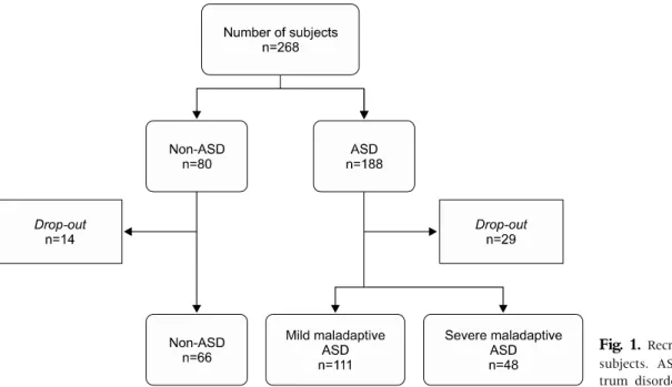
We recruited 268 subjects, of which 225 completed the study (Fig. 1). We obtained data from 66 healthy controls, 111 children with mild maladaptive ASD, and 48 children with severe maladaptive ASD.
Subject characteristics at inclusion are shown in Table 1.
Gastro-intestinal symptoms
GI symptoms were reported in 14/66 (21.2%) chil- dren in the control group and in 36/159 (22.6%) in ASD children, of whom 22/111 (19.8%) had mild
Fig. 1. Recruitment of study subjects. ASD: autism spec- trum disorders.
Table 1.Subject Characteristics Non-ASD
(n=66)
Mild maladaptive ASD (n=111)
Severe maladaptive ASD (n=48) Sex
Male 37 99 42
Female 29 12 6
Age (yr) 5.0 (2-10) 5.0 (2.1-10) 6.4 (2.1-10) Diagnosis
PDD-NOS – 52 10
Autism – 53 37
Asperger’s syndrome – 6 1
Nutritional status 38 86 30
Undernourished 3 11 4
Well-nourished 31 61 19
Overnourished 4 14 7
Values are presented as number only or median (range).
ASD: autism spectrum disorders, PDD-NOS: pervasive develop- mental disorders not otherwise specified.
maladaptive ASD and 14/48 (29.1%) had severe mal- adaptive ASD (not significant). The median GI symptom severity index was 0 in all groups (p=0.35) (Fig. 2).
Inflammation
Fecal calprotectin was measured in 60 healthy controls, 102 children with mild maladaptive ASD,
and 44 with severe maladaptive ASD. The median (range) fecal calprotectin excretion was 108.1 (5.3 to 3,595) in the control group, 100.54 (5.3 to 3,536) in the group with mild maladaptive ASD and 56.37 (5.3 to 2,778) μg/g feces in the group with severe malad- aptive ASD (p=0.80).
Gastro-intestinal microbiota
Urinary D-lactate was measured in 66 controls, 109 subjects with mild maladaptive ASD, and 47 with severe maladaptive ASD. Median (range) uri- nary D-lactate was 100.11 (41.87 to 777.48) nmol/mg creatinine in the control group, 98.7 (34.86 to 1,441.14) nmol/mg creatinine in the mild malad- aptive ASD group, and 89.26 (3.1 to 1,767.32) nmol/mg creatinine in the severe maladaptive ASD group (p=0.35).
Intestinal permeability
Urinary lactulose-mannitol excretion ratio was measured in 62 control children, 102 children with mild maladaptive ASD, and 45 children with severe maladaptive ASD. The median (range) urinary lactu- lose-mannitol excretion ratio was 3.63 (0 to 100) in the control, 3.05 (0 to 100) in the mild maladaptive
Fig. 2. Gastrointestinal disturbance, stool calprotectine level, urine intestinal fatty acids binding protein and lactulose excretion ratio in the autism spectrum disorders (ASD).
ASD, and 2.53 (0 to 43.75) in the severe maladaptive ASD subjects (p=0.45).
Opioid peptide was absent in all urine samples.
Enterocyte damage
Urinary I-FABP was measured in 64 controls, 109 children with mild maladaptive ASD and 47 patients with severe maladaptive ASD. Median (range) uri- nary I-FABP was 69.63 (21.74 to 250.82) pg/mL in the controls, 76.02 (13.24 to 930.38) pg/mL in the mild maladaptive ASD and 109.08 (29.26 to 1,454.98) pg/mL in the severe maladaptive ASD group (p=
0.01). The difference in urinary I-FABP was sig- nificant between the severe maladaptive ASD and the control groups (p=0.04) and between the severe
and mild maladaptive ASD groups (p=0.019). There was no significant difference in urinary I-FABP be- tween the mild maladaptive ASD and the control groups (p=0.94).
DISCUSSION
The goal of this study was to evaluate if an associa- tion could be found between maladaptive behavior in children with ASD children and GI symptoms, in- testinal inflammation, intestinal bacterial products, increased intestinal permeability, and abnormal ex- cretion of opioid peptides and enterocyte damage. We found that urinary I-FABP, a parameter evaluating enterocyte damage, was significantly increased in
children with ASD and severe maladaptive behavior.
Urinary I-FABP was the only parameter that was significantly different between controls, children with mild maladaptive ASD and the group with se- vere maladaptive ASD. We found a significant differ- ence between the severe maladaptive ASD and con- trol groups (p=0.04) and between the severe and mild maladaptive ASD groups (p=0.02). The differ- ence was not significant between the mild malad- aptive ASD and the control groups (p=0.94). An in- creased I-FABP reflects the presence of enterocyte impairment or damage [20]. It is elevated in the early stages of enterocyte damage and normalizes as cell regeneration occurs [20-22]. A significantly higher urinary I-FABP without increased lactulose-man- nitol excretion ratio suggests an enterocyte damage which is mild enough to preserve normal intestinal permeability.
We found that GI symptoms were present in about 20% of all children, without any difference between the healthy controls and the ASD children (p=0.35).
In contrast to our study, Smith et al. [6] showed that children with ASD presented significantly more GI symptoms when compared to normal children, but reported a comparable prevalence in children with ASD children and children with other special-need.
Another study did not find a significant difference in the incidence of constipation between children with and without ASD [8]. These results suggest that the presence of GI symptoms in children with ASD may be the result of a neurobehavioral artefact. At least three independent studies found the rs1858830 C variant in the MET gene promoter to be associated with autism [23]. The MET rs1858830 C allele was associated with both ASD and GI conditions. Dis- rupted MET signaling may contribute to increased risk for ASD that includes familial GI dysfunction [24].
Fecal calprotectin was not significantly different between controls, children with mild and severe maladaptive ASD (p=0.800). The median levels of calprotectin were similar in the three groups and in each group there were some children with clearly elevated levels. Age may be one of the contributory
factors as infants and young children below 2 years of age have been reported with increased fecal cal- protectin concentrations in the absence of any dis- ease [25]. Also, major differences exist between spe- cificity and absolute values between different tests [26]. Other factors which may explain the wide range of fecal calprotectin may be related to the han- dling of the samples and the fact that a number of these children may have chronic intestinal in- flammation caused by different microorganisms.
Anyway, these results suggest that maladaptive be- havior in children with ASD is not associated with intestinal inflammation. Similar results have been reported in a study that failed to find elevated fecal calprotectin and rectal nitric oxide in children with autism [27,28]. However, other investigators have found endoscopic and laboratory evidence of in- testinal inflammation in children with ASD [29].
The lack of a significant difference in urinary D-lactate between controls and patients with mild and severe maladaptive ASD subjects (p=0.35) sug- gests that there is no increase in bacteria or bacterial products in the GI tract of children with ASD.
Similarly, another study did not find a difference in intestinal microflora between ASD children with and without GI symptoms and controls [30].
The urinary lactulose-mannitol excretion ratio was not significantly different between controls and subjects with mild and severe maladaptive ASD (p=0.45). These results suggest that maladaptive be- havior in children with ASD is not associated with impaired intestinal permeability. Similar results have been reported previously in ASD children [13].
Acute diarrhea and malnutrition may impair in- testinal permeability and act as confounders [31]. In our study, malnutrition was present in only a few patients.
No subject showed opioid peptide excretion in the urine. These results are in contrast with those re- ported by some other investigators [13,26]. However, these studies used the standard high-performance liquid chromatography technique what may be sub- ject to false positive results as this method is unable to differentiate spectrometry peaks caused by opi-
oids and other substances [10]. We used the more specific UPLC-MS method. Two other studies using the same method did not find opioid peptides in the urine of ASD children [31,32].
Gut and dietary factors are reported to possibly worsen or improve transiently behavioral symptoms in a subset of persons with ASD. Propionic acid, a major short chain fatty acid produced by ASD-asso- ciated GI bacteria (clostridia, Bacteroides, Desulfovi- brio) and also a common food preservative, can pro- duce reversible behavioral, electrographic, neuroin- flammatory, metabolic, and epigenetic changes clo- sely resembling those found in ASD when admini- stered to rodents. More studies on the impact of the microbiota on behavioral changes are needed [33].
In conclusion, according to our results malad- aptive behavior in ASD children seems not asso- ciated with GI symptoms, intestinal inflammation, microbiota and intestinal permeability. However, we found arguments for a more important enterocyte damage in ASD children with severe maladaptive be- havior than in ASD children with mild adaptive be- havior disturbace and normal children. There is no evidence of urinary opioid peptide excretion in ASD and non-ASD children. Further research is needed on the pathogenesis of enterocyte damage in ASD children.
REFERENCES
1. Steyaert JG, De la Marche W. What's new in autism?
Eur J Pediatr 2008;167:1091-101.
2. Autism and Developmental Disabilities Monitoring Network Surveillance Year 2008 Principal Investiga- tors; Centers for Disease Control and Prevention.
Prevalence of autism spectrum disorders--Autism and Developmental Disabilities Monitoring Network, 14 sites, United States, 2008. MMWR Surveill Summ 2012;61:1-19.
3. Stilp RL, Gernsbacher MA, Schweigert EK, Arneson CL, Goldsmith HH. Genetic variance for autism screen- ing items in an unselected sample of toddler-age twins.
J Am Acad Child Adolesc Psychiatry 2010;49:267-76.
4. Hertz-Picciotto I, Croen LA, Hansen R, Jones CR, van de Water J, Pessah IN. The CHARGE study: an epi- demiologic investigation of genetic and environmental
factors contributing to autism. Environ Health Per- spect 2006;114:1119-25.
5. Currenti SA. Understanding and determining the etiol- ogy of autism. Cell Mol Neurobiol 2010;30:161-71.
6. Smith RA, Farnworth H, Wright B, Allgar V. Are there more bowel symptoms in children with autism com- pared to normal children and children with other devel- opmental and neurological disorders?: A case control study. Autism 2009;13:343-55.
7. Gorrindo P, Williams KC, Lee EB, Walker LS, McGrew SG, Levitt P. Gastrointestinal dysfunction in autism:
parental report, clinical evaluation, and associated factors. Autism Res 2012;5:101-8.
8. Ibrahim SH, Voigt RG, Katusic SK, Weaver AL, Barbaresi WJ. Incidence of gastrointestinal symptoms in children with autism: a population-based study.
Pediatrics 2009;124:680-6.
9. Mouridsen SE, Rich B, Isager T. A longitudinal study of gastrointestinal diseases in individuals diagnosed with infantile autism as children. Child Care Health Dev 2010;36:437-43.
10. Reichelt KL, Knivsberg AM. Can the pathophysiology of autism be explained by the nature of the discovered urine peptides? Nutr Neurosci 2003;6:19-28.
11. Chaidez V, Hansen RL, Hertz-Picciotto I. Gastrointes- tinal problems in children with autism, developmental delays or typical development. J Autism Dev Disord 2014;44:1117-27.
12. Wang LW, Tancredi DJ, Thomas DW. The prevalence of gastrointestinal problems in children across the United States with autism spectrum disorders from families with multiple affected members. J Dev Behav Pediatr 2011;32:351-60.
13. Kemperman RF, Muskiet FD, Boutier AI, Kema IP, Muskiet FA. Brief report: normal intestinal perme- ability at elevated platelet serotonin levels in a sub- group of children with pervasive developmental dis- orders in Curaçao (The Netherlands antilles). J Autism Dev Disord 2008;38:401-6.
14. Handen BL, Melmed RD, Hansen RL, Aman MG, Burnham DL, Bruss JB, et al. A double-blind, place- bo-controlled trial of oral human immunoglobulin for gastrointestinal dysfunction in children with autistic disorder. J Autism Dev Disord 2009;39:796-805.
15. American Psychiatric Association. Diagnostic and stat- istical manual of mental disorders IV-TR. Washington DC: American Psychiatric Association; 2000.
16. Cohen IL, Sudhalter V. PDD behavior inventory. Pro- fessional manual. Florida: Psychological Assessment Resources, Inc.; 2005.
17. Centers for Disease Control and Prevention, National
Center for Health Statistics. Clinical Growth Chart:
United States [Internet]. Atlanta (GA): Centers for Disease Control and Prevention; 2000 [cited 2012 June 1]. Available from: http://www.cdc.gov/growthcharts.
18. Sjarif DR, Lestari ED, Mexitalia M, Nasar SS. Textbook of pediatric nutrition and metabolic diseases. Jakarta:
Indonesian Pediatric Society; 2011:36-48.
19. Waterlow JC. Classification and definition of pro- tein-calorie malnutrition. Br Med J 1972;3:566-9.
20. Vreugdenhil AC, Wolters VM, Adriaanse MP, Van den Neucker AM, van Bijnen AA, Houwen R, et al. Additio- nal value of serum I-FABP levels for evaluating celiac disease activity in children. Scand J Gastroenterol 2011;46:1435-41.
21. Grootjans J, Thuijls G, Verdam F, Derikx JP, Lenaerts K, Buurman WA. Non-invasive assessment of barrier integrity and function of the human gut. World J Gastrointest Surg 2010;2:61-9.
22. Dettmer K, Hanna D, Whetstone P, Hansen R, Ham- mock BD. Autism and urinary exogenous neuropep- tides: development of an on-line SPE-HPLC-tandem mass spectrometry method to test the opioid excess theory. Anal Bioanal Chem 2007;388:1643-51.
23. Jackson PB, Boccuto L, Skinner C, Collins JS, Neri G, Gurrieri F, et al. Further evidence that the rs1858830 C variant in the promoter region of the MET gene is as- sociated with autistic disorder. Autism Res 2009;2:
232-6.
24. Campbell DB, Buie TM, Winter H, Bauman M, Sutcliffe JS, Perrin JM, et al. Distinct genetic risk based on asso- ciation of MET in families with co-occurring autism and gastrointestinal conditions. Pediatrics 2009;123:1018- 24.
25. Savino F, Castagno E, Calabrese R, Viola S, Oggero R, Miniero R. High faecal calprotectin levels in healthy, ex- clusively breast-fed infants. Neonatology 2010;97:
299-304.
26. Prell C, Nagel D, Freudenberg F, Schwarzer A, Koletzko S. Comparison of three tests for faecal calpro- tectin in children and young adults: a retrospective monocentric study. BMJ Open 2014;4:e004558.
27. Fernell E, Fagerberg UL, Hellström PM. No evidence for a clear link between active intestinal inflammation and autism based on analyses of faecal calprotectin and rectal nitric oxide. Acta Paediatr 2007;96:1076-9.
28. Buie T, Campbell DB, Fuchs GJ 3rd, Furuta GT, Levy J, Vandewater J, et al. Evaluation, diagnosis, and treat- ment of gastrointestinal disorders in individuals with ASDs: a consensus report. Pediatrics 2010;125 Suppl 1:S1-18.
29. de Magistris L, Familiari V, Pascotto A, Sapone A, Frolli A, Iardino P, et al. Alterations of the intestinal barrier in patients with autism spectrum disorders and in their first-degree relatives. J Pediatr Gastroenterol Nutr 2010;51:418-24.
30. Gondalia SV, Palombo EA, Knowles SR, Cox SB, Meyer D, Austin DW. Molecular characterisation of gastro- intestinal microbiota of children with autism (with and without gastrointestinal dysfunction) and their neuro- typical siblings. Autism Res 2012;5:419-27.
31. Boaz RT, Joseph AJ, Kang G, Bose A. Intestinal perme- ability in normally nourished and malnourished chil- dren with and without diarrhea. Indian Pediatr 2013;
50:152-3.
32. Cass H, Gringras P, March J, McKendrick I, O'Hare AE, Owen L, et al. Absence of urinary opioid peptides in chil- dren with autism. Arch Dis Child 2008;93:745-50.
33. MacFabe DF. Enteric short-chain fatty acids: microbial messengers of metabolism, mitochondria, and mind:
implications in autism spectrum disorders. Microb Ecol Health Dis 2015;26:28177.