ABSTRACT
Purpose: Non-coding RNA activated by DNA damage (NORAD) has been reported to be a cancer-related long non-coding RNA (lncRNA) implicated in the progression of several cancers; however, its role in breast cancer (BC) has not yet been clarified.
Methods: Quantitative real-time polymerase chain reaction was used to examine NORAD, microRNA (miR)-155-5p, and suppressor of cytokine signaling 1 (SOCS1) mRNA expression levels. Western blotting was used to analyze SOCS1 protein expression. The malignancy of BC cells was assessed using the cell counting kit-8 (CCK-8), BrdU, and Transwell assays.
Bioinformatics analysis, RNA immunoprecipitation assay, and dual-luciferase reporter gene assays were used to verify the targeted relationship between NORAD and miR-155-5p.
Additionally, the regulatory effects of NORAD and miR-155-5p on SOCS1 expression were determined by western blotting.
Results: NORAD expression was significantly reduced in BC cell lines and tissues, and its low expression was associated with poor tumor tissue differentiation. NORAD overexpression repressed BC cell proliferation, migration, and invasion, whereas its knockdown produced the opposite effects. Additionally, miR-155-5p was found to be a target of NORAD, and the biological functions of miR-155-5p and NORAD were counteractive. MiR-155-5p was confirmed to target SOCS1, and SOCS1 was found to be positively regulated by NORAD.
Conclusion: NORAD suppresses miR-155-5p to upregulate SOCS1, thereby repressing the proliferation, migration, and invasion of BC cells.
Keywords: Breast neoplasms; Cell proliferation; Metastasis
INTRODUCTION
Breast cancer (BC) is one of the most common causes of cancer-related deaths among women [1]. Although radical surgery, radiotherapy, chemotherapy, targeted-therapy, and endocrine therapy have reduced the mortality of patients with BC, treatment failures are still common in clinical practice [2,3]. Therefore, there is an urgent need to decipher the mechanism of BC progression and identify novel therapeutic targets.
Original Article
Received: Sep 28, 2020 Revised: May 9, 2021 Accepted: May 30, 2021 Correspondence to
Aijun Chen
Department of General Surgery, The First College of Clinical Medical Science, China Three Gorges University, Yichang Central People's Hospital, No. 183 Yiling Avenue, Wujiagang District, Yichang 443000, China.
E-mail: [email protected]
© 2021 Korean Breast Cancer Society This is an Open Access article distributed under the terms of the Creative Commons Attribution Non-Commercial License (https://
creativecommons.org/licenses/by-nc/4.0/) which permits unrestricted non-commercial use, distribution, and reproduction in any medium, provided the original work is properly cited.
ORCID iDs
Weipeng Liu
https://orcid.org/0000-0001-5783-600X Xin Zhou
https://orcid.org/0000-0001-7403-1481 Yuanqiang Li
https://orcid.org/0000-0002-8262-2983 Hong Jiang
https://orcid.org/0000-0002-1814-2184 Aijun Chen
https://orcid.org/0000-0002-0372-902X
Conflict of InterestThe authors declare that they have no competing interests.
Weipeng Liu , Xin Zhou , Yuanqiang Li , Hong Jiang , Aijun Chen
Department of General Surgery, The First College of Clinical Medical Science, China Three Gorges University, Yichang Central People's Hospital, Yichang, China
Long Non-Coding RNA NORAD Inhibits Breast Cancer Cell Proliferation and Metastasis by Regulating miR-155-5p/
SOCS1 Axis
Author Contributions
Conceptualization: Liu W, Chen A; Data curation: Liu W, Chen A; Formal analysis:
Zhou X; Investigation: Zhou X, Li Y, Jiang H;
Methodology: Zhou X, Li Y, Jiang H; Validation:
Li Y; Visualization: Jiang H; Writing - original draft: Liu W, Zhou X, Chen A; Writing - review
& editing: Liu W, Chen A.
Long non-coding RNAs (lncRNAs) are transcripts longer than 200 nucleotides in length that do not encode proteins [4]. Over the past few years, several studies have indicated that lncRNAs are vital in regulating tumor growth and metastasis [5]. For instance, lncRNA TUC338 accelerates lung cancer progression by regulating the mitogen-activated protein kinase pathway [6]. Furthermore, lncRNA MALAT1 inhibits BC metastasis, while lncRNA ITGB2-AS1 facilitates BC cell metastasis by upregulating ITGB2 [7,8]. As a member of the lncRNA family, non-coding RNA activated by DNA damage (NORAD) is associated with the progression of multiple cancers, such as gastric, pancreatic, and thyroid cancers [9].
However, the role of NORAD in BC remains controversial, as high NORAD expression was reported to predict poor prognosis in patients with BC and activate the transforming growth factor (TGF)-β pathway to drive the metastasis of BC cells [10]; whereas another study reported that NORAD functioned as a tumor suppressor in BC by repressing S100P-elicited pro-metastatic signaling [11]. Therefore, its role in BC is not fully understood.
In this study, we demonstrated that NORAD was downregulated in BC tissues and cell lines, and functionally, NORAD suppressed the malignancy of BC cells, which indicated that NORAD functions as a tumor-suppressor. Mechanistically, NORAD functions as a competing endogenous RNA (ceRNA) for miR-155-5p, reduces its expression, and positively regulates suppressor of cytokine signaling 1 (SOCS1).
METHODS
Tissue samples and ethics statement
BC and adjacent tissue samples were obtained from 46 patients who underwent surgery.
The tissues were immediately stored in liquid nitrogen following resection. The collection and use of human tissue samples were authorized by the Institutional Review Board (IRB) of Yichang Central People's Hospital (IRB approval number: 2017036). All participants provided informed consent.
Cell culture
The normal mammary epithelial cell line MCF-10A and BC cell lines HCC70, MCF-7, SK- BR-3, and T-47D were purchased from the Cell Center of the Chinese Academy of Sciences (Shanghai, China). These cells were cultured in RPMI-1640 medium (Hyclone, South Logan, USA) supplemented with 100 U/mL penicillin, 100 μg/mL streptomycin (Invitrogen, Carlsbad, USA), and 10% fetal bovine serum (FBS) (Invitrogen) in a thermostatic incubator perfused with 5% CO
2at 37°C.
Cell transfection
MCF-7 and HCC70 cells in the logarithmic growth phase were selected. After trypsinization,
the cell suspension was plated in 6-well plates (1 × 10
5cells/well) and cultured for 24 hours
in 5% CO
2at 37°C. Subsequently, the cells were transfected with pcDNA-NORAD, NORAD
small interfering RNA (siRNA; si-NORAD), miR-155-5p mimics/inhibitors, and their negative
controls (Biosettia Inc., San Diego, USA) using Lipofectamine
®3000 transfection reagent
(Invitrogen) according to the manufacturer's instructions. At 48 hours after transfection,
the transfection efficiency was validated by quantitative real-time polymerase chain reaction
(qRT-PCR).
qRT-PCR
Total RNA was extracted from the cells using TRIzol reagent (Invitrogen), and the PrimeScript RT Reagent Kit (TaKaRa, Dalian, China) was used to synthesize cDNA. The mRNA expression levels of NORAD, miR-155-5p, and SOCS1 were determined using SYBR
®Premix Ex Taq™
(TaKaRa). The relative expression levels of these genes were normalized to U6 or GAPDH using the 2
−ΔΔCTmethod. The primer sequences used in this study are listed in Table 1.
Cell counting kit-8 (CCK-8) assay
The transfected HCC70 and MCF-7 cells were transferred to 96-well plates (2,000 cells/
well) and cultured at 37°C. Cell viability was evaluated every 24 hours using the CCK-8 Kit (Dojindo Molecular Technologies, Inc., Kumamoto, Japan) according to the manufacturer's instructions. Four days later, the proliferation curves were plotted based on the cell viability for each day.
BrdU assay
In brief, cells were transferred into a culture dish (35 mm; with a cover slip in each well), and after culturing for 1 day, they were synchronized with medium containing 4% FBS for 3 days. Then, BrdU solution (BD Pharmingen, San Diego, USA) was added, and the cells were cultured for 4 hours at 37°C. Next, the culture solution was discarded, the cover slips were rinsed with phosphate-buffered saline (PBS) thrice, and the cells were fixed with methanol and air dried. The cells were blocked with rabbit serum. Subsequently, the slides were washed with PBS, and anti-BrdU antibody (Abcam, Shanghai, China) was added to the cells and incubated at 37°C for 2 hours. DAPI staining solution (BD Pharmingen) was then added to stain the nuclei. Five fields of view (× 200), randomly selected from each slide, were observed under a fluorescence microscope, and the percentage of BrdU-positive cells was calculated.
Transwell assays
For the migration assay, transfected HCC70 and MCF-7 cells were harvested, resuspended in serum-free medium, and the cell concentration was adjusted to 1.0 × 10
5cells/mL. Next, 200 μL of the cell suspension was added to the upper chamber of a Transwell
®system (Corning, Beijing, China), and the lower chamber was replenished with medium containing 10% FBS. After 24 hours of culture, cotton swabs were used to remove the cells remaining in the upper chamber. The cells on the lower surface of the Transwell membrane were fixed with 4% paraformaldehyde and stained with 0.1% crystal violet solution. The membranes were washed with tap water, dried, and the cells were then observed and counted under a microscope. For the invasion assay, the Transwell
®membranes were precoated with a layer
Table 1. Sequences used for qRT-PCRName Primer sequences
NORAD Forward: 5′-TGATAGGATACATCTTGGACATGGA-3′
Reverse: 5′-AACCTAATGAACAAGTCCTGACATACA-3′
miR-155-5p Forward: 5′-UUAAUGCUAAUUGUGAUAGGGGU-3′
Reverse: 5′-CCCUAUCACGAUUAGCAUUAAUU-3′
U6 Forward: 5′-GAGGGTTAATGCTAATCGTGATAGG-3′
Reverse: 5′-GCACAGAATCAACACGACTCACTAT-3′
SOCS1 Forward: 5′-GGTCCCCCTGGTTGTTGTA-3′
Reverse: 5′-GTTGGTTGGTGTGTGGGAGC-3′
GAPDH Forward: 5′-TAGGAGGTGCGAGTTCAGGT-3′
Reverse: 5′-GGCTGTTGTCATACTTCTCATGG-3′
qRT-PCR = quantitative real-time polymerase chain reaction; NORAD = non-coding RNA activated by DNA
damage; miR = microRNA; SOCS1 = suppressor of cytokine signaling 1; GAPDH = glyceraldehyde 3-phosphate
dehydrogenase.
of Matrigel (BD Biosciences, Franklin Lakes, USA). The remaining steps in the protocol were identical to those used in the migration assay.
Terminal deoxynucleotidyl transferase (TdT) dUTP Nick-End Labeling (TUNEL) assay
Apoptotic cells were determined by TUNEL staining using an In Situ Cell Death Detection Kit (Roche Diagnostics GmbH, Mannheim, Germany), according to the manufacturer's instructions. Images were captured with an Olympus BX41 fluorescence microscope equipped with an OlympusDP72 color digital camera. Five photos (magnification ×250) were randomly captured in each sample. The percentage of TUNEL-positive cells in the total number of cells was used to calculate the apoptotic rate.
Dual-luciferase reporter assay
StarBase (http://starbase.sysu.edu.cn/index.php) was used to predict the binding site between NORAD and miR-155-5p, and PCR was used to amplify the binding fragments between miR-155-5p and NORAD. Subsequently, the amplified products were inserted into pGL3-promoter plasmid vector (Promega, Madison, USA) to construct a wild-type (WT) reporter. Site-directed mutagenesis of the binding fragments was performed to construct a NORAD mutant (MUT) reporter. Next, 293T cells were co-transfected with recombinant reporter plasmids and miR-155-5p mimics or miR-control. At 48 hours after transfection, the cells were collected, and a Dual-luciferase Reporter Kit (Promega) was used to detect luciferase activity. The firefly luciferase activity was normalized to that of the renilla.
RNA immunoprecipitation (RIP) assay
The direct interaction between miR-155-5p and NORAD was examined using the Magna RIP™ RNA-Binding Protein Immunoprecipitation Kit (Millipore, Billerica, USA). RIP lysis buffer was used to lyse the BC cells. Next, the lysate was incubated with anti-argonaute 2 (Ago2) or control anti-immunoglobulin G (IgG) antibodies conjugated to magnetic beads for 6 hours at 4°C. After the mixtures were incubated with proteinase K, RNA was extracted from the immunoprecipitates using the TRIzol method. qRT-PCR was then performed to detect the expression of NORAD.
Western blot analysis
Radioimmunoprecipitation assay (RIPA) lysis buffer (Beyotime, Shanghai, China) was used to lyse BC cells and tissues, and a BCA Protein Assay Kit (Bio-Rad, Hercules, USA) was used to determine the protein concentration. The protein samples were separated by sodium dodecyl sulfate polyacrylamide gel electrophoresis (SDS-PAGE), and then transferred onto polyvinylidene fluoride (PVDF) membranes (Beyotime). The membranes were then blocked with 5% non-fat milk followed by incubation with primary anti-SOCS1 (1:1,000, ab62584, Abcam) and anti-γH2AX (1:1,000, ab81299, Abcam) antibodies for 12 h at room temperature. Next, the membranes were washed with Tris-buffered saline containing 0.1%
Tween 20 (TBST), and incubated for 1 h with horseradish peroxidase (HRP) conjugated secondary antibodies at room temperature. An Amersham Imager 600 instrument (GE Healthcare, Chicago, USA) was used to visualize the protein bands developed using the ECL Kit (Beyotime).
Statistical analysis
Data are presented as the mean ± standard deviation (SD). Statistical analysis was performed
using GraphPad Prism 8.0 Software (GraphPad Inc., San Diego, USA). Student's t-test was
used to analyze the differences between two groups, and Pearson's correlation analysis was conducted to examine the correlation between NORAD and miR-155-5p expression.
Statistical significance was set at p < 0.05.
RESULTS
NORAD expression is downregulated in BC tissues and cells
The StarBase database revealed that NORAD expression was markedly reduced in BC samples compared to that in normal samples (Figure 1A). Next, we used qRT-PCR to analyze NORAD expression in 46 paired BC and adjacent tissue samples. Consistent with the StarBase data, qRT-PCR results also showed that NORAD expression was lower in BC tissues compared to that in adjacent tissues (Figure 1B). Subsequently, NORAD expression was analyzed in a normal mammary epithelial cell line (MCF-10A) and four different BC cell lines (HCC70, MCF-7, T-47D, and SK-BR-3). The results showed that NORAD expression was also reduced in BC cell lines (Figure 1C). Additionally, NORAD expression was found to be reduced in patients with poor tumor tissue differentiation (Figure 1D).
NORAD inhibits BC cell proliferation and metastasis
Next, pcDNA-NORAD and si-NORAD were transfected into MCF-7 and HCC70 cells, respectively and qRT-PCR confirmed that NORAD overexpression and knockdown were effective (Figure 2A). Subsequently, CCK-8 and BrdU assays revealed that NORAD
A B
*
C D
Inc -NORAD expr ession l ev el 0 0.5 1.0 1.5
*
MCF -7 MCF -10AHCC 70
T-4 7D SK-BR -3
†
†
‡
Relativ e e xpr ession of NORAD 0 0.2 0.6 1.0
0.4 0.8
Poor
differentiation High differentiation
‡
Inc -NORAD expr ession l ev el 0 0.5 1.5
1.0
Adjacent Cancer Expr ession l ev el lo g2 (FPKM + 0 .01 )
4.5 6.5 8.5
5.5 7.5
Cancer
NORAD with 1,104 cancer and 113 normal samples in BRCA
Normal
Figure 1. NORAD is downregulated in BC and negatively correlates with tumor differentiation in patients with BC.
(A) Analysis of the StarBase database to examine NORAD expression in normal and BC tissues. (B) qRT-PCR analysis of the relative expression of NORAD in 46 pairs of BC and matched normal tissues. (C) qRT-PCR analysis of the relative expression of NORAD in four BC cell lines and the normal breast cell line, MCF-10A. (D) qRT-PCR analysis of NORAD expression in BC tissues with different differentiation status.
NORAD = non-coding RNA activated by DNA damage; BC = breast cancer; qRT-PCR = quantitative real-time polymerase chain reaction.
*p < 0.05, †p < 0.01, and ‡p < 0.001.
overexpression suppressed MCF-7 cell proliferation, whereas NORAD knockdown promoted HCC70 cell proliferation (Figure 2B and C). In addition, the Transwell assay indicated that NORAD knockdown markedly promoted BC cell migration and invasion, whereas NORAD overexpression produced the opposite effect (Figure 2D). Furthermore, western blotting showed that overexpression of NORAD increased the expression of γH2AX, a marker of DNA damage, whereas NORAD knockdown inhibited γH2AX expression (Figure 3A). TUNEL assay revealed that NORAD overexpression significantly promoted the apoptosis of BC cells, whereas its knockdown inhibited apoptosis (Figure 3B). These findings confirm that NORAD suppresses malignancy of BC cells.
Cell number
0 300 600 900 1,200
si-NORAD si-Vector
Migration Invasion
‡
†
Cell number
0 100 200
300 pcDNA-NORAD
Vector
Migration Invasion
†
MCF-7 MCF-7
Migr ation In vasion
Vector pcDNA-NORAD
D HCC70 HCC70
Migr ation In vasion
si-Vector si-NORAD
†
MCF-7
Br dU DAPI Mer ge
C
Vector pcDNA-NORAD
HCC70
Br dU DAPI Mer ge
si-Vector si-NORAD
Br dU-positiv e cells (%)
si-Vector si-NORAD
*
Br dU-positiv e cells (%)
0 20 40 80 60
0 20 40 80 60
Vector pcDNA-NORAD
‡
MCF-7 HCC70
A
Inc -NORAD e xpr ession l ev el
0 2 6
4
Vector pcDNA-NORAD
‡
MCF-7
Inc -NORAD e xpr ession l ev el
0 0.5 1.5
1.0
si-Vector si-NORAD
‡
†
HCC70 B
Optical density at 450 nm
0
240.4 0.2 0.8 0.6
Time (day)
48 72 96MCF-7
Inc -NORAD e xpr ession l ev el
0
10.4 0.2 1.0 0.8 0.6
Time (day)
2 3 4HCC70
pcDNA-NORAD †
Vector
si-NORAD si-Vector
Figure 2. NORAD suppresses BC cell proliferation, migration, and invasion.
(A) qRT-PCR was used to verify the relative expression of NORAD in MCF-7 and HCC70 cells transfected with pcDNA-NORAD or siRNA targeting NORAD. (B and C) BrdU and CCK-8 assays were conducted to analyze the proliferation of BC cells. (D) Transwell assays were used to analyze migration and invasion of BC cells.
NORAD = non-coding RNA activated by DNA damage; BC = breast cancer; qRT-PCR = quantitative real-time polymerase chain reaction; siRNA = small interfering RNA; CCK-8 = cell counting kit-8; DAPI = 4′,6-diamidino-2-phenylindole.
*p < 0.05, †p < 0.01, and ‡p < 0.001.
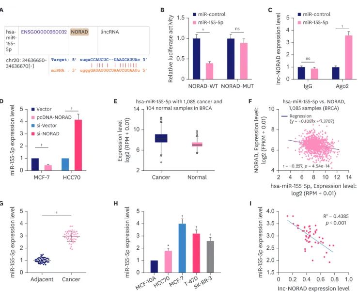
NORAD serves as a molecular sponge of miR-155-5p
The “ceRNA network” is a well-known classic regulatory mechanism by which lncRNAs regulate various biological processes. LncRNAs can sponge miRNAs to repress their expression and regulate their effect on target mRNAs. The StarBase database suggested a potential binding site between miR-155-5p and NORAD (Figure 4A). To confirm this, we performed a dual-luciferase reporter assay. The reporter assay indicated that miR-155-5p inhibited luciferase activity of NORAD WT reporter but not that of the MUT reporter (Figure 4B). Furthermore, RIP assay indicated that in contrast with control IgG, NORAD and miR-155-5p were preferentially enriched in Ago2-containing microribonucleoproteins (Figure 4C). Moreover, NORAD overexpression reduced miR-155-5p expression in MCF-7 cells, whereas NORAD knockdown increased miR-155-5p expression in HCC70 cells (Figure 4D). Notably, the data from the StarBase database suggested that compared to normal tissues, miR-155-5p expression was higher in BC tissues (Figure 4E), and the expression of NORAD and miR-155-5p were negatively correlated with each other in these samples (Figure 4F). Consistently, qRT-PCR showed that compared to normal controls, miR-155-5p expression was markedly enhanced in both BC tissues and cells (Figure 4G and H). Furthermore, miR-155-5p expression was negatively correlated with NORAD expression in BC tissues (Figure 4I). These data suggest that miR-155- 5p is a downstream target of NORAD in BC.
MCF-7 HCC70
Relativ e e xpr ession of γH 2AX pr ot ein 0 1 3 2 4 5
si-NC NC
si-NORAD pcDNA-NORAD γH2AX
*GAPDH
MCF-7 HCC70
A
*
si-NC
NC pcDNA
-NORAD si-NORAD
TUNEL
DAPI
Merge B
si-NC
MCF-7 HCC70
Apopt osis r at e (%)
0 20 10 30
40
**
NC pcDNA-NORAD si-NORAD
si-NC NC
pcDNA -NORAD si-NORAD
Figure 3. NORAD promotes DNA damage and cell apoptosis.
(A) Western blotting was used to detect the expression of the DNA damage marker γH2AX, following overexpression or knockdown of NORAD. (B) TUNEL assay was used to detect apoptotic cells.
NORAD = non-coding RNA activated by DNA damage; TUNEL = terminal deoxynucleotidyl transferase dUTP Nick-End Labeling; GAPDH = glyceraldehyde 3-phosphate dehydrogenase; DAPI = 4′,6-diamidino-2-phenylindole; NC = negative control.
*p < 0.001.
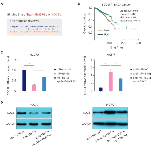
Tumor suppressor SOCS1 is regulated by the NORAD/miR-155-5p axis SOCS1 is a target gene of miR-155-5p (Figure 5A) [12]. Importantly, survival analysis in the StarBase database indicated that the overall survival rate of patients with BC with high SOCS1 expression was higher than that of the low expression group (Figure 5B). Moreover, western blotting and qRT-PCR showed that NORAD enhanced SOCS1 expression and reversed the miR-155-5p-induced SOCS1 downregulation in HCC70 cells. Conversely, NORAD knockdown reduced SOCS1 expression and attenuated miR-155-5p antagonist-induced SOCS1 upregulation in MCF-7 cells (Figure 5C and D). These data suggest that the NORAD/miR-155- 5p axis regulates BC progression by modulating the expression of SOCS1.
† ns
NORAD-MUT
Relativ e l ucifer ase activity
0 0.5 1.5
1.0
miR-control miR-155-5p
B
NORAD-WT
ns
†
Inc -NORAD e xpr ession l ev el Ago2 0 1 5
3 4
2
miR-control miR-155-5p
C
IgG E
Expr ession l ev el lo g2 (RPM + 0 .01 ) 2 6 14
10
Cancer
hsa-miR-155-5p with 1,085 cancer and 104 normal samples in BRCA
Normal A
HCC70
miR -155 -5 p e xpr ession l ev el
0 1 3 5
2
4 si-Vector Vector pcDNA-NORAD si-NORAD
†
D
†
MCF-7 hsa- miR-
155- 5p
ENSG00000260032
NORAD lincRNA
chr20: 34636650-
34636670[-]
Target: 5’ uugaCCAUCUC--UAAGCAUUAc 3’miRNA : 3’ ugggGAUAGUGCUAAUCGUAAUu 5’
G
†
miR -155 -5 p e xpr ession l ev el
0 5 4 3 2 1
Adjacent Cancer
H
miR -155 -5 p e xpr ession l ev el
0 1 3 5
2 4
*
MCF -7 MCF -10AHCC 70 T-4 7D
SK-BR -3
†
†
†
F
NORAD , Expr ession l ev el: lo g2 (FPKM + 0 .01 )
hsa-miR-155-5p, Expression level:
log2 (RPM + 0.01)
Inc-NORAD expression level
4 2 4 6 8 10 12 14
6 10
8
hsa-miR-155-5p vs. NORAD, 1,085 samples (BRCA)
0 0.2 0.4 0.6 0.8 1.0
I
miR -155 -5 p e xpr ession l ev el
1.5 2.0 3.0 4.0
2.5 3.5
r = −0.227, p = 4.24e−14
R
2= 0.4385
p < 0.001 Regression(y = −0.1081x + 7.3707)
Figure 4. NORAD targets miR-155-5p and negatively regulates its expression.
(A) Bioinformatics analysis was used to predict the binding site between NORAD and miR-155-5p. Dual-luciferase reporter assay (B), and RIP assay (C) were conducted to validate the interaction between miR-155-5p and NORAD in BC cells. (D) Relative miR-155-5p expression in BC cells transfected with NORAD- overexpression/control plasmids and si-NORAD/control plasmids. Analysis of the StarBase database to examine miR-155-5p expression (E), and the correlation between miR-155-5p and NORAD in BC (F). qRT-PCR was conducted to analyze miR-155-5p expression in BC tissues (G), and cell lines (H). (I) Analysis of the correlation between NORAD and miR-155-5p in BC tissues.
NORAD = non-coding RNA activated by DNA damage; miR = microRNA; RIP = RNA immunoprecipitation; BC = breast cancer; WT = wild-type; MUT = mutant; IgG
= immunoglobulin G; Ago2 = argonaute 2; ns, no statistical significance.
*p < 0.01 and †p < 0.001.
Overexpression of NORAD/SOCS1 reverses the promotional effects of miR- 155-5p on BC cells
To further investigate the significance of the NORAD/miR-155-5p/SOCS1 axis in regulating BC progression, NORAD or SOCS1 overexpression plasmids and miR-155-5p mimics were co-transfected into HCC70 cells. Similarly, NORAD or SOCS1 siRNA and miR-155-5p inhibitors were co-transfected into MCF-7 cells (Figure 6A). Transfection with miR-155-5p mimics promoted the proliferation, migration, and invasion of BC cells, whereas inhibition of miR-155-5p reduced the malignancy of BC cells (Figure 6B-D). However, NORAD/
SOCS1 overexpression and knockdown counteracted the effects of miR-155-5p mimics and inhibitors, respectively (Figure 6B-D). These findings imply that NORAD inhibits the malignant behaviors of BC cells partly by repressing miR-155-5p/SOCS1.
A B SOCS1 in BRCA cancer
Log-rank p = 0.03 Low num = 541 High num = 541 Hazard ratio = 0.70
Ov er all survival
Time (mo)
0 100 200 300
0.2 0.4 1.0 0.8 0.6
HCC70
SOCS 1 mRNA e xpr ession l ev el
0 0.5 1.0 1.5
miR-155-5p +pcDNA-NORAD miR-control miR-155-5p
*
SOCS1 GAPDH
SOCS1 GAPDH
HCC70 MCF-7
C
D
*
MCF-7
SOCS 1 mRNA e xpr ession l ev el
0 2 4 8 6
anti-miR-155-5p +si-NORAD anti-miR-NC anti-miR-155-5p
* *
anti-miR -NC anti-miR
-155 -5p miR -contr
ol miR -155 -5p
miR -155 -5p +pcDNA
-NORAD anti-miR
-155 -5p +si-NORAD Binding Site of hsa-miR-155-5p on SOCS1:
chr16: 11348669-11348676[-]
Target: 5’ cgCCGUG-CACG--CAGCAUUAa 3’
miRNA : 3’ ugGGGAUAGUGCUAAUCGUAAUu 5’
Low
High
Figure 5. NORAD sponges miR-155-5p to elevate SOCS1 expression.
(A) StarBase database was used to examine binding site for miR-155-5p in the SOCS1 3′-UTR. (B) StarBase database was used to analyze the relationship between the overall survival of patients with BC and SOCS1 expression. qRT-PCR (C) and western blotting (D) were performed to analyze SOCS1 expression in BC following NORAD and miR-155-5p regulation.
NORAD = non-coding RNA activated by DNA damage; miR = microRNA; SOCS1 = suppressor of cytokine signaling 1; UTR = untranslated region; BC = breast cancer; qRT-PCR = quantitative real-time polymerase chain reaction; GAPDH = glyceraldehyde 3-phosphate dehydrogenase.
*p < 0.001.
DISCUSSION
LncRNAs are crucial regulators of tumorigenesis and progression. Small nucleolar RNA host gene 16 (SNHG16) is upregulated in cervical cancer tissues and cells, and knockdown A
miR -155 -5 p e xpr ession l ev el miR -155 -5 p e xpr ession l ev el
0 1 5
3 4
2
‡
HCC70
†
†
†
†
‡
miR-155-5p+pcDNA-NORAD miR-control
miR-155-5p miR-155-5p+SOCS1
0 0.5 1.5
1.0
MCF-7
anti-miR-155-5p+si-NORAD anti-miR-155-5p+si-SOCS1 anti-miR-NC
anti-miR-155-5p
C
Br dU positiv e cells (%) Br dU positiv e cells (%)
0 120
60 90
30
†
HCC70
*
†
†
†
†
miR-155-5p+pcDNA-NORAD miR-control
miR-155-5p miR-155-5p+SOCS1
0 40 20 80 60
MCF-7
anti-miR-155-5p+si-NORAD anti-miR-155-5p+si-SOCS1 anti-miR-NC
anti-miR-155-5p
D
Cell number Cell number
0 1,200
600 900
300
HCC70
‡ †
†
‡ *
‡
miR-155-5p+pcDNA-NORAD miR-control
miR-155-5p miR-155-5p+SOCS1
0 100 300
200
MCF-7
anti-miR-155-5p+si-NORAD anti-miR-155-5p+si-SOCS1 anti-miR-NC
anti-miR-155-5p
B
Optical density at 450 nm Optical density at 450 nm
†
0
10.6 0.3 1.5 1.2 0.9
0 0.4 0.2 0.8 0.6
Time (day)
2 3 4HCC70
1
Time (day)
2 3 4MCF-7
miR-155-5p+pcDNA-NORAD miR-control
miR-155-5p miR-155-5p+SOCS1
anti-miR-155-5p+si-NORAD anti-miR-155-5p+si-SOCS1 anti-miR-NC
anti-miR-155-5p
† ‡ *† †
Migration Invasion
‡
†
†
†
†
†
Migration Invasion
Figure 6. NORAD counteracts the effects of miR-155-5p in BC cells.(A) HCC70 cells were transfected with control miRNA, miR-155-5p mimics, miR-155-5p mimics + NORAD overexpression plasmid, or miR-155-5p mimics + SOCS1 overexpression plasmid. MCF-7 cells were transfected with miRNA inhibitor control, miR-155-5p inhibitors, miR-155-5p inhibitors + NORAD siRNA, or miR-155-5p inhibitors + SOCS1 siRNA. Then, qRT-PCR was used to analyze the expression of miR-155-5p in each group of cells. (B) CCK-8, (C) BrdU, and (D) Transwell assays were conducted to analyze proliferation, migration, and invasion of BC cells.
NORAD = non-coding RNA activated by DNA damage; miR = microRNA; BC = breast cancer; siRNA = small interfering RNA; SOCS1 = suppressor of cytokine signaling 1; qRT-PCR = quantitative real-time polymerase chain reaction; CCK-8 = cell counting kit-8.
*p < 0.05, †p < 0.01, and ‡p < 0.001.
of SNHG16 suppresses cancer cell proliferation and epithelial-mesenchymal transition (EMT) by modulating the cell cycle and apoptosis [13]. In BC, the upregulation of basal- like breast cancer-associated transcript 1, resulting from hypomethylation at CpG islands in the promoter, reduces apoptosis and promotes the invasion of cancer cells [14]. Nuclear transcription factor nuclear factor (NF)-κB-interacting lncRNA represses TGF-β-induced EMT by blocking NF-κB signaling in BC [15]. LncRNA cancer susceptibility candidate 9 functions as an oncogene in BC by sponging the miR-195/497 cluster and promoting BC cell proliferation and survival [16]. NORAD is dysregulated in various tumors. However, its role in BC remains controversial. We demonstrated that NORAD is downregulated in BC tissues and cells, and that the downregulation of NORAD is associated with unfavorable pathological characteristics of the patients. Loss- and gain-of-function experiments revealed that NORAD repressed the malignancy of BC cells. Our data support that NORAD functions as a tumor suppressor, and not an oncogene, in BC.
MiRNAs are non-coding RNAs 21–25 nucleotides in length that are involved in the
pathogenesis of many human diseases [17]. They are also key regulators of BC tumorigenesis and progression. MiR-330-3p promotes BC metastasis by targeting collagen and calcium- binding epidermal growth factor domain-1 [18]. Another study showed that miR-193b inhibits metastasis and angiogenesis by suppressing dimethylarginine dimethylaminohydrolase 1 in BC [19]. MiR-155-5p has also drawn a lot of attention in cancer research. MiR-155-5p promotes cervical cancer cell metastasis by targeting tumor protein p53-inducible nuclear protein 1 [20]. MiR-155-5p is pivotal in cancer-associated fibroblast-induced tumor angiogenesis via inhibition of SOCS1 expression and activation of Janus kinase (JAK2)/signal transducers and activators of transcription (STAT3) signaling pathway in melanoma [12]. Importantly, miR- 155-5p is markedly upregulated in the plasma of patients with BC [21]. Functionally, miR-155- 5p promotes BC cell proliferation and migration by targeting matrix metallopeptidase 16 and SOCS1 [22]. In the present work, we also validated that miR-155-5p had the properties of an oncomiR, was upregulated in BC, and promoted the malignant behaviors of BC cells.
LncRNAs serve as ceRNAs to regulate the translation of mRNAs by competing for shared miRNAs [23]. This mechanism has been shown to be a crucial process during cancer progression. For example, lncRNA small nucleolar RNA host gene 5 facilitates BC cell proliferation by sponging miR-154-5p and upregulating proliferating cell nuclear antigen [24]. LINC01116 upregulates estrogen receptor 1 expression by competing with miR-145, thus facilitating BC progression [25]. MiR-155-5p is also involved in ceRNA networks.
The lncRNA, myocardial infarction-associated transcript, facilitates BC progression and functions as a ceRNA by sponging miR-155-5p [26]. In this study, NORAD was identified as a ceRNA for miR-155-5p, and NORAD positively regulated SOCS1 by sponging miR-155.
Importantly, NORAD or SOCS1 overexpression counteracted the tumor-promoting effects of miR-155-5p in BC. Therefore, our results support the view that NORAD/miR-155-5p/SOCS1 is a novel ceRNA network in BC progression.
As a member of the SOCS family, SOCS1 participates in multiple biological processes, including tumorigenesis [27]. SOCS1 contains an SH2 domain, a C-terminal SOCS box motif, and an N-terminal variable sequence. Based on its protein structure, SOCS has two main functions: first, SOCS1 regulates cell proliferation and apoptosis as a signal inhibitor;
second, SOCS1 regulates cell growth by participating in ubiquitination and mediating the
degradation of ubiquitination substrate [28]. With the C-terminal SOCS box motif, SOCS1
can function as a tumor suppressor by reducing oncoproteins via ubiquitination mechanism
[29]. SOCS1 directly or indirectly regulates many cancer-related molecules or pathways, including cyclin D1, cyclin-dependent kinase 4 (CDK4), mitogen-activated protein kinase (MAPK)/p38, p53, programmed cell death receptor ligand 1 (PD-L1), STAT3, STAT1, STAT6, etc. [27]. In previous studies, SOCS1 was shown to be regulated by miRNAs. For example, miR-221 represses SOCS1 expression, thereby facilitating prostate cancer cell proliferation and metastasis [30]. SOCS1 is targeted and negatively regulated by miR-155-5p [12]. The present work demonstrated that low expression of SOCS1 was associated with poor prognosis of patients with BC, suggesting a tumor-suppressor role of SOCS1 in BC. Moreover, we validated that the NORAD/miR-155-5p axis regulates the expression of SOCS1. Our findings provide a novel explanation for the dysregulation of SOCS1 in cancers.
In summary, this study revealed that low expression of NORAD indicates poor prognosis for patients with BC. NORAD contributes to BC cell proliferation and high potential of metastasis by modulating the miR-155-5p/SOCS1 axis. However, further studies, including experiments based on animal models, are needed to validate the tumor-suppressor functions of NORAD in BC.
ACKNOWLEDGMENTS
We thank Hubei Yican Health Industry Co., Ltd. (Luminescience) for its linguistic assistance during the preparation of this manuscript.
REFERENCES
1. Fahad Ullah M. Breast cancer: current perspectives on the disease status. Adv Exp Med Biol 2019;1152:51-64.
PUBMED | CROSSREF
2. Song JL, Chen C, Yuan JP, Sun SR. Progress in the clinical detection of heterogeneity in breast cancer.
Cancer Med 2016;5:3475-88.
PUBMED | CROSSREF
3. Yang J, Cheng L, Zhang J, Chen L, Wang D, Guo X, et al. Predictive value of circulating cell-free DNA in the survival of breast cancer patients: a systemic review and meta-analysis. Medicine (Baltimore) 2018;97:e11417.
PUBMED | CROSSREF
4. Wei GH, Wang X. lncRNA MEG3 inhibit proliferation and metastasis of gastric cancer via p53 signaling pathway. Eur Rev Med Pharmacol Sci 2017;21:3850-6.
PUBMED
5. Lei T, Lv ZY, Fu JF, Wang Z, Fan Z, Wang Y. LncRNA NBAT-1 is down-regulated in lung cancer and influences cell proliferation, apoptosis and cell cycle. Eur Rev Med Pharmacol Sci 2018;22:1958-62.
PUBMED | CROSSREF
6. Zhang YX, Yuan J, Gao ZM, Zhang ZG. LncRNA TUC338 promotes invasion of lung cancer by activating MAPK pathway. Eur Rev Med Pharmacol Sci 2018;22:443-9.
PUBMED | CROSSREF
7. Kim J, Piao HL, Kim BJ, Yao F, Han Z, Wang Y, et al. Long noncoding RNA MALAT1 suppresses breast cancer metastasis. Nat Genet 2018;50:1705-15.
PUBMED | CROSSREF
8. Liu M, Gou L, Xia J, Wan Q, Jiang Y, Sun S, et al. LncRNA ITGB2-AS1 could promote the migration and invasion of breast cancer cells through up-regulating ITGB2. Int J Mol Sci 2018;19:1866.
PUBMED | CROSSREF
9. Yang Z, Zhao Y, Lin G, Zhou X, Jiang X, Zhao H. Noncoding RNA activated by DNA damage (NORAD):
Biologic function and mechanisms in human cancers. Clin Chim Acta 2019;489:5-9.
PUBMED | CROSSREF
10. Zhou K, Ou Q, Wang G, Zhang W, Hao Y, Li W. High long non-coding RNA NORAD expression predicts poor prognosis and promotes breast cancer progression by regulating TGF-β pathway. Cancer Cell Int 2019;19:63.
PUBMED | CROSSREF
11. Tan BS, Yang MC, Singh S, Chou YC, Chen HY, Wang MY, et al. LncRNA NORAD is repressed by the YAP pathway and suppresses lung and breast cancer metastasis by sequestering S100P. Oncogene 2019;38:5612-26.
PUBMED | CROSSREF
12. Zhou X, Yan T, Huang C, Xu Z, Wang L, Jiang E, et al. Melanoma cell-secreted exosomal miR-155-5p induce proangiogenic switch of cancer-associated fibroblasts via SOCS1/JAK2/STAT3 signaling pathway. J Exp Clin Cancer Res 2018;37:242.
PUBMED | CROSSREF
13. Wu W, Guo L, Liang Z, Liu Y, Yao Z. Lnc-SNHG16/miR-128 axis modulates malignant phenotype through WNT/β-catenin pathway in cervical cancer cells. J Cancer 2020;11:2201-12.
PUBMED | CROSSREF
14. Han YJ, Boatman SM, Zhang J, Du XC, Yeh AC, Zheng Y, et al. LncRNA BLAT1 is upregulated in basal-like breast cancer through epigenetic modifications. Sci Rep 2018;8:15572.
PUBMED | CROSSREF
15. Wu W, Chen F, Cui X, Yang L, Chen J, Zhao J, et al. LncRNA NKILA suppresses TGF-β-induced epithelial- mesenchymal transition by blocking NF-κB signaling in breast cancer. Int J Cancer 2018;143:2213-24.
PUBMED | CROSSREF
16. Shao G, Wang M, Fan X, Zhong L, Wang Z, Zhang P, et al. lncRNA CASC9 positively regulates CHK1 to promote breast cancer cell proliferation and survival through sponging the miR-195/497 cluster. Int J Oncol 2019;54:1665-75.
PUBMED | CROSSREF
17. McGuire A, Brown J A L, Kerin MJ. Metastatic breast cancer: the potential of miRNA for diagnosis and treatment monitoring. Cancer Metastasis Rev 2015;34:145-55.
PUBMED | CROSSREF
18. Mesci A, Huang X, Taeb S, Jahangiri S, Kim Y, Fokas E, et al. Targeting of CCBE1 by miR-330-3p in human breast cancer promotes metastasis. Br J Cancer 2017;116:1350-7.
PUBMED | CROSSREF
19. Hulin JA, Tommasi S, Elliot D, Hu DG, Lewis BC, Mangoni AA. MiR-193b regulates breast cancer cell migration and vasculogenic mimicry by targeting dimethylarginine dimethylaminohydrolase 1. Sci Rep 2017;7:13996.
PUBMED | CROSSREF
20. Li N, Cui T, Guo W, Wang D, Mao L. MiR-155-5p accelerates the metastasis of cervical cancer cell via targeting TP53INP1. Onco Targets Ther 2019;12:3181-96.
PUBMED | CROSSREF
21. Khalighfard S, Alizadeh AM, Irani S, Omranipour R. Plasma miR-21, miR-155, miR-10b, and Let-7a as the potential biomarkers for the monitoring of breast cancer patients. Sci Rep 2018;8:17981.
PUBMED | CROSSREF
22. Zhang W, Chen CJ, Guo GL. MiR-155 promotes the proliferation and migration of breast cancer cells via targeting SOCS1 and MMP16. Eur Rev Med Pharmacol Sci 2018;22:7323-32.
PUBMED | CROSSREF
23. Gao C, Li H, Zhuang J, Zhang H, Wang K, Yang J, et al. The construction and analysis of ceRNA networks in invasive breast cancer: a study based on The Cancer Genome Atlas. Cancer Manag Res 2018;11:1-11.
PUBMED | CROSSREF
24. Chi JR, Yu ZH, Liu BW, Zhang D, Ge J, Yu Y, et al. SNHG5 promotes breast cancer proliferation by sponging the miR-154-5p/PCNA axis. Mol Ther Nucleic Acids 2019;17:138-49.
PUBMED | CROSSREF
25. Hu HB, Chen Q, Ding SQ. LncRNA LINC01116 competes with miR-145 for the regulation of ESR1 expression in breast cancer. Eur Rev Med Pharmacol Sci 2018;22:1987-93.
PUBMED | CROSSREF
26. Luan T, Zhang X, Wang S, Song Y, Zhou S, Lin J, et al. Long non-coding RNA MIAT promotes breast cancer progression and functions as ceRNA to regulate DUSP7 expression by sponging miR-155-5p.
Oncotarget 2017;8:76153-64.
PUBMED | CROSSREF
27. Sharma J, Larkin J 3rd. Therapeutic implication of SOCS1 modulation in the treatment of autoimmunity and cancer. Front Pharmacol 2019;10:324.
PUBMED | CROSSREF
28. Ying J, Qiu X, Lu Y, Zhang M. SOCS1 and its potential clinical role in tumor. Pathol Oncol Res 2019;25:1295-301.
PUBMED | CROSSREF
29. Lessard F, Saint-Germain E, Mignacca L, Ferbeyre G. SOCS1: phosphorylation, dimerization and tumor suppression. Oncoscience 2019;6:386-9.
PUBMED | CROSSREF
30. Shao N, Ma G, Zhang J, Zhu W. miR-221-5p enhances cell proliferation and metastasis through post- transcriptional regulation of SOCS1 in human prostate cancer. BMC Urol 2018;18:14.
PUBMED | CROSSREF