서론
눈물흘림은 안과에서 흔히 볼 수 있는 증상으로 눈물분비 와 눈물배출의 상호작용에 의해 발생한다. 따라서 진단 시 에는 눈물의 과다분비나 안구의 자극으로 인한 과도한 눈 물흘림과 눈물길의 배출장해로 인한 눈물흘림을 감별하는 것이 중요하다. 눈물배출장해가 있는 경우에는 코눈물관막 힘과 같은 구조적 이상과 눈물배출펌프 기능이상을 감별해 야 한다.
눈물배출 경로인 눈물길은 눈물점부터 눈물소관, 눈물주
머니 및 코눈물관으로 이루어진 연속적인 해부학적 구조물 로서 부분적, 또는 완전한 막힘이 발생하는 경우 눈물흘림의 증상이 발생하게 된다. 눈물배출 이상이 의심되는 경우에는 눈물소관 관류술 및 더듬자검사법, 형광염색소실검사, 눈물 주머니조영술, 눈물길신티그라피, 컴퓨터단층촬영 등의 검 사를 적절히 시행하여 눈물흘림 증상을 감별하고 눈물길질 환을 정확히 진단할 수 있다.
눈물길질환의 치료는 급성염증이 있거나 부분 눈물길막힘 초기에는 약물치료를 시행할 수 있지만 대부분의 경우에는 수술적 치료가 필요하게 된다. 본 논문에서는 다양한 임상양 상의 눈물길질환의 진단 및 치료에 대해 소개하고 최신 치 료기법의 발전현황을 문헌고찰을 통해 알아보고자 하였다.
눈물길의 해부와 기능
눈물길은 눈물점, 눈물소관, 눈물주머니, 코눈물관으로 구
눈물길질환의 정의와 치료
이 정 규 | 중앙대학교 의과대학 중앙대학교병원 안과
Definition and treatment of lacrimal drainage disease
Jeong Kyu Lee, MD
Department of Ophthalmology, Chung-Ang University Hospital, Chung-Ang University College of Medicine, Seoul, Korea
Received: July 24, 2017 Accepted: August 8, 2017 Corresponding author: Jeong Kyu Lee
E-mail: lk1246@cau.ac.kr
© Korean Medical Association
This is an Open Access article distributed under the terms of the Creative Commons Attribution Non-Commercial License (http://creativecommons.
org/licenses/by-nc/3.0) which permits unrestricted non-commercial use, distribution, and reproduction in any medium, provided the original work is properly cited.
Epiphora is one of the most common problems in ophthalmological practice, and is caused by dysfunction of the lacrimal pathway. The lacrimal drainage system is a continuous anatomical structure consisting of the lacrimal punctum, inferior and superior canaliculi, common canaliculus, lacrimal sac, and nasolacrimal duct. Lacrimal disease can be medically treated in case of acute inflammation or partial obstruction at the beginning of treatment, but surgical treatment is necessary in most cases. This paper discusses the etiology, clinical features, diagnosis, and surgical treatment of various lacrimal diseases through a selective review of the relevant literature. Advances in lacrimal duct surgery can now be performed in such a way that the structural integrity and normal physiological function of the entire efferent lacrimal pathway is preserved.
Key Words: Lacrimal apparatus diseases; Lacrimal duct obstruction; Dacryocystorhinostomy;
Conjunctivodacryocystorhinostomy
성되어 있다. 눈물샘과 덧눈물샘, 결막에서 생성된 눈물은 눈의 안쪽구석에 모여 위아래 눈물점을 통과한 후 눈물소관, 눈물주머니, 코눈물관을 통하여 코안으로 배출된다. 눈물배 출의 기전은 아직 정확히 알려져 있지 않지만 눈꺼풀의 눈둘 레근이 눈물주머니를 당겨서 눈물배출펌프의 작동에 의해 눈물이 배출되는 것으로 이해되고 있으며 이 과정에서 눈꺼 풀깜박임은 눈물길 내에 음압 형성에 관여하여 눈물배출을 돕는다[1]. 눈물주머니 및 코눈물관에 나선형으로 배열된 결 합조직과 수축혈관계인 해면체 역시 눈물의 배출에 관여한 다고 보고되고 있다[2].
눈물길에는 눈물소관에서 눈물주머니, 코눈물관 및 코안 으로 진행하는 과정에서 특징적인 굴곡이 있으면서 반경이 좁아지고 점막주름이 있는 해부학적 구조물들이 있다. 이들 은 각각 로센뮐러판막(valve of Rosenmüller), 크라우제판 막(valve of Krause), 하스너판막(valve of Hasner)으로 불 리우며 눈물이 역류되는 것을 방지하는 기능이 있다. 이 기 능이 약화되면 코를 풀 때 공기가 눈물점을 통해 눈으로 나 오게 된다. 이들 판막들은 눈물길막힘이 흔하게 발생하는 위 치이기도 하다.
눈물길질환의 원인
눈물길은 감염, 반흔성 질환, 외상, 만성염증, 약물, 종 양 등 다양한 원인에 의해 질환이 발생할 수 있다. 상부눈 물길인 눈물점과 눈물소관을 침범하는 가장 흔한 감염원 은 단순포진바이러스이며 방사성요오드 치료나 docetaxel, 5-fluorouracil과 같은 항암화학요법도 눈물길막힘을 유 발할 수 있다[3,4]. 눈물점의 발적 및 부종과 농성분비 물을 동반한 눈물소관염은 주로 혐기성 세균인 방선균 (Actinomyces)에 의한 만성감염에 기인하며, 때로 만성결막 염으로 오인되어 진단이 늦어지는 경우가 있다[5]. 최근에는 안구건조증을 치료할 목적으로 삽입한 눈물소관마개에 의한 이차적 염증이 보고되고 있기도 하다[6].
하부눈물길인 코눈물관의 막힘은 주로 40대 이상 여자에 서 호발하며 대부분은 발병기전을 정확히 알 수 없는 원발후
천코눈물관막힘이다. 눈물길의 감염이나 염증, 호르몬 불균 형 등으로 인해 눈물길 상피세포 및 결합조직에 염증 및 부 종이 발생하고, 진행하여 섬유화로 인한 기계적 폐쇄가 발생 하는 것으로 생각되고 있다[7]. 코눈물관막힘으로 인해 눈물 주머니내 눈물흐름이 정체된 상태에서 감염이 되면 급성눈 물주머니염이 발생하는 경우도 있다. 가장 흔한 원인균은 폐 렴구균이며 초기에는 약물치료를 하고 증상이 완화되면 눈 물주머니코안연결술을 시행하여 치료한다[8].
눈물길질환의 진단
눈물흘림 환자를 검사할 때는 자극으로 인한 눈물의 과다 분비와 눈물배출장애로 인한 눈물흘림을 감별하는 것이 가 장 중요하다. 눈물흘림이 지속적이고 한쪽 눈이 증상이 더 심하거나, 눈곱 등의 증상이 동반된다면 눈물길막힘의 가능 성이 높다.
눈물길막힘을 진단할 때 가장 중요한 진단방법은 눈물소 관 관류술 및 더듬자검사법이다. 눈물소관 관류술은 외래에 서 간단히 시행할 수 있으며 눈물배출계의 협착 유무 및 협 착부위를 판단할 수 있는 좋은 검사법이다. 그러나 검사과정 에서 높은 압력으로 생리식염수가 주입되면서 부분 막힘을 감지하지 못할 수 있고 기능적 폐쇄 여부를 감별할 수 없는 단점이 있다. 눈물소관 관류술로 눈물길막힘이 확인되면 더 듬자검사법을 시행하여 눈물점으로부터 막힘이 있는 부위까 지 거리를 측정하여 수술방법을 결정할 수 있다.
눈물길질환을 진단하는 영상의학적 검사는 눈물주머니조 영술과 눈물길신티그래피가 널리 사용된다(Figure 1). 눈 물주머니조영술은 X-ray 검사 하에서 조영제를 눈물소관 으로 주사하여 눈물길 내 흐름을 관찰하는 것으로 눈물길 의 해부학적 구조와 폐쇄 양상을 관찰할 수 있다[9]. 눈물길 신티그래피는 방사성 동위원소(technetium 99m pertech- netate)를 점안한 후 감마카메라로 눈물내 흐름을 관찰하 는 것으로 생리적인 정상환경에서 눈물의 배출을 관찰할 수 있어서 주로 눈물길의 기능적 폐쇄를 진단하는데 도움이 된다[10].
눈물길질환의 치료
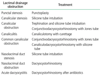
눈물길질환은 폐쇄나 염증이 발생한 위치에 따라 다양한 수술적 방법으로 치료가 가능하다(Table 1). 눈물점협착 초 기에는 눈물점확장기로 넓혀주는 것만으로도 치료가 되지만
협착이 심한 경우에는 눈물점성형술이 필요하다(Figure 2).
눈물점성형술은 눈물점에 수직으로 절개를 해서 눈물점을 넓히는 것으로 1-3개의 절개를 상황에 따라 시행할 수 있으 며 71-94%까지 성공률이 보고되고 있다[11,12]. 눈물점의 재협착을 방지하기 위해 실리콘관삽입술을 시행하여 성공률 이 더 높았다는 보고도 있다[13].
눈물소관막힘이 있는 경우에는 불완전막힘의 경우에는 실리콘관삽입술을 시행할 수 있지만 완전막힘이 있는 경우
에는 trephine을 이용하여 개통후 실리 콘관삽입술을 시행하게 된다. 그러나 개 통이 불가능한 경우에는 Jones관을 이 용하여 눈물이 결막낭에서 코로 직접 내 려가게 하는 결막눈물주머니코안연결술 을 시행하게 된다. 저자에 따라 다르지 만 최종성공률은 약 90% 이상으로 알려 져 있다[14,15]. 결막눈물주머니코안연 결술에서 가장 중요한 것은 Jones관의 위치이며 관이 코선반, 코사이막에 닿지 않아야 하며 코안의 외측 벽 속에 파묻 히지 않도록 해야 한다.
코눈물관막힘 환자에서는 눈물주머니코안연결술이 가장 널리 사용되고 있다. 눈물주머니코안연결술은 1904년에 처 음 소개되었으며 피부절개후 눈물주머니오목에 골공을 만들 고 눈물주머니와 코점막 피판을 연결하여 눈물주머니에서
Table 1. Treatments of lacrimal drainage obstruction Lacrimal drainage
obstruction Treatment
Punctal stenosis Punctoplasty Canalicular stenosis Silicone tube intubation Canalicular
obstruction
Trephination and silicone tube intubation Conjunctivodacryocystorhinostomy with Jones tube Canaliculitis Canaliculotomy with curettage
Common canalicular obstruction
Conjunctivodacryocystorhinostomy with Jones tube Canaliculodacryocystorhinostomy with silicone tube
Nasolacrimal duct stenosis
Silicone tube intubation Nasolacrimal duct
obstruction Dacryocystorhinostomy
Acute dacryocystitis Dacryocystorhinostomy after antibiotics
Figure 1. Representative images of Digital subtraction dacryocystography and lacrimal scintigraphy. (A) Dacryocystography reveals partial and (B) complete nasolacrimal duct obstruction. (C) Lacrimal scintigraphy shows normal pathway and (D) pre-sac obstruction in both eyes.
B A
D C
Figure 2. Slit lamp examination of punctum. (A) Punctal stenosis in left lower lid is noted. (B) Dilated punctum after punctoplasty is observed.
B A
코 안쪽으로 직접 눈물이 배출되는 통로를 만드는 방법이다 [16]. 피부접근이 아닌 코내시경을 이용하여 눈물주머니코안 연결술을 시행할 수도 있는데 초기에는 수술방법이 까다로 워 성공률이 낮았다. 그러나 내시경 및 수술기구가 발달하면 서 피부경유 수술과 유사한 82-95%의 높은 성공률이 보고 되고 있으며 피부 반흔이 없고 회복이 빠른 장점이 있어 점 차 선호도가 높아지고 있다[17,18].
코눈물길의 부분막힘이 있을 때는 실리콘관삽입술을 고 려해볼 수도 있다. 실리콘관삽입술은 1969년에 처음 소개 되었으며[19], 술기가 간편하여 유아의 선천 코눈물관막힘 에서부터 성인의 후천코눈물관막힘, 상부 눈물길의 불완전 막힘, 기능적 폐쇄에 이르기까지 여러 적응증에서 시술 되 고 있다. 수술 성공률은 51.6%에서 74%까지 다양하게 보 고되고 있으며 눈물주머니코안연결술이나 결막눈물주머니 코안연결술에 비해 성공률이 낮은 것으로 알려져 있으나 최근 눈물길 내시경이 보급되면서 직접 폐쇄부위를 확인하 면서 개통하는 시술이 발전하면서 점차 적응증이 확대되고 있다[20,21].
내시경이 점차 소형화되고 카메라기법이 발전되면서 최근 에는 눈물점을 통하여 내시경을 삽입하여 눈물길의 점막과 형태적 변화를 직접 관찰하고 치료까지 할 수 있게 되었다 (Figure 3). Sasaki 등[22]은 코눈물관막힘 환자에서 눈물 길내시경을 이용하여 진단과 동시에 실리콘관삽입술을 시행 하여 87.5%의 성공률을 보고하였다. 향후 눈물길 수술은 레 이저 등 수술기구의 발전과 동반하여 지속적으로 덜 침습적 이고 눈물길의 정상적인 생리기능을 유지하거나 회복시키는 수술로 발전할 것으로 기대되고 있다.
소아에서의 눈물길질환
소아에서 관찰되는 눈물길질환은 태생 학적으로 눈물길이 발아되어 발달하는 과정에서 무형성, 페쇄, 샛길, 게실형성 등 다양한 기형발생에 기인한다. 가장 흔 한 눈물길질환은 코눈물관 말단의 하스 너판막의 막이 생후에도 지속적으로 개 통되지 않는 선천코눈물관폐쇄로 국내에 서도 약 7% 유병률이 보고된 바 있다[23]. 하스너판막은 생 후 6개월 이내에 대부분 저절로 개방되지만 폐쇄가 지속시 에는 더듬자검사를 이용하여 폐쇄부위를 뚫어줄 수도 있으 며 실리콘관삽입술로 치료하기도 한다.
결론
눈물흘림은 안과에서 매우 흔한 증상이며 눈물 배출경로 의 장애로 인해 발생한다. 진단 시에는 안구의 과도한 자극 으로 인한 눈물흘림과 감별하는 것이 중요하다. 눈물길질환 은 초기에는 약물치료를 고려할 수 있지만 대부분의 경우에 수술적 치료가 필요하며 눈물길의 폐쇄나 염증이 발생한 위 치에 따라 다양한 수술적 방법이 가능하다. 내시경 및 레이 저 등이 발전하면서 향후 눈물길 수술은 덜 침습적이고 눈물 길의 정상적인 생리기능을 유지하거나 회복시키는 수술로 발전할 것으로 기대되고 있다.
찾아보기말: 눈물흘림; 눈물길막힘; 눈물주머니코안연결술;
결막눈물주머니코안연결술
ORCID
Jeong Kyu Lee, http://orcid.org/0000-0001-9797-9945
REFERENCES
1. Jones LT. An anatomical approach to problems of the eyelids Figure 3. Dacryoendoscopic view. Canaliculus (A), lacrimal sac, and entrance of nasolacrimal duct (B) is
observed.
B A
and lacrimal apparatus. Arch Ophthalmol 1961;66:111-124.
2. Paulsen FP, Thale AB, Hallmann UJ, Schaudig U, Tillmann BN. The cavernous body of the human efferent tear ducts:
function in tear outflow mechanism. Invest Ophthalmol Vis Sci 2000;41:965-970.
3. Yoshida M, Hondo R. Transmission of herpes simplex virus infection via lacrimal canaliculi. Ophthalmologica 1992;204:
101-102.
4. Kim DG, Lee JJ, Ham DS, Yang JW. Surgical outcomes of dacryocystorhinostomy in lacrimal drainage obstruction after systemic chemotherapy or radioactive iodine therapy. J Korean Ophthalmol Soc 2016;57:1684-1690.
5. Kim SD, Koh SI, Kim JD. Diagnosis and therapy of canali- culitis. J Korean Ophthalmol Soc 1998;39:2207-2210.
6. Ahn SM, Kim HC, Jang JW, Kim SJ. Treatment of the SmartPLUG-related canaliculitis. J Korean Ophthalmol Soc 2009;50:1768-1773.
7. Linberg JV, McCormick SA. Primary acquired nasolacrimal duct obstruction: a clinicopathologic report and biopsy tech- nique. Ophthalmology 1986;93:1055-1063.
8. Cahill KV, Burns JA. Management of acute dacryocystitis in adults. Ophthal Plast Reconstr Surg 1993;9:38-41.
9. Higashi H, Tamada T, Mizukawa K, Ito K. MR dacryocysto- graphy: comparison with dacryoendoscopy in positional diagnosis of nasolacrimal duct obstruction. Radiol Med 2016;
121:580-587.
10. Joo KS, Lee JK. Comparison of lacrimal scintigraphy and fluorescein dye disappearance test in functional blockage of lacrimal system. J Korean Ophthalmol Soc 2011;52:1013-1018.
11. Jeong S. A simple test for epiphora caused by punctal stenosis.
J Korean Ophthalmol Soc 1998;39:2864-2866.
12. Kwon JK, Chang MW, Baek SH, Lee TS. Punctoplasty using a radiofrequency surgical unit for punctal stenosis. J Korean Ophthalmol Soc 2012;53:1727-1731.
13. Choi CU, Seo SW, Kim SD. The comparison of punctoplasty and silicone tube intubation in patients with punctal obstruc- tion. J Korean Ophthalmol Soc 2008;49:543-547.
14. Lee TS, Lee H. Purse-string suture technique for jones tube fixation in conjunctivodacryocystorhinostomy. J Korean Oph- thalmol Soc 2008;49:1553-1558.
15. Rose GE, Welham RA. Jonesʼ lacrimal canalicular bypass tubes:
twenty-five yearsʼ experience. Eye (Lond) 1991;5(Pt 1):13-19.
16. Davies DL. Lacrymal obstruction: the results of the anas-
tomotic method of treatment (Toti). Proc R Soc Med 1921;
14(Sect Ophthalmol):59-64.
17. Weidenbecher M, Hosemann W, Buhr W. Endoscopic endo- nasal dacryocystorhinostomy: results in 56 patients. Ann Otol Rhinol Laryngol 1994;103(5 Pt 1):363-367.
18. Shin IH, Lim HB, Lee JJ, Lee SB. Prognostic factors for successful endonasal dacryocystorhinostomy. J Korean Oph- thalmol Soc 2015;56:1661-1666.
19. Keith CG. Lacrimal obstruction. Trans Ophthalmol Soc U K 1969;88:519-522.
20. Kim HD, Jeong SK. Silicone tube intubation in acquired nasolacrimal duct obstruction. J Korean Ophthalmol Soc 2000;41:327-331.
21. Kwon YH, Lee YJ. Long-term results of silicone tube intu- bation in incomplete nasolacrimal duct obstruction (NLDO).
J Korean Ophthalmol Soc 2008;49:190-194.
22. Sasaki T, Nagata Y, Sugiyama K. Nasolacrimal duct obstruc- tion classified by dacryoendoscopy and treated with infe- rior meatal dacryorhinotomy: part II. Inferior meatal dacry- orhinotomy. Am J Ophthalmol 2005;140:1070-1074.
23. Oh HS, Ahn Y. The incidence and medical treatment of con- genital nasolacrimal duct obstruction in Korean infants. J Korean Ophthalmol Soc 1995;36:1007-1013.
Peer Reviewers’ Commentary
본 논문은 임상에서 흔히 접할 수 있는 눈물흘림증에 대해 병인, 임상양상, 진단적 접근법, 치료방법(수술법) 등을 적절한 문헌고 찰과 함께 체계적으로 기술한 종설 논문이다. 눈물배출의 기전에 서는 다양한 배출이론을 소개하였으며, 눈물길질환의 원인과 진 단부분에서는 임상적으로 만날 수 있는 모든 원인과 이를 진단할 수 있는 방법이 순서대로 소개되어 있다. 눈물길의 해부학적 설 명과 함께 눈물길 질환의 원인을 해부학적 위치에 따라 감별하고 각 원인에 따른 치료법을 제시하여 실제 임상에서 눈물흘림증 환 자를 진료함에 있어 매우 유용한 논문이라 판단된다.
[정리: 편집위원회]