www.krspine.org
The Changes in Neural Foramen Shown on Computed Tomography Depending on the Changes in the Height of Intervertebral Disc after Anterior Cervical Discectomy
and Fusion (ACDF)
Young-Sang Lee, M.D., Woo-Suk Song, M.D., Joon-Cheol Choi, M.D., Woo-Sung Kim, M.D.
Hwa-Yeop Na, M.D., Yu-Hoon Jung, M.D., Kook-Hee Cho, M.D., Tae-Hoon Park, M.D.
Dae Hyeon Kim, M.D., Heui-Jeon Park, M.D.*
J Korean Soc Spine Surg 2011 Sep;18(3):96-102.
Originally published online September 30, 2011;
http://dx.doi.org/10.4184/jkss.2011.18.3.96
Korean Society of Spine Surgery
Department of Orthopedic Surgery, Inha University School of Medicine
#7-206, 3rd ST. Sinheung-Dong, Jung-Gu, Incheon, 400-711, Korea Tel: 82-32-890-3044 Fax: 82-32-890-3467
©Copyright 2011 Korean Society of Spine Surgery pISSN 2093-4378 eISSN 2093-4386
The online version of this article, along with updated information and services, is located on the World Wide Web at:
http://www.krspine.org/DOIx.php?id=10.4184/jkss.2011.18.3.96
This is an Open Access article distributed under the terms of the Creative Commons Attribution Non-Commercial License (http://
creativecommons.org/licenses/by-nc/3.0) which permits unrestricted non-commercial use, distribution, and reproduction in any medium, provided the original work is properly cited.
Journal of Korean Society of
Spine Surgery
The Changes in Neural Foramen Shown on Computed Tomography Depending on the Changes in the Height of Intervertebral Disc after Anterior Cervical Discectomy and Fusion (ACDF)
Young-Sang Lee, M.D., Woo-Suk Song, M.D., Joon-Cheol Choi, M.D., Woo-Sung Kim, M.D.
Hwa-Yeop Na, M.D., Yu-Hoon Jung, M.D., Kook-Hee Cho, M.D., Tae-Hoon Park, M.D.
Dae Hyeon Kim, M.D., Heui-Jeon Park, M.D.
*Department of Orthopedic Surgery, Bundang Jesaeng General Hospital, Daejin Medical Center,Sungnam , Gyounggi, Korea Department of Orthopedic Surgery, Yonsei University Wonju College of Medicine, Wonju Christian Hospital*
Study Design: A prospective radiological assessment.
Objectives: Changes in the height, area, and width--captured using computed tomography (CT)--of the neural foramen with respect to changes in the intervertebral disc height, after undergoing an anterior cervical disc removal and fusion procedure.
Summary of Literature Review: The multiple authors of this study, by obtaining central canal and area of neural foramen by increasing the disc spacing height and area of the neural foramen, attempted to assess the height increase of disc spacing. It is necessary to consider the synergistic effects of decompression through dissection of the posterior longitudinal ligament (PLL).
Materials and Methods: The authors studied 17 patient cases that underwent one segment anterior cervical discectomy and fusion (ACDF) for degenerative cervical disease from June 2006 to March 2007. All patient cases underwent autogenous iliac bone graft or cage insertion with plate fixation procedure. We measured the areas of the neural foramen, heights of the vertebra body above and below the removed intervertebral disc with CT before and after ACDF. Radiographic measurements were averaged.
Results: Among the 17 cases, the height of the cervical disc increased in 15 cases and decreased in 2 cases. The heights of the neural foramen increased in 19 cases and showed no changes in 13 cases. The areas of the neural foramen increased in 23 cases and decreased in 6 cases. The heights of vertebral body above and below the removed disc increased by 5.4% (p=0.734), and the heights of the neural foramen increased by 13.3% (p=0.002). The area of the neural foramen increased by 13.6% (p=0.192). The widths of the neural foramen increased by 2.3% (p=0.586). The intervertebral disc height, neural foramen height, and neural foramen area increased by 39.6%, 8.4%, and 17.9%, respectively, after a 2mm lengthening of bone transplant. The intervertebral disc height, neural foramen height, and neural foramen area increased by 59.8%, 22.9%, and 10.3%, respectively, after a 3mm lengthening of bone transplant. The height and area of neural foramen increased by 18.3% and 18.2%, respectively, after the PLL removal and dissection.
Conclusions: The follow-up observations of the intervertebral disc height, neural foramen height, and neural foramen area showed increases after one segment ACDF in cervical disease cases, when compared to the preoperative radiographic findings. As the height of bone transplant increased, the intervertebral disc height, neural foramen height, and neural foramen area increased. The neural foramen height and neural foramen area significantly increased, when PLL was dissected.
Key Words: Intervertebral disc, Neural foramen, Anterior interbody fusion, Cage
Received: May 4, 2009 Revised: June 10, 2011 Accepted: July 12, 2011
Published Online: September 30, 2011 Corresponding author: Woo-Suk Song, M.D.
Department of Orthopedic Surgery, Bundang Jesaeng General Hospital, Daejin Medical Center,#255-2,Seohyun-dong,Bundang-gu,Sungnam-Si,Gyounggi- Do,Korea,
TEL: 82-31-779-0175, FAX: 82-31-779-0176 E-mail: [email protected]
“This is an Open Access article distributed under the terms of the Creative Commons Attribution Non-Commercial License (http://
creativecommons.org/licenses/by-nc/3.0/) which permits unrestricted non-commercial use, distribution, and reproduction in any medium, provided the original work is properly cited.”
INTRODUCTION
The anterior cervical discectomy and fusion (ACDF) using the Smith-Robinson method1,2) is one of the widely-used treatment methods for degenerative cervical spine disease. This surgical technique, by directly removing lesions and by transplanting anterior trabecular bone, can attain postoperative stability, increase intervertebral disc height, and indirectly expand neural foramen; on the other hand however, it has disadvantages of
The Changes of Neural Foramen Following the Dimension of Disc after ACDF using by CT
Journal of Korean Society of Spine Surgery
www.krspine.org 97 difficulty in directly identifying the nerve roots and inability
in directly determining increase in the neural foramen after trabecular bone transplant.2-9)
Although a number of studies have progressed and reported in regard to the anatomical and clinical relationships among the intervertebral discs, neural foramen and nerve root in the lumbar, as far as the cervical vertebrae is concerned they are confined to a few research bodies and it’s rare to see studies done with real CT imaging of before-after 45 degree angle views of neural foramen.1-13)
As such, the authors of this study attempted to evaluate the changes in the height of intervertebral disc -- after undergoing ACDF for degenerative cervical disease -- and the accompanying interrelationship through radiological measurements.
RESEARCH SUBJECTS AND METHODS
From the intervertebral discs of the 17 patients who underwent ACDF at our hospital from June 2006 to March 2007 for cervical spinal disease, the neural foramen of the 32 cases of which the pre- and post-operative measurements were feasible were used as study subjects. The average age was 40.06-years (range of 36-65 years).
In all cases, the standard left-side Smith-Robinson method was used in surgery.14,15) Among these, 5 spinal segments underwent conventional palliative disc removal procedure without the aid of a microscope or loupe; 12 spinal segments underwent palliative disc removal procedure with the aid of a microscope. Autogenous iliac bone graft was done on 7 spinal segments, and cage was used in 10 spinal segments. Regarding the surgical sites, 2 segments were C3-4; 2 segments were C4-5; 8 segments were C5-6; 5 segments were C6-7. Of the total 17 segments, patients with posterior longitudinal ligament removed or dissected were 12 segments in 23 cases of neural foramen; patients who preserved posterior longitudinal ligament were 5 segments in 9 cases of neural foramen. The left-side Smith-Robinson method was used to remove intervertebral disc and anterior osteophyte, and afterwards endplate cartilage was
removed, and calipers were used to measure disc spacing and a transplant material that is 1-2mm or 3mm larger than the original disc was inserted.
Radiographic changes were measured using the axial cervical spine computer plots taken at every 1mm and CT imaging including the 45-degree slope and the side reconstructions (SIEMENS-somatom sensation 64®) that were taken before surgery and within 1 week after surgery, for the distance between adjacent vertebrae, neural foramen height, neural foramen width, and neural foramen area. In order to find out the height of the vertebral disc, the distance between the upper endplates of the upper vertebral body and the lower endplates of the lower vertebral body of the area proximal to the vertebral disc removal site was measured (Fig. 1A). The reason for measuring the disc spacing by including the adjacent upper and lower intervertebral discs was because the endplate cartilage of the intervertebral disc and subchondral bone were partially removed during surgery, i.e., measuring only the disc spacing before-after surgery could indicate inaccurate values.
The average of the measurements by two spine surgeons was used as the measurement. The statistical significance of the measurements of the changes in the neural foramen height and area with respect to the changes in the height of the intervertebral disc was evaluated.
Statistical processing was done with the SPSS ver. 15.0; the Mann-Whitney test and Wilcoxon test were used.
RESULTS
Among the total of 17 segmental surgery cases, 15 segments showed a post-operative increase in the distance between adjacent vertebrae; there were no cases of a decrease; 2 segments showed no changes. 19 cases showed a post-operative increase in the neural foramen height; there were 13 cases of neural foramen with no changes in height; there were no segments that showed a decrease in height. The neural foramen area increased in 23 cases; 3 cases showed no changes; 6 cases showed a decrease.
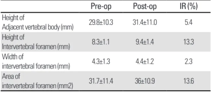
The distance between adjacent vertebrae showed a 5.4%
increase by going from the average 29.8 ± 10.3mm to average 31.4 ± 11.0mm; the neural foramen height showed a 13.3%
increase by going from 8.3 ± 1.1mm to 9.3 ± 1.4mm. The neural foramen area showed a 13.6% increase by going from the average 31.7 ± 11.4mm2 to 36 ± 10.9mm2 (Table 1).
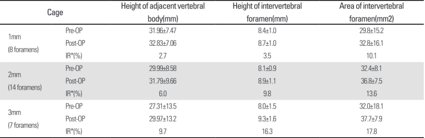
The cases of inserting transplant materials that were 1mm, 2mm and 3mm larger than the intervertebral disc height measured during surgery were separated and measured, and the results of which showed that the case with transplant that was 3mm larger showed the largest increases in the disc
Table 1. preop baseline measurement and postop change in intervertebral disc and foramen of total 17 segments
Pre-op Post-op IR (%)
Height of
Adjacent vertebral body (mm) 29.8±10.3 31.4±11.0 5.4 Height of
Intervertebral foramen (mm) 8.3±1.1 9.4±1.4 13.3 Width of
intervertebral foramen (mm) 4.3±1.3 4.4±1.2 2.3 Area of
intervertebral foramen (mm2) 31.7±11.4 36±10.9 13.6
*IR=increased rate
Fig. 1. (A) Height of intervertebral disc,(B) Height of intervertebral foramen. (C) Width of intervertebral foramen
The Changes of Neural Foramen Following the Dimension of Disc after ACDF using by CT
Journal of Korean Society of Spine Surgery
www.krspine.org 99 spacing, neural foramen height and neural foramen area: a
directly-proportional trend with increasing transplant size was indicated (Table 2).
In the cases where posterior longitudinal ligament was preserved, the neural foramen height average stayed the same, going from 8.7 ± 0.7mm to 8.7 ± 0.8mm; the neural foramen area increased by 2.2%, going from 32.4 ± 7.9mm2 to 33.1 ± 6.7mm2. However, in the cases where posterior longitudinal ligament was removed or dissected, the neural foramen height average increased by 18.3%, going from 8.2 ± 1.1mm to 9.7 ± 1.3mm; the neural foramen area increased by 18.2%, going from 31.4 ± 12.6mm2 to 37.1 ± 12.1mm2 (Table 3). These increase
were statistically significant (p <0.05).
We attempted to ascertain whether the increase in the intervertebral disc height and the change in the neural foramen area had any effect on the adjacent segments of the area proximal to surgery, by measuring the neural foramen height area and width of the upper segment and lower segment around the surgery site, however, there were no significant changes (Table 4).
DISCUSSION
The size of the cervical neural foramen is related not only to the intervertebral height but also to the cervical lordosis angle; an Table 2. Changes in height of intervertebral disc, Height of intervertebral foramen and area of intervertebral foramen with graft height difference
Cage Height of adjacent vertebral body(mm)
Height of intervertebral foramen(mm)
Area of intervertebral foramen(mm2) 1mm
(8 foramens)
Pre-OP 31.96±7.47 8.4±1.0 29.8±15.2
Post-OP 32.83±7.06 8.7±1.0 32.8±16.1
IR*(%) 2.7 3.5 10.1
2mm (14 foramens)
Pre-OP 29.99±8.58 8.1±0.9 32.4±8.1
Post-OP 31.79±9.66 8.9±1.1 36.8±7.5
IR*(%) 6.0 9.8 13.6
3mm (7 foramens)
Pre-OP 27.31±13.5 8.0±1.5 32.0±18.1
Post-OP 29.97±13.2 9.3±1.6 37.7±7.9
IR*(%) 9.7 16.3 17.8
*IR=increased rate
Table 3. Changes in intervertebral foramen. Between PLL released group and PLL preserved group
Height of Intervertebral foramen (mm)** Area of intervertebral foramen (mm2)***
PLL released (23 foramens)
Pre-OP 8.2±1.1 31.4±12.6
Post-OP 9.7±1.3 37.1±12.1
IR(%) 18.3 18.2
PLL preserved (9 foramens)
Pre-OP 8.7±0.7 32.4±7.9
Post-OP 8.7±0.8 33.1±6.7
IR(%) 0 2.2
p=0.00 p=0.028
*IR=increased rate, **p < 0.05, ***p < 0.05
Table 4. preop baseline measurement and postop change in adjacent intervertebral disc and foramen of total 17 segments
Pre-op Post-op IR (%) P-value
Height of Intervertebral foramen (mm) 9.78±1.14 10.26±1.39 4.8 P=0.23
Width of intervertebral foramen (mm) 4.5±0.8 4.7±1.1 4.7 P=0.653
Area of intervertebral foramen (mm2) 36.0±11.4 36.6±10.9 1.67 P=0.575
*IR=increased rate
increase in the cervical lordosis angle decreases reduces the neural foramen area.1,16,17)
The cervical neural foramen is a 4-5mm bony tube that is 45-degree angle to the coronal plane toward the anterior direction and 10-degree angle below the horizontal plane.16) The neural foramen height is 8-9mm; within the neural foramen, the nerve roots are below the intervertebral disc; the dorsal nerve ganglion is close to the upper directional joint condoyle; the ventral nerve root is close to the lower side of the neural foramen and the uncinate process.18) As such, due to close association with the nerve roots within the neural foramen, the nerve roots are susceptible to degenerative changes. In other words, the mechanical stress due to hypertrophies of the facet joint and uncovertebral joint and a decrease in intervertebral disc height can cause cervical radiculopathy.19-21) ACDF, with improved safety from its direct approach to lesion and high fusion rate, prevents hypertrophy the facet joint, and promotes increase in disc spacing, thereby resulting in indirect decompression; for these reasons, it has recently been used widely as an effective treatment for cervical degenerative disease.
Bayley et al.22) stated that with only an increase in the intervertebral height without removing bony spurs, by increasing the areas of the central canal and neural foramen, indirect decompression effect can be achieved. In our study, we attempted see the effects of indirect decompression on the nerve roots by measuring the changes in the intervertebral discs and neural foramen using CT scans.
The results of our study showed that the neural foramen height and area increased with respect to the actual changes in the adjacent vertebrae; this was shown as being statistically significant. However, the neural foramen width change was not statistically significant, the cause of which can be surmised as attributable to the fact that the cervical neural foramen is comprised of the upper vertebrae notch and lower vertebrae notch, and this does not overlap in the space that forms the neural foramen, and the changes in the neural foramen height and changes in the anterior lordosis angle and posterior lordosis angle would need to be to some extent constant. Therefore, in the cases of those patients who developed neurological
symptoms due to, according to the preoperative radiological observation, the protrusion of bony spurs causing the narrowing of the neural foramen width, an indirect decompression would have limitations; a direct decompression procedure would need to be attempted.
There have been numerous studies about the postoperative appropriate height of disc spacing; according to Olsewski et al.23) it was recorded that increasing the height of the intervertebral disc by more than 3mm could lead to increased pressure on the transplant material, thereby leading to a collapse. And An et al.
stated in their study that a 2mm increase of the intervertebral disc led to the largest increase in the neural foramen area and height.1) In our study, in regard to selecting the height of transplant material, 3mm increase rather than 2mm showed as having meaningful impact on increasing the intervertebral disc height, neural foramen height and neural foramen area; even in the cases where the increase was more than 3mm, a collapse of the transplant material was not observed. However, these observations were made 1-week after the surgery, therefore, a continual follow-up future observations are necessary.
In our study, in the cases where the posterior longitudinal ligament was dissected, statistically significant amounts of increases were shown for the neural foramen height and area; this would be attributable to the fact that dissecting of the posterior longitudinal ligament can act as an important ingredient to indirectly decompressing the neural foramen.
The reason why the increase in the intervertebral disc height was showing small, compared to the amount of increase in the transplant material, was thought to be due to the potential
Fig. 2. Foraminal area of 54 years old female patient decreased by in- creasing lordosis and facet orientation change after ACDF.
The Changes of Neural Foramen Following the Dimension of Disc after ACDF using by CT
Journal of Korean Society of Spine Surgery
www.krspine.org 101 association with the soft tissues proximal to the cervical discs;
as such, there can be some cases where there is no increase in the neural foramen height. This can be supporting evidence that, during the anterior decompression, there is a higher possibility of an intervertebral disc height increase for patients who underwent posterior longitudinal ligament removal.
In this study, there were 6 cases of decrease in the neural foramen area, the cause of which can be considered as the fact that the transplant material was positioned only in the anterior part of the vertebrae while in a state of incomplete decompression to the posterior part of the vertebrae, thereby causing only a local change and reduction in the neural foramen area(Fig.2).
In addition, although it was predicted that the increase in the neural foramen area would affect the adjacent segments, based on the results of this study, no statistically significant changes were observed(Table 4).
In this study, the post-ACDF changes in the neural foramen were radiographically measured, but a clinical comparative research of the patients with foraminal stenosis with actual clinical symptoms is needed; since these would be preoperative and postoperative measurements, follow-up observations would be needed for changes due to the sedimentation of the endplates after fusion; the fact that the anterior lordosis angle was not taken into consideration can be limiting; a study based on many more patients is needed.
CONCLUSION
Based on the results of studying the follow-up observations of patients who underwent one-segment ACDF for cervical spine disease, the height of the intervertebral disc and neural foramen height and neural foramen area were observed to have increased after operation compared to before operation. The height of the intervertebral disc and neural foramen height and neural foramen area increased proportionally with the increase in the height of the transplant material used in the operation. In the case where the posterior longitudinal ligament was removed, the neural foramen height and neural foramen area showed
significant increase as well.
REFERENCES
1. An HS, Evanich CJ, Nowicki BH, Haughton VM. Ideal thickness of Smith-Robinson graft for anterior cervical fusion. A cadaveric study with computed tomographic correlation. Spine (Phila Pa 1976). 1993;18:2043-7.
2. Robinson, Robert A., George W. Anterolateral cervical Disc removal and interbody fusion for cervical disc syndrome.
Bull Johns Hopkins Hosp. 1955;95:223-4.
3. Aronson N, Filtzer DL, Bagan M. Anterior cervical fusion by the Smith-robinson approach. J Neurosurg.
1968;29:396-404.
4. BAILEY RW, BADGLEY CE. Stabilization of the cervical spine by anterior fusion. J Bone Joint Surg Am.
1960;42:565-94.
5. Bernard TN Jr, Whitecloud TS 3rd. Cervical spondylotic myelopathy and myeloradiculopathy. Anterior decompression and stabilization with autogenous fibula strut graft. Clin Orthop Relat Res. 1987;(221):149-60.
6. Emery SE, Bohlman HH, Bolesta MJ, Jones PK. Anterior cervical decompression and arthrodesis for the treatment of cervical spondylotic myelopathy. Two to seventeen-year follow-up. J Bone Joint Surg Am. 1998;80:941-51.
7. Hasegawa T, An HS, Haughton VM, Nowicki BH.
Lumbar foraminal stenosis: critical heights of the intervertebral discs and foramina. A cryomicrotome study in cadavera. J Bone Joint Surg Am. 1995;77:32-8.
8. Song KJ, Choi BW, Park HJ. Anterior Cervical Decompression and Fusion for the Treatment of Cervical Spondylotic Myelopathy. J Korean Orthop Assoc.
2002;37:787-94.
9. Song KJ, Kim HJ, Kang HK. Anterior Decompression and Fusion for the Surgical Treatment of Cervical Spondylotic Myelopathy. J Korean Soc Spine Surg. 2000;7:439-47.
10. Ullrich CG, Binet EF, Sanecki MG, Kieffer SA. Quantitative assessment of the lumbar spinal canal by computed tomography. Radiology. 1980;134:137-43.
11. Humphreys SC, Hodges SD, Patwardhan A, Eck JC, Covington LA, Sartori M. The natural history of the cervical foramen in symptomatic and asymptomatic individuals aged 20-60 years as measured by magnetic resonance imaging. A descriptive approach. Spine (Phila Pa 1976). 1998;23:2180- 4.
12. Jenis LG, Banco S, Jacquemin JJ, Lin KH. The effect of
posterior cervical distraction on foraminal dimensions utilizing a screw-rod system. Spine (Phila Pa 1976).
2004;29:763-6.
13. Kim NH, Kim HK, Suh JS. A computed tomographic analysis of changes in the spinal canal after anterior lumbar interbody fusion. Clin Orthop Relat Res. 1993;(286):180- 91.
14. Bohlman HH, Emery SE, Goodfellow DB, Jones PK.
Robinson anterior cervical discectomy and arthrodesis for cervical radiculopathy. Long-term follow-up of one hundred and twenty-two patients. J Bone Joint Surg Am.
1993;75:1298-1307.
15. SMITH GW, ROBINSON RA. The treatment of certain cervical-spine disorders by anterior removal of the intervertebral disc and interbody fusion. J Bone Joint Surg Am. 1958;40:607-24.
16. HADLEY LA. Intervertebral joint subluxation, bony impingement and foramen encroachment with nerve root changes. Am J Roentgenol Radium Ther. 1951;65:377- 402.
17. Yoo JU, Zou D, Edwards WT, Bayley J, Yuan HA. Effect of cervical spine motion on the neuroforaminal dimensions of
human cervical spine. Spine (Phila Pa 1976). 1992;17:1131- 6.
18. Pech P, Daniels DL, Williams AL, Haughton VM. The cervical neural foramina: Correlation of microtomy and CT anatomy. Radiology. 1985;155:143-6.
19. Ebraheim NA, An HS, Xu R, Ahmad M, Yeasting RA.
The quantitative anatomy of the cervical nerve root groove and the intervertebral foramen. Spine (Phila Pa 1976).
1996;21:1619-23.
20. Hinkle D.E., W. Wiersma, S.G. Jurs. Applied statistics for the behavioral sciences. Houghton-Mifflin. 1979;84-5.
21. Tanaka N, Fujimoto Y, An HS, Ikuta Y, Yasuda M. The anatomic relation among the nerve roots, intervertebral foramina, and intervertebral discs of the cervical spine. Spine (Phila Pa 1976). 2000;25:286-91.
22. Bayley JC, Yoo JU, Kruger DM, Schlegel J. The Role of distraction in Improving the space available for the cord in cervical spondylosis. Spine (Phila Pa 1976). 1995;20:771-5.
23. Olsewski JM, Garvey TA, Schendel MJ. Biomechanical analysis of facet and graft loading in a Smith-Robinson type cervical spine model. Spine (Phila Pa 1976). 1994;19:
2540-4.
경추 전방 추간판 제거술 및 유합술 후 CT 상에서 추간판 높이 변화에 따라 나타나는 추간공의 변화
이영상 • 송우석 • 최준철 • 김우성 • 나화엽 • 정유훈 • 조국희 • 박태훈 • 김대현 • 박희전*
분당제생병원 정형외과, 연세대학교 원주의과대학 정형외과학교실*
연구계획 : 전향적 연구
연구목적: 경추부 전방 추간판 제거술, 유합술을 시행받은 후 추간판의 높이 변화에 따라 추간공의 높이와 넓이, 너비 변화를 컴퓨터 단층 촬영 상에서 측정하였다.
선행문헌의 요약 : 여러 저자들은 추간공의 높이와 넓이를 증가시켜 중심관과 신경공의 너비를 확보하여 추간판 간격의 높이 증가를 다루고 있다.
후방 종축 인대의 유리를 통하여 감압의 상승효과에 대해 고려할 필요가 있다.
대상 및 방법: 2006년 6월부터 2007년 3월까지 경추부 퇴행성 질환으로 단분절 전방 추간판 제거술과 유합술을 시행받은 환자 17례를 대상으로 하였 다. 전례에서 자가 장골 이식술 혹은 케이지를 삽입하였고 금속판을 이용한 고정술을 병행하였다.
수술 전후 컴퓨터 단층 촬영 상의 추간판을 제거한 부위의 위아래 추체의 높이, 추간공의 높이, 너비, 넓이를 측정하였다. 방사선학적 계측치를 계산하 여 평균치로 나타내었다.
결과: 총 17명의 환자 중 수술 후 추체의 높이가 증가한 환자가 15명이었으며, 2명은 높이가 감소하였다. 수술 후 추간공의 높이가 증가한 환자는 19례 이었고 높이 변화가 없는 환자가 13례이었으며, 감소한 환자는 없었다. 추간공의 넓이는 23례가 증가하였으며, 6례는 넓이가 감소하였다.
추간판 제거술을 시행한 부위 위아래 추체 높이는 약 5.4%의 증가율을 보였으며(p=0.734), 추간공 높이는 13.3%(p=0.002), 추간공 넓이는 13.6%(p=0.192), 추간공 너비는 2.3%(p=0.586) 증가하였다.
삽입된 이식물의 높이가 2mm 증가했을 때 추간판 높이는 39.6%, 추간공 높이는 8.4%, 넓이는 17.9% 증가하였다. 골 이식물 길이가 3 mm 증가했을 때 추간판 높이는 59.8%, 추간공 높이는 22.9%, 넓이는 10.3% 증가하였다. 후방 종축 인대를 제거, 유리한 경우 추간공 높이는 약 18.3 %, 추간공 넓이 는 18.2 % 증가하였다.
결론: 추시 결과, 추간판의 높이, 추간공의 높이, 넓이는 수술 전에 비해 증가된 소견을 보였다. 골 이식물의 높이가 증가함에 따라 추간판의 높이와 추간 공의 높이, 넓이도 증가하였다. 후방종축인대를 유리한 경우 추간공 높이, 넓이 또한 유의한 증가율을 보였다.
색인단어 : 추간판, 추간공, 전방 유합술, 금속판 고정, 케이지 약칭제목 : 전방 유합술 후 추간공의 변화