http://dx.doi.org/10.3988/jcn.2013.9.3.165 J Clin Neurol 2013;9:165-175
Multimarker Approach in Discriminating Patients with Symptomatic and Asymptomatic Atherosclerotic Carotid Artery Stenosis
Piotr Musialek,a,c Wieslawa Tracz,a,c Lukasz Tekieli,a,c Piotr Pieniazek,a,c Anna Kablak-Ziembicka,a,c Tadeusz Przewlocki,a,c Ewa Stepien,d Przemyslaw Kapusta,c Rafal Motyl,c,e
Jakub Stepniewski,a,c Anetta Undas,b,c Piotr Podoleca,c
aJagiellonian University Department of Cardiac and Vascular Diseases, Krakow, Poland
bJagiellonian University Department of Cardiac and Vascular Surgery and Transplantation-Division of Experimental Cardiology, Krakow, Poland
cJohn Paul II Hospital, Krakow, Poland
dJagiellonian University Department of Clinical Biochemistry, Krakow, Poland
eCenter for Clinical Neurology, Krakow, Poland
Received November 5, 2012 Revised March 27, 2013 Accepted March 27, 2013 Correspondence Piotr Musialek, MD, DPhil Jagiellonian University
Department of Cardiac and Vascular Diseases, John Paul II Hospital, ul. Pradnicka 80, Krakow 31-202, Poland
Tel +48-12-6142287 Fax +48-12-4234376 E-mail pmusialek@szpitaljp2.
krakow.pl
Background and PurposezzSeveral circulating biomarkers have been implicated in carotid atherosclerotic plaque rupture and thrombosis; however, their clinical utility remains unknown.
The aim of this study was to determine the role of a large biomarker panel in the discrimination of symptomatic (S) vs. asymptomatic (A/S) subjects in a contemporary population with carotid artery stenosis (CS).
MethodszzProspective sampling of circulating cytokines and blood lipids was performed in 300 unselected, consecutive patients with ≥50% CS, as assessed by duplex ultrasound (age 47- 83 years; 110 with A/S and 190 with S) who were referred for potential CS revascularization.
ResultszzCS severity and pharmacotherapy did not differ between the A/S and S patients. The median values of total cholesterol, low-density lipoprotein cholesterol, and lipoprotein(a) did not differ, but high-density lipoprotein (HDL) cholesterol was significantly higher (p<0.001) and triglycerides were lower (p=0.03) in the A/S-CS group than in the S-CS group. Interleukin-6 (IL-6) and high-sensitivity C-reactive protein were higher (p=0.04 and p=0.07, respectively) in the S-CS group. Circulating visfatin, soluble CD 40 receptor ligand, soluble vascular cell adhe- sion molecule, leptin, adiponectin, IL-1β, IL-8, IL-18, monocyte chemoattractant protein-1, my- eloperoxidase, matrix metalloproteinases-8, -9, and -10, and fibrinogen were similar, but tissue inhibitor of matrix metalloproteinases-1 (TIMP) was reduced in S-CS compared to A/S-CS (p=0.02). Nevertheless, incorporation of TIMP and IL-6 did not improve the HDL-cholesterol receiver operating characteristics for S-CS status prediction. S-CS status was unrelated to angio- graphic stenosis severity or plaque burden, as assessed by intravascular ultrasound (p=0.16 and p=0.67, respectively). Multivariate logistic regression analysis revealed low HDL-cholesterol to be the only independent predictor of CS symptoms, with an odds ratio of 1.81 (95% confidence interval=1.15-2.84, p=0.01) for HDL <1.00 mmol/L (first quartile) vs. >1.37 (third quartile). In S-CS, osteoprotegerin and lipoprotein-associated phospholipase A2 (Lp-PLA2) were elevated in those with recent vs. remote symptoms (p=0.01 and p=0.02, respectively).
ConclusionszzIn an all-comer CS population on contemporary pharmacotherapy, low HDL- cholesterol (but not other previously implicated or several novel circulating biomarkers) is an independent predictor of S-CS status. In addition, an increase in circulating osteoprotegerin and Lp-PLA2 may transiently indicate S transformation of the carotid atherosclerotic plaque.
J Clin Neurol 2013;9:165-175 Key Wordszz carotid artery stenosis, biomarkers, circulating cytokines, risk factors, stroke risk,
HDL-cholesterol.
Open Access
cc This is an Open Access article distributed under the terms of the Creative Commons Attribution Non-Commercial License (http://creativecommons.org/
licenses/by-nc/3.0) which permits unrestricted non-commercial use, distribution, and reproduction in any medium, provided the original work is properly cited.
Introduction
Risk stratification in asymptomatic subjects with atheroscle- rotic extracranial internal carotid artery (ICA) stenosis (CS) is a major challenge in contemporary neurology and vascular medicine. In the general population, as many as 10-15% in- dividuals aged over 55-60 years have significant (≥50%) CS.1 Carotid plaque destabilization and rupture with thrombus formation is associated with 20-25% of ischemic strokes th- rough embolization to the ipsilateral intracranial arteries and/
or an increase in stenosis severity resulting in hemodynamic compromise.1,2 Although CS is a well-documented and mod- ifiable risk factor for ischemic stroke,1 population screening for CS is not recommended1 because of the difficulty identi- fying those asymptomatic individuals who would benefit from carotid plaque removal (endarterectomy) or plaque sealing (stent) to reduce the stroke risk.1 For lesions with stenosis of
≥50%, two large randomized trials (Asymptomatic Carotid Atherosclerosis Study, and Asymptomatic Carotid Surgery Trial) found no relationship between the stenosis severity and the risk of CS-associated stroke.1,3 Although the conversion of asymptomatic CS to symptomatic CS occurs relatively infre- quently (≈0.3-2.0% per year),1,3 ≈80% of disabling strokes occur without any warning sign,1 indicating that for stroke- affected patients with CS, any mechanical revascularization of CS (if offered) is already “too late”. Finally, routine diag- nostic tools (such as duplex ultrasound, magnetic resonance angiography, or computed tomography angiography) remain largely ineffective for stratifying asymptomatic CS subjects according to stroke risk.1 Therefore, at present, interventional treatment of asymptomatic CS remains a statistical risk (with the number-needed-to-treat to prevent one stroke over 10 years being as high as 20-100)1,3 rather than a risk-assessment- based treatment of those who are likely to have a stroke de- spite currently optimal medical management.
Several circulating biomarkers have been implicated in symptomatic transformation of the atherosclerotic carotid plaque through their association with plaque erosion, rupture, and thrombosis,4-10 and it has been proposed that such bio- markers could play an important part in identifying those as- ymptomatic subjects with CS who would benefit from carotid plaque removal or sealing.11,12 Since individual biomarkers may lack a sufficient discriminating power to impact clinical decision-making, it has been suggested that a “multimarker approach” will provide more powerful and clinically useful information.12,13 The value of multimarker analysis involving blood lipids and a large panel of circulating cytokines in pa- tient discrimination according to CS-symptomatic status was evaluated in an all-comer population of CS subjects on con- temporary pharmacotherapy.
Methods
Study subjects
This study involved an all-comer population of 300 consecu- tive subjects with CS referred to a tertiary referral center14 for carotid artery revascularization decision-making during 2008- 2011. CS was at least 50%, as assessed by duplex ultrasound velocity; this was further confirmed by conventional angiog- raphy. The patients consulted by an independent neurologist were classified either as “symptomatic” if they had a history of symptoms attributable to atherosclerotic CS, or “asympto- matic” in the absence of such neurological symptoms. Based on the last occurrence of neurological symptoms, the symp- tomatic subjects were further classified as those with last- symptom occurrence during the preceding 6 months (labeled
“recently” symptomatic) or those with a last-symptom episode more than 6 months prior (labeled “remotely” symptomatic).
Patients with restenotic or nonatherosclerotic carotid dis- ease (e.g., Takayasu arteritis), known or suspected infection, chronic inflammatory disease, congestive heart failure (New York Heart Association class III/IV), or on renal replacement therapy were excluded. The following additional exclusion criteria were also applied: stroke or acute coronary syndrome during the preceding 2 weeks (to minimize the confounding effect of a temporary biomarker elevation as a result of the ischemic event), critical limb ischemia, inability to evaluate CS plaque burden with intravascular ultrasound (e.g., string- sign ICA stenosis on noninvasive imaging), and a potential cause for past or future neurological symptoms other than ath- erosclerotic carotid disease2 (e.g., atrial fibrillation, thrombo- philia, or documented intracranial atherosclerosis). The dis- tributions of classic risk factors (diabetes, hyperlipidemia, ar- terial hypertension, and smoking) were evaluated, and detailed medical treatment was recorded on admission.
The study protocol was approved by the institutional Ethi- cal Committee, and the patients gave informed written con- sent to participate.
Laboratory data
Fasting venous blood was drawn between 7 and 9 a.m. from the antecubital vein with minimal stasis. Ethylenediaminetet- raacetic acid-anticoagulated plasma and serum samples were centrifuged at 1600×g (at 4°C for 20 minutes for plasma and at 20°C for 10 minutes for serum), and the obtained aliquots were stored at -80°C before being analyzed. Lipid profile and creatinine were assayed by routine laboratory techniques.
High-sensitivity C-reactive protein (hsCRP) was determined by an immunoturbidimetric assay (Roche). Fibrinogen was measured according to the Clauss method (Instrumentation Laboratory). Biomarkers were evaluated according to manu-
facturer’s reagents and standards by using commercially av- ailable high-sensitivity ELISA kits from the following manu- facturers: total adiponectin and total leptin-ALPCO Diagno- stics; soluble CD 40 receptor ligand (CD 40L), interleukin (IL)- 1β, IL-6, IL-8, and IL-18, matrix metalloproteinase (MMP)- 8, MMP-9, and MMP-10-R&D Systems; lipoprotein-associat- ed phospholipase A2 (Lp-PLA2)-diaDexus; monocyte chemo- attractant protein (MCP), myeloperoxidase (MPO), tissue in- hibitor of matrix metalloproteinases-1 (TIMP), and soluble vascular cell adhesion molecule (sVCAM)-Bender MedSys- tems; and total human lipoprotein a [Lp(a)]-BIOTEK; osteo- protegerin (OPG) and visfatin-MBL International. All mea- surements were performed in duplicate by technicians blind- ed to the sample status, and the average value was used for an- alysis. The intra- and interassay coefficients of variation were
≤6.4% and ≤8.1%, respectively.
Imaging
A CS severity of ≥50% was determined by duplex sonogram (Toshiba Aplio PowerVision ultrasound machine equipped with a 4- to 11-MHz linear-array transducer) using the classic velocity criteria of peak-systolic velocity ≥125 cm/s, end-di- astolic velocity ≥40 cm/s, and a visible plaque,1,14-16 and was further confirmed by catheter angiography.14 Of the initial 303 screened patients, 3 were excluded because the catheter angio- gram did not confirm a carotid lesion severity with a diameter stenosis of at least 50%. The presence of symptomatic periph- eral arterial occlusive disease (PAD) was verified by noninva- sive imaging (duplex ultrasound or computed tomography an- giography) or a history of surgical or endovascular PAD in- terventions. Consistent with our previously reported proto- col,14 coronary angiography was performed routinely, and coronary artery disease was diagnosed from a history of cor- onary revascularization or the presence of at least one signifi- cant stenosis in a major branch on a coronary angiogram. To evaluate the burden of CS atheroma, intravascular ultrasound (IVUS) images were acquired with a commercially available rapid-exchange phased-array scanner (Eagle Eye, ChromaFlo application from Volcano Corp for improved vessel lumen/
plaque interface determination)17 in 293 subjects (97.7%). In seven patients (2.3%) the angiographically detected stenosis was considered too severe to attempt lesion crossing with an IVUS probe without predilatating the lesion.
Statistical analysis
Data were evaluated with Statistica 10.0. The distributions of all continuous variables were assessed using the Shapiro-Wilk test. Continuous data are expressed as median (first quartile- third quartile) values and differences between groups were analyzed using a parametric t-test or Mann-Whitney test, as
applicable. The categorical data are presented as the percent- age and number of patients in the groups, and were compared using the χ2 test or the Fisher exact test. Biomarkers that were not interrelated were entered into receiver operating charac- teristics (ROC) analysis for CS-symptomatic status predic- tion. The biomarker cutoff values were calculated by using the Youden index. In addition, univariate and multivariate lo- gistic regression analyses (including data log-transformation as necessary) were performed to identify the independent biomarkers with a discriminating power. All tests were two- tailed, and the significance level was defined as p<0.05.
Results
Clinical presentation, carotid stenosis severity, and medical treatment
Patients with symptomatic ICA stenosis (n=190, 63.3%) had a history of cerebral stroke (n=127), retinal embolization (n=
5), cerebral transient ischemic attack (TIA, n=79), or transient ocular blindness (n=23). The asymptomatic and symptomatic patient groups did not differ with respect to clinical character- istics and carotid stenosis severity, as assessed by duplex so- nogram (Table 1). Medical treatment on index hospital admis- sion included the use of angiotensin-converting enzyme in- hibitors or angiotensin receptor inhibitors (89.1% vs. 90.5%
in asymptomatic vs. symptomatic patients, respectively), β-bl- ockers (70.1% vs. 67.8%), calcium-channel blockers (35.5%
vs. 32.1%), and diuretics (38.1% vs. 35.7%; p>0.1 for all). Ne- arly all patients were on a statin (asymptomatic, 99.1%; symp- tomatic, 100%). The groups were similar with respect to ator- vastatin/simvastatin use (65.5%/34.5% and 71.1%/28.9%) and the proportion of patients on different statin doses (20-80 mg, p=0.26 for asymptomatic vs. symptomatic, and p=0.45 for recently vs. remotely symptomatic). Concomitant fibrate use also did not differ between asymptomatic (4.5%) and symptomatic (3.7%) patients. All subjects were receiving an- tiplatelet treatment with aspirin and/or thienopyridine. The absence of intergroup differences in clinical characteristics and medical therapy enabled biomarker profile comparisons with respect to CS symptoms.
The degree of angiographic ICA diameter stenosis did not differ between asymptomatic and symptomatic CS (medians of 67.4% vs. 65.7%, p=0.18). Within the group of symptom- atic lesions, median angiographic CS severity by diameter stenosis was 66.5% in those with symptoms last symptom occurrence ≤6 months and 63.9% in those with last symp- tom occurrence >6 months (p=0.08). As measured by IVUS, there was no overall difference in the atheroma burden be- tween the asymptomatic and symptomatic subjects, with me- dian (range) values of 83.0% (77.1-87.6%) vs. 82.7% (76.2-
88.8%; p=0.87). However, the atheroma burden was signifi- cantly (p=0.03) higher in those with recent symptoms of CS (84.6%; 76.9-89.8%) than in those with remote symptoms of CS (80.1%; 73.1-86.0%), consistent with inward plaque remo- deling following its symptomatic rupture.18
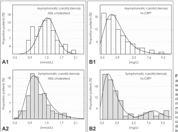
Circulating biomarkers in asymptomatic and symptomatic patients with carotid stenosis Circulating cytokines and blood lipid levels in the study gr- oups are given in Table 2. While the levels of total cholesterol, low-density lipoprotein (LDL) cholesterol, and Lp(a) did not differ, those of high-density lipoprotein (HDL) cholesterol were significantly higher in the CS-asymptomatic subjects [1.30 mmol/L (range=1.1-1.5 mmol/L) vs. 1.08 mmol/L (ran- ge=0.9-1.3 mmol/L), p<0.001]. The level of triglycerides (TGs) was significantly higher in the CS-symptomatic pa- tients [1.31 mmol/L (range=0.9-1.7 mmol/L) vs. 1.42 mmol/L (range=1.1-1.9 mmol/L), p=0.03]. The distribution of HDL- cholesterol levels (Fig. 1A) indicates a clear shift toward lower HDL-cholesterol values in those with symptomatic CS.
In addition, the LDL-/HDL-cholesterol ratio was significantly higher in the CS-symptomatic patients [2.35 (range=1.7-2.9) vs. 2.05 (range=1.6-2.6), p=0.008]. However, there were no differences in the lipid profile between those with last symp- tom occurrence ≤6 months and those with the last episode
>6 months (Table 2), consistent with the concept that lipids are associated with a long-term (chronic) rather than acute risk of CS-symptomatic transformation.
The IL-6 level was significantly higher in the symptomatic patients [3.69 pg/mL (range=1.44-6.81 pg/mL) vs. 2.44 pg/mL (range=1.07-5.41 pg/mL, p=0.04)], but no between-group difference was found for the other studied interleukins (IL- 1β, IL-8, and IL-18) (Table 2). Although the median level of hsCRP was higher in the symptomatic subjects [2.11 mg/L (range=1.4-6.1 mg/L) vs. 2.08 mg/L (range=1.2-3.4 mg/L)], the overall difference did not reach statistical significance (p=
0.07). Exclusion of subjects with an hsCRP exceeding 10 mg/L19 (n=2, 1.8%, in the asymptomatic group; n=8, 6.7% in the symptomatic ≤6 months group; n=6, 8.4%, in the symptom- atic >6 months group; in all cases, hsCRP values were ≤15 mg/L) had no effect on the hsCRP analysis outcome (Table 2).
Nevertheless, analysis of hsCRP distribution (Fig. 1B) indi- cated that while both asymptomatic and symptomatic sub- jects exhibited a prevalence peak at ≈2 mg/L, the symptom- atic subjects exhibited a bimodal distribution with a nadir at
≈5.2 mg/L and a second, smaller prevalence peak at ≈7 mg/
L. When this nadir was taken as a cutoff, there were 11.2%
asymptomatic subjects vs. 27.7% symptomatic subjects with hsCRP ≥5.2 mg/L [odds ratio (OR)=3.06, 95% confidence in- terval (95%CI)=1.51-6.22, p=0.002], indicating that hsCRP levels exceeding 5.2 mg/L may be associated with a signifi- cant increase in the likelihood of CS-symptomatic status.
However, hsCRP ≥5.2 mg/L could not differentiate between those with recent vs. remote symptoms of CS (p=0.727), con- sistent with the potential chronic rather than acute association between hsCRP and the risk of CS-symptomatic transforma- Table 1. Demographic and clinical characteristics of the study group, and index internal carotid artery (ICA) Doppler velocities
Asymptomatic patients
(n=110)
Symptomatic patients
(n=190)
p value
Symptomatic patients
p value
≤6 months (n=119)
>6 months (n=71)
Age, years 67 (60-71) 66 (60-72) 0.96 67 (61-73) 64 (59-69) 0.09
Gender: men, % (n) 59.1 (65) 67.4 (128) 0.15 68.1 (81) 66.2 (47) 0.79
Arterial hypertension, % (n) 86.4 (95) 90.5 (172) 0.20 89.9 (107) 91.5 (65) 0.84
Diabetes, % (n) 27.3 (30) 35.3 (67) 0.15 37.8 (45) 30.9 (22) 0.34
On insulin, % (n) 8.2 (9) 11.6 (22) 0.35 10.9 (13) 12.7 (9) 0.72
h/o MI 29.1 (32) 23.2 (44) 0.24 17.6 (21) 32.4 (23) 0.02
Smoking (current or past), % (n) 56.4 (62) 55.3 (105) 0.89 53.8 (64) 57.8 (41) 0.64
CAD, % (n) 67.3 (74) 66.3 (126) 0.87 61.3 (73) 74.7 (53) 0.09
PAD, % (n) 17.3 (19) 11.6 (22) 0.17 10.9 (13) 12.7 (9) 0.70
BMI 27.8 (25.9-30.2) 27.7 (25.5-30.1) 0.95 27.7 (25.5-30.1) 27.7 (25.5-30.1) 0.95
BMI ≥30 kg/m2, % (n) 28.2 (31) 27.9 (53) 0.98 28.6 (34) 26.7 (19) 0.89
Creatinine (µmol/L) 87.0 (74-103) 85.0 (74-99) 0.66 83 (74-97) 87 (76-104) 0.21
eGFR <60 mL/min (MDRD), % (n) 24.5 (27) 20.5 (39) 0.47 19.5 (23) 22.5 (16) 0.36
Index ICA peak systolic velocity, m/s 2.64 (2.02-3.5) 2.55 (1.9-3.3) 0.22 2.62 (1.9-3.5) 2.44 (1.9-3.2) 0.61 Index ICA end-diastolic velocity, m/s 0.87 (0.7-1.2) 0.86 (0.7-1.2) 0.86 0.87 (0.7-1.3) 0.84 (0.6-1.2) 0.56 Continuous data are median (Q1-Q3); categorical data are % (n).
BMI: Body Mass Index, CAD: coronary artery disease, eGFR: estimated Glomerular filtration rate, h/o MI: history of myocardial infarct, MDRD: modification of died in renal disease formula, PAD: peripheral arterial occlusive disease.
tion. In addition, there was a weak but statistically significant negative correlation between HDL and hsCRP (r=-0.32, p<
0.0001).
The plasma levels of the MMPs (MMP-8, MMP-9, and MMP-10) did not differ between the study groups (Table 2).
In contrast, circulating TIMP was significantly lower in the CS- symptomatic patients [130.0 ng/mL (range=7.3-166.5 ng/mL) vs. 146.2 ng/mL (range=115.3-177.9 ng/mL), p=0.02], indi- cating a shift toward reduced metalloproteinase inhibition in subjects with symptomatic CS. The overall levels of fibrino- gen, visfatin, CD40L, sVCAM, leptin, adiponectin (including leptin/adiponectin ratio), LP-PLA2, MCP-1, MPO, and OPG also did not differ between the symptomatic and asymptom-
atic patients. However, when the symptomatic patients were divided into those with recent vs. remote symptoms, the levels of LP-PLA2 and OPG were significantly higher in the recently CS-symptomatic subjects [0.33 µg/mL (range=0.3-0.4 µg/mL) vs. 0.31 µg/mL (range=0.2-0.4 µg/mL), p=0.02; 4.70 pg/mL (range=3.6-6.3 pg/mL) vs. 4.14 pg/mL (range=2.8-5.5 pmol/L), p=0.02].
Since patients for whom >6 months has elapsed since their last episode of CS-attributable neurological symptoms (i.e., those “remotely” symptomatic by the current study definition) are often classified as “asymptomatic”,1,16,20 the effect of merg- ing this subset (n=71) with the a priori asymptomatic group (n=190) on the biomarker data was tested. Incorporation in Table 2. Laboratory characteristics of the study group
Asymptomatic*
patients (n=110)
Symptomatic*
patients (n=190)
p value
Symptomatic* patients
p value
≤6 months (n=119)
>6 months (n=71)
Total cholesterol (mmol/L) 4.5 (3.8-5.3) 4.4 (3.9-5.0) 0.31 4.38 (3.9-4.9) 4.43 (3.9-5.2) 0.42 LDL-cholesterol (mmol/L) 2.58 (2.0-3.2) 2.51 (2.1-3.0) 0.37 2.46 (2.0-2.9) 2.54 (2.1-3.1) 0.46 HDL-cholesterol (mmol/L) 1.30 (1.1-1.5) 1.08 (0.9-1.3) <0.001 1.07 (0.9-1.3) 1.11 (0.9-1.3) 0.74
LDL/HDL ratio 2.05 (1.6-2.6) 2.35 (1.7-2.9) 0.008 2.24 (1.7-2.8) 2.37 (1.9-3.0) 0.41
Triglycerides (mmol/L) 1.31 (0.9-1.7) 1.42 (1.1-1.9) 0.03 1.41 (0.9-1.8) 1.42 (1.2-2.0) 0.12 Lp(a) (mg/dL) 9.27 (4.1-17.3) 9.79 (4.1-23.6) 0.25 10.11 (4.1-27.1) 9.48 (4.0-19.4) 0.75
hs-CRP (mg/L) 2.08 (1.2-3.4) 2.11 (1.4-6.1) 0.07 2.37 (1.4-5.5) 1.93 (1.2-6.4) 0.47
hs-CRP (≤10)† (mg/L) 1.99 (1.2-3.3) 2.0 (1.2-3.9) 0.28 2.17 (1.3-3.9) 1.77 (1.1-2.8) 0.27
Fibrinogen (g/L) 4.44 (3.6-5.0) 4.11 (3.3-4.9) 0.19 3.96 (3.4-5.2) 4.13 (3.3-4.8) 0.62
IL-1β (pg/mL) 0.14 (0.1-0.2) 0.13 (0.1-0.2) 0.87 0.13 (0.1-0.2) 0.12 (0.1-0.2) 0.68
IL-6 (pg/mL) 2.44 (1.1-5.4) 3.69 (1.4-6.8) 0.04 3.41 (1.5-6.7) 4.16 (1.3-7.3) 0.94
IL-8 (pg/mL) 8.21 (5.3-11.4) 8.73 (5.8-11.9) 0.49 8.73 (6.3-12.0) 8.73 (4.9-13.3) 0.67
IL-18 (µg/mL) 0.33 (0.3-0.4) 0.32 (0.2-0.4) 0.78 0.33 (0.2-0.4) 0.32 (0.3-0.4) 0.47
sVCAM (mg/mL) 0.87 (0.7-1.1) 0.91 (0.7-1.2) 0.28 0.86 (0.7-1.1) 0.94 (0.7-1.2) 0.43
CD40L (ng/mL) 0.31 (0.2-0.5) 0.35 (0.2-0.5) 0.63 0.37 (0.2-0.5) 0.31 (0.2-0.4) 0.67
Lp-PLA2 (ng/mL) 326.9 (260-381) 319.7 (266-373) 0.65 328.8 (274-407) 310.4 (247-338) 0.02
Visfatin (ng/mL) 0.32 (0.2-0.6) 0.25 (0.1-0.5) 0.25 0.26 (0.2-0.6) 0.22 (0.1-0.5) 0.39
MCP-1 (ng/mL) 0.22 (0.2-0.3) 0.25 (0.2-0.3) 0.25 0.26 (0.2-0.3) 0.24 (0.2-0.3) 0.34
MPO (ng/mL) 45.4 (28.9-79.4) 48.3 (29.6-77.8) 0.82 49.1 (29.6-78.6) 47.5 (29.0-76.9) 0.88 MMP-8 (ng/mL) 20.0 (10.6-32.3) 20.2 (11.3-37.5) 0.42 19.7 (9.9-35.0) 24.5 (12.4-39.1) 0.40
MMP-9 (µg/mL) 0.13 (0.1-0.2) 0.14 (0.1-0.2) 0.36 0.13 (0.1-0.2) 0.16 (0.1-0.2) 0.29
MMP-10 (µg/mL) 0.66 (0.4-0.8) 0.58 (0.5-0.8) 0.63 0.56 (0.5-0.8) 0.66 (0.5-0.8) 0.12
TIMP (ng/mL) 146.2 (115-178) 130.7 (108-176) 0.02 130.6 (101-166) 131 (112-165) 0.75
Leptin (ng/mL) 12.15 (4.9-27.2) 11.37 (5.1-19.5) 0.57 11.06 (5.1-19.8) 12.18 (5.8-19.2) 0.70 Adiopnectin (μg/mL) 4.13 (2.9-5.9) 3.96 (2.7-6.6) 0.64 3.56 (2.3-6.7) 4.42 (3.0-6.5) 0.24 Leptin/adiponectin ratio 3.01 (1.0-6.3) 1.91 (0.9-5.6) 0.36 1.81 (0.9-5.4) 2.19 (0.9-7.3) 0.61
OPG (pmol/L) 4.28 (2.9-5.0) 4.45 (3.3-5.9) 0.21 4.71 (3.6-6.3) 4.13 (2.8-5.4) 0.01
Data are shown as median (Q1-Q3).
*The terms “symptomatic” or “asymptomatic” refer to neurological symptoms attributable to carotid artery stenosis by an indepen- dent neurologist, †Subjects with hsCRP ≥10 mg/L excluded (n=2 with asymptomatic carotid stenosis, n=8 symptomatic ≤6 months and n=6 with last symptoms >6 months; NB. the peak hsCRP level was 13.86 mg/L).
CD40L: soluble CD 40 receptor ligand, HDL: high-density lipoprotein, hs-CRP: high sensitivity C-reactive protein, IL: interleukin, LDL: low- density lipoprotein, Lp(a): lipoprotein a, Lp-PLA2: lipoprotein-associated phospholipase A2, MCP: monocyte chemoattractant protein, MMP: matrix metalloproteinase, MPO: myeloperoxidase, OPG: osteoprotegerin, sVCAM: soluble vascular cell adhesion molecule, TIMP: tissue inhibitor of matrix metalloproteinases-1.
the asymptomatic cohort of the patients with the last neuro- logical symptoms occurring >6 months prior to biomarker sampling blunted the asymptomatic vs. symptomatic differ- ence in HDL-cholesterol and removed the differences in LDL/
HDL-cholesterol, TGs, TIMP, and IL-6, indicating that sub- jects with prior symptoms of CS remain distinct from the as- ymptomatic group.
ROC analysis for CS-symptomatic status prediction
Receiver operating characteristics analysis included HDL, TIMP, and IL-6 (but not TGs, which showed a weak though significant negative correlation with the HDL level; r=-0.28, p<0.001). The highest area under the curve (AUC) was found for HDL-cholesterol (AUC=0.70, 95%CI=0.63-0.76, cut- off=1.16 mmol/L, positive predictive value=0.78, negative pre- dictive value=0.52) (Fig. 2A). IL-6 paired with TIMP performed similarly to HDL-cholesterol taken alone (Fig. 2B), but com- bining all three biomarkers (i.e., HDL+IL-6+TIMP) did not surpass the diagnostic accuracy of HDL-cholesterol (Fig. 2C).
Lipoprotein-associated phospholipase A2 and OPG were significantly higher in those with CS symptoms occurring ≤6 months vs. >6 months (Table 2) and were not interrelated (r=
-0.09, p=0.32), and so their individual vs. combined power in the prediction of CS-recently vs. CS-remotely symptomatic sta- tus was tested. A greater power was found for Lp-PLA2+OPG (AUC=0.70, 95%CI=0.60-0.78, p<0.01) when compared to the ROC for Lp-PLA2 alone (AUC=0.63, 95%CI=0.53-0.72, p=0.019, cutoff=361 ng/mL) or OPG alone (AUC=0.62, 95%
CI=0.52-0.72, p=0.02, cutoff=3.73 pmol/L), consistent with the idea that Lp-PLA2 and OPG may affect plaque biology via different mechanisms.
Univariate and multivariate logistic regression models
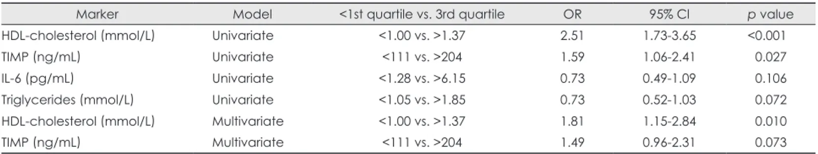
Univariate and multivariate logistic regression analyses were performed to assess the role of angiographic stenosis severity and atheroma burden on intravascular ultrasound, and of HDL- cholesterol, TIMP, IL-6, and TG level in predicting CS-symp- tomatic status. In the univariate model, HDL-cholesterol and TIMP (but not IL-6 or TG) predicted the CS-symptomatic status (OR=2.51, 95%CI=1.73-3.65, p<0.001 for HDL-cho- lesterol <1 mmol/L vs. >1.37 mmol/L; and OR=1.59, 95%CI=
1.06-2.41, p=0.027 for TIMP <111 ng/mL vs. >204 ng/mL) (Table 3). The angiographic stenosis severity and atheroma burden as assessed by IVUS were not predictive of CS symp- toms (p=0.16 and p=0.68, respectively). In a multivariate mo- del, a low HDL-cholesterol level was the sole predictor of CS- symptomatic status (OR=1.81, 95%CI=1.15-2.84 for HDL- cholesterol <1 mmol/L vs. >1.37 mmol/L, p=0.01) (Table 3).
Discussion
The key finding of this study, which evaluated the largest up- to-date panel of circulating biomarkers in subjects with CS, is that low HDL-cholesterol was the sole independent predictor of CS-symptomatic status in a contemporary CS population.
In particular, we found that the likelihood of CS-associated
18 12 6
18 12 6
Proportion patients (%)Proportion patients (%)
0.5 2.9 5.2 7.6 9.2 (mg/L)
0.5 2.9 5.2 7.6 9.2 (mg/L)
Asymptomatic carotid stenosis hs-CRP*
Symptomatic carotid stenosis hs-CRP*
B1
B2
Fig. 1. Distributions of HDL-cholesterol and hsCRP* in patients with carotid st- enosis according to the presence or ab- sence of a history of carotid stenosis- associated symptoms. A: Displays HDL- cholesterol level distribution (asympto- matic carotid stenosis top, A1; sympto- matic carotid stenosis bottom, A2). B:
hsCRP level distribution (asymptomatic carotid stenosis top, B1; symptomatic carotid stenosis bottom, B2). HDL: high- density lipoprotein, hsCRP: high sensi- tivity C-reactive protein.
18 15 11 7 4
18 15 11 7 4 Proportion patients (%)Proportion patients (%)
0.5 0.9 1.3 1.7 2.1 (mmol/L)
0.5 0.9 1.3 1.7 2.1 (mmol/L)
Asymptomatic carotid stenosis HDL-cholesterol
Symptomatic carotid stenosis HDL-cholesterol
A1
A2
symptoms increased by a factor of 1.81 (95%CI=1.15-2.84) between the first and fourth HDL-cholesterol quartiles. De- spite statistically significant differences in IL-6, TIMP, and TG level between the CS-symptomatic and CS-asymptomatic subjects, in the multivariable model these biomarkers were not independent predictors of CS symptoms. Moreover, their ad- dition to HDL-cholesterol failed to provide any incremental value over HDL-cholesterol alone in the ROC analysis (Fig. 2).
Thus, the findings of the present study in an unselected pop- ulation of CS subjects on contemporary pharmacotherapy are unable to confirm the clinical utility of several circulating
biomarkers that have previously been suggested to indicate the symptomatic status of CS, including hsCRP,4,5 IL-6,4,21 Lp(a),4 and MMPs.4,6 Moreover, this analysis did not demonstrate the utility of several other classic (e.g., CD40L, sVCAM, fi- brinogen, IL-1β, IL-8, IL-18, MPO, and MCP-1)4,11 and novel (e.g., visfatin and leptin/adiponectin)7,9,22 circulating biomark- ers of atherosclerotic plaque destabilization and rupture.
Subjects with symptomatic CS demonstrated a reduced le- vel of circulating TIMP, consistent with dysregulation of the MMP/TIMP balance in patients with symptomatic CS, result- ing in decreased endogenous inhibition of MMPs.23 Although Fig. 2. ROC curves for determining the symptomatic status of CS.
Biomarker(s) sensitivity is demonstrated as a function of 1-specificity based on a logistic model incorporating three uncorrelated biomark- ers (HDL-cholesterol, TIMP, and IL-6) whose levels differed signifi- cantly between symptomatic and asymptomatic subjects. A: Contri- bution of each biomarker taken alone; the AUC was highest for HDL- cholesterol. B: Contributions of paired biomarkers, indicating that neither of the pairs has a higher diagnostic power than HDL-choles- terol alone (cf. A). C: ROC curve for combined HDL-cholesterol+IL- 6+TIMP, indicating that this was not higher than for HDL-cholesterol alone (cf. A). AUC: area under the curve, CS: carotid stenosis, HDL:
high-density lipoprotein, IL-6: interleukin-6, ROC: receiver operating characteristics, TIMP: tissue inhibitor of matrix metalloproteinases-1.
1.0
0.8
0.6
0.4
0.2
0.0
1.0
0.8
0.6
0.4
0.2
0.0
1.0
0.8
0.6
0.4
0.2
0.0
Sensitivity Sensitivity
Sensitivity
0.0 0.2 0.4 0.6 0.8 1.0 0.0 0.2 0.4 0.6 0.8 1.0
0.0 0.2 0.4 0.6 0.8 1.0 HDL TIMP IL-6
HDL+TIMP TIMP+IL-6 IL-6+TIMP
HDL+IL-6+TIMP
1-specificity 1-specificity
1-specificity
AUC 95%CI Cutoff Sensitivity Specificity PPV NPV
HDL 0.70 0.63-0.76 1.16
mmol/L 0.62 0.71 0.78 0.52
TIMP 0.60 0.52-0.68 122
ng/mL 0.45 0.72 0.72 0.45
Il-6 0.59 0.50-0.67 3.66
pg/mL 0.51 0.67 0.72 0.45
AUC 95%CI Sensitivity Specificity PPV NPV
HDL+TIMP 0.67 0.59-0.74 0.69 0.58 0.72 0.54
HDL+IL-6 0.66 0.58+0.73 0.58 0.65 0.73 0.49
Il-6+TIMP 0.70 0.64-0.76 0.62 0.73 0.79 0.53
AUC 95%CI Sensitivity Specificity PPV NPV
HDL+IL-6+TIMP 0.67 0.60-0.75 0.55 0.72 0.74 0.52
A
C
B
the overall levels of Lp-PLA2 and OPG did not differ between those with and without CS symptoms, these two biomarkers were significantly elevated in subjects with recent vs. remote CS symptoms. Both Lp-PLA2 and OPG promote plaque in- stability and rupture.4,8 The present finding is in line with re- cent histological evidence of significantly increased Lp-PLA2
and OPG expression in carotid plaques in patients neurologi- cally symptomatic during the preceding 1.5-4 months,4,8 and OPG prediction of premature atherosclerosis progression in asymptomatic normotensive individuals.24 Furthermore, the absence of a correlation between levels of Lp-PLA2 and OPG (r=-0.09, p=0.32) is consistent with their independent action in promoting plaque instability. Incorporation of both Lp- PLA2 and OPG in the ROC analysis increased their individu- al AUC in the determination of recently symptomatic CS sta- tus (AUC=0.63 for Lp-PLA2, 0.62 for OPG, and 0.70 for Lp- PLA2+OPG).
“Symptomatic” status of the carotid stenosis There is currently no single, generally applied definition of symptomatic CS with respect to the time elapsed since the last symptom episode. For example, the term “asymptomatic”
has been used not only to label patients with no history of ipsi- lateral symptoms,10,25 but also to those with prior symptoms who have been free of neurological events for a period from 1-4 months8 to 12 months,6 with the cutoff for the CS symp- tom-free period frequently taken as 6 months.1,16,20 With these varying definitions, subjects with a history of CS symptoms during the previous 1-12 months have been classified in dif- ferent previous studies either as “symptomatic” or “asymp- tomatic.”8,20,26,27 Data from these studies,8,20,26,27 when consid- ered on aggregate, suggest that the use of different definitions of CS “symptomatic” status could have a significant impact on the findings. For clarity of analysis, in the present study all patients with history of CS symptoms were considered “symp- tomatic”. Moreover, the applied cutoff of 6 months for distin- guishing those with recent vs. remote symptoms is consistent with recent histological evidence that most carotid plaques stabilize within 6 months after the neurological event.25
There is experimental evidence4,10,25 that certain biomarkers have a chronic effect on carotid plaque stability (e.g., low HDL,
low TIMP, and elevated IL-6), whereas others (possibly LP- PLA2 and OPG) have a more transient impact or acutely re- flect plaque destabilization. Several previous studies23,28 have interpreted the serum biomarker level in the context of plaque histology rather than of the neurological symptoms of carotid plaque rupture and thrombosis. Indeed, it is well known that carotid plaques can undergo several episodes of rupture and thrombosis that may remain clinically silent, although they usually lead to plaque progression.29,30 Our finding that inclu- sion of the remotely symptomatic subjects in the asymptom- atic group reduced or abolished the between-group differences in circulating biomarkers is consistent with the concept that the circulating biomarker profile of subjects with remote symp- toms of CS may remain at least partially different from that of never-symptomatic subjects.
Systemic biomarker concentration vs. in-situ plaque destabilization and rupture
In an ideal setting of the assessment of circulating biomarkers in relation to CS clinical symptoms, either the release of the biomarker from the index lesion should be sufficiently high to affect its systemic level or a “causative” circulating bio- marker released elsewhere should exclusively affect stability of the index-in this case carotid-lesion under consideration.
This is not necessarily the case because instability of the ath- erosclerotic lesion(s) in one vascular bed (e.g., carotid) is of- ten associated with instability of atherosclerotic lesion(s) in other beds (e.g., coronary)30 through “vulnerable blood”
mechanisms.31,32 The available data suggest that atheroscle- rotic plaque destabilization and rupture in one specific vascu- lar territory involves an interplay between the local (in situ) factors7,33-35 that make a particular plaque prone to erosion or rupture (“vulnerable plaque”) and systemic factors (“vulner- able blood”).31,32 Thus, the circulating biomarker level actually reflects the net effect of 1) biomarker production and release in the “target” atherosclerotic lesion, with a local release pos- sibly too small to be detected systemically6,27,34 and/or occur- ring only transiently;25 2) the contribution of biomarker release from atherosclerotic plaques in other vascular territories; and 3) for some biomarkers, their production elsewhere (e.g., the liver in the case of fibrinogen or hsCRP).22,26,36
Table 3. Univariate and multivariate logistic regression analysis for the prediction of carotid artery stenosis symptomatic status
Marker Model <1st quartile vs. 3rd quartile OR 95% Cl p value
HDL-cholesterol (mmol/L) Univariate <1.00 vs. >1.37 2.51 1.73-3.65 <0.001
TIMP (ng/mL) Univariate <111 vs. >204 1.59 1.06-2.41 0.027
IL-6 (pg/mL) Univariate <1.28 vs. >6.15 0.73 0.49-1.09 0.106
Triglycerides (mmol/L) Univariate <1.05 vs. >1.85 0.73 0.52-1.03 0.072
HDL-cholesterol (mmol/L) Multivariate <1.00 vs. >1.37 1.81 1.15-2.84 0.010
TIMP (ng/mL) Multivariate <111 vs. >204 1.49 0.96-2.31 0.073
HDL: high-density lipoprotein, IL: Interleukin, OR: odds ratio, TIMP: tissue inhibitor of matrix metalloproteinases-1.
Several previous studies of the relationship between circu- lating biomarkers and carotid atherosclerosis have either ex- cluded patients with atherosclerosis in other vascular territo- ries35 or have used combined end points (e.g., death/TIA/st- roke/myocardial infarction, or any revascularization and/or carotid plaque progression) to identify a biomarker utility in relation to CS-symptomatic status.9,13,15,26,32 In addition, con- temporary pharmacotherapy, which involves a high propor- tion of statin and angiotensin-converting enzyme inhibitor use, may play an important role in reducing the level of inflamma- tory markers and/or weaken the relationship between biomark- ers and the risk of symptomatic plaque transformation.37-39 Low HDL-cholesterol as an independent predictor of CS-symptomatic status
High-density lipoprotein-cholesterol protects against athero- sclerosis via several anti-inflammatory, antioxidant, and anti- thrombotic effects, including reverse cholesterol transport in the liver, prostacyclin release, and the inhibition of endotheli- al adhesion molecule expression, monocyte chemotactic ac- tivity, and LDL oxidation.40 Our novel finding of HDL-chole- sterol as an independent predictor of CS-symptomatic status in patients with established carotid atherosclerosis is consis- tent with the association between low HDL-cholesterol with the unstable carotid plaque phenotype on conventional his- tology.30 It is also consistent with the finding of low HDL-ch- olesterol more frequent in stroke and TIA patients with ath- erosclerotic large-vessel stenosis than in those with stroke/TIA in the absence of atherosclerotic large-vessel stenosis.41 The failure of previous studies to identify a link between low HDL- cholesterol and the neurological symptoms of carotid athero- sclerosis may have been due to the inclusion of relatively small samples and/or the classification of the patients with last symptoms of CS >3-6 months as asymptomatic,8 which would blunt any potential differences between the truly asymptom- atic vs. those with prior symptoms of CS. Previous work sug- gested a protective role for high HDL-cholesterol levels ag- ainst the progression of carotid atherosclerosis rather than its symptomatic conversion;4 there is also recent prospective evi- dence that increased HDL-cholesterol protects against the pro- gression of intracranial atherosclerosis.43
The CS severity was similar in the symptomatic and as- ymptomatic subjects included in the present study, which sup- ports the concept of a protective role played by high HDL-cho- lesterol through reducing the risk of symptomatic transform- ation of the carotid plaque. A recent study of left main co- ronary artery atherosclerosis44 indicated that a low HDL/LDL- cholesterol ratio may be related to an increased lipid content and smaller fibrous content observed on the plaque radiofre- quency IVUS imaging, possibly rendering the atherosclerotic
plaque more amenable to symptomatic transformation. Such an association is yet to be evaluated for carotid bifurcation atherosclerotic disease.
Limitations
While this study employed the most extensive up-to-date pa- nel of biomarkers in patients with carotid atherosclerosis, the discriminative power of remnant lipoprotein cholesterol,43,45 which was shown recently to be a risk factor for large-artery atherosclerotic stroke,46 was not evaluated. Secondly, due to the natural history of carotid atherosclerosis, the CS cohort labeled “asymptomatic” is known to include up to ≈10-15%
of subjects whose CS is likely to turn symptomatic over the subsequent 10 years.3 This natural presence of future-symp- tomatic subjects in the thus-far-asymptomatic group (both labeled “asymptomatic”) may obscure biomarker profile dif- ferences in a cross-sectional study. Moreover, in search of patient characteristics that might aid clinical decision-making in an all-comer population with CS, we deliberately avoided any preselection of study subjects.47 Thus, the present study cohort included a sizeable proportion of patients with estab- lished (and/or symptomatic) atherosclerosis in other vascular territories (Table 1). This is likely to blunt any differences in the circulating biomarker profiles between the study groups.
Finally, the finding that circulating OPG and Lp-PLA2 might transiently label symptomatic transformation of the carotid atherosclerotic plaque warrants further, prospective48 valida- tion in a large “natural history” study with 1) repeated (serial) biomarker sampling to enable a change in biomarker level
“just” prior to symptom occurrence, and 2) ideally, repeated carotid plaque imaging. However, due to the relatively low event rate in subjects with asymptomatic carotid stenosis on contemporary pharmacotherapy and the unavoidable cross- over of some patients to mechanical revascularization, such a large longitudinal study is unlikely to be conducted.
Conclusions
The present analysis in patients with CS subjected to con- temporary pharmacotherapy found that among several circu- lating biomarkers that have previously been implicated in in- dicating carotid plaque destabilization and symptomatic tr- ansformation, low HDL-cholesterol was the only independent predictor of neurological symptoms. This finding is consistent with the detrimental effect of low HDL-cholesterol in patients with clinical manifestation of atherosclerosis in some other vascular beds,39 indicating that the HDL-cholesterol level sh- ould be considered in the routine risk stratification of asymp- tomatic carotid stenosis. The findings also suggest that low HDL-cholesterol could constitute an important therapeutic target in subjects with carotid stenosis. Nevertheless, the ex-
tents to which a low HDL-cholesterol level is a causal factor versus an epiphenomenon have not been determined.40 More- over, there is recent evidence that forced elevation of defective HDL-cholesterol is clinically ineffective.40 Therefore, whether (and which) pharmacologic interventions aimed at increasing HDL-cholesterol would actually reduce the risk of carotid pla- que symptomatic transformation has to be demonstrated.
Conflicts of Interest
The authors have no financial conflicts of interest.
Acknowledgements
We thank Ms. Justyna Stefaniak of Data Management and Statistical Analysis (DMSA), Krakow, for data processing.
This work was supported by grants from the Ministry of Science and Higher Education, Poland (no. N402184234), Polish Cardiac Society/Ser- vier (2007), Polish Cardiac Society/Adamed (2008), and the “For Heart”
Foundation in Krakow, Poland.
REFERENCES
1. Goldstein LB, Bushnell CD, Adams RJ, Appel LJ, Braun LT, Ch- aturvedi S, et al. Guidelines for the primary prevention of stroke: a guideline for healthcare professionals from the American Heart As- sociation/American Stroke Association. Stroke 2011;42:517-584.
2. Kim JT, Yoo SH, Kwon JH, Kwon SU, Kim JS. Subtyping of ischemic stroke based on vascular imaging: analysis of 1,167 acute, consecutive patients. J Clin Neurol 2006;2:225-230.
3. Halliday A, Harrison M, Hayter E, Kong X, Mansfield A, Marro J, et al. 10-year stroke prevention after successful carotid endarterectomy for asymptomatic stenosis (ACST-1): a multicentre randomised trial.
Lancet 2010;376:1074-1084.
4. Koenig W, Khuseyinova N. Biomarkers of atherosclerotic plaque in- stability and rupture. Arterioscler Thromb Vasc Biol 2007;27:15-26.
5. Alvarez Garcia B, Ruiz C, Chacon P, Sabin JA, Matas M. High-sensi- tivity C-reactive protein in high-grade carotid stenosis: risk marker for unstable carotid plaque. J Vasc Surg 2003;38:1018-1024.
6. Heo SH, Cho CH, Kim HO, Jo YH, Yoon KS, Lee JH, et al. Plaque rupture is a determinant of vascular events in carotid artery athero- sclerotic disease: involvement of matrix metalloproteinases 2 and 9.
J Clin Neurol 2011;7:69-76.
7. Dahl TB, Yndestad A, Skjelland M, Øie E, Dahl A, Michelsen A, et al.
Increased expression of visfatin in macrophages of human unstable ca- rotid and coronary atherosclerosis: possible role in inflammation and plaque destabilization. Circulation 2007;115:972-980.
8. Mannheim D, Herrmann J, Versari D, Gössl M, Meyer FB, McConnell JP, et al. Enhanced expression of Lp-PLA2 and lysophosphatidylcho- line in symptomatic carotid atherosclerotic plaques. Stroke 2008;39:
1448-1455.
9. Urbonaviciene G, Frystyk J, Flyvbjerg A, Henneberg EW, Lindholt JS.
Association of serum adiponectin with risk for cardiovascular events in patients with peripheral arterial disease. Atherosclerosis 2010;210:
619-624.
10. Golledge J, McCann M, Mangan S, Lam A, Karan M. Osteoprote- gerin and osteopontin are expressed at high concentrations within symptomatic carotid atherosclerosis. Stroke 2004;35:1636-1641.
11. Bornstein N, Korczyn A. Asymptomatic carotid artery stenosis (AC- AS). J Neural Transm 2011;118:629.
12. Cola C, Clementi E, Biondi-Zoccai G, Sangiorgi G. From carotid plaque biology to serologic markers of vulnerability to predict the risk of cerebrovascular events. Acta Chir Belg 2007;107:129-142.
13. Ikonomidis I, Stamatelopoulos K, Lekakis J, Vamvakou GD, Kremas-
tinos DT. Inflammatory and non-invasive vascular markers: the multi- marker approach for risk stratification in coronary artery disease. Ath- erosclerosis 2008;199:3-11.
14. Pieniazek P, Musialek P, Dzierwa K, Motyl R, Trystuła M, Przewlocki T, et al. Flow reversal for proximal neuroprotection during endovascu- lar management of critical symptomatic carotid artery stenosis coex- isting with ipsilateral external carotid artery occlusion. J Endovasc Ther 2009;16:744-751.
15. Kablak-Ziembicka A, Przewlocki T, Sokołowski A, Tracz W, Podolec P. Carotid intima-media thickness, hs-CRP and TNF-α are indepen- dently associated with cardiovascular event risk in patients with ath- erosclerotic occlusive disease. Atherosclerosis 2011;214:185-190.
16. European Stroke Organisation, Tendera M, Aboyans V, Bartelink ML, Baumgartner I, Clément D, et al. ESC Guidelines on the diagnosis and treatment of peripheral artery diseases: document covering athero- sclerotic disease of extracranial carotid and vertebral, mesenteric, re- nal, upper and lower extremity arteries: the Task Force on the Diag- nosis and Treatment of Peripheral Artery Diseases of the European Society of Cardiology (ESC). Eur Heart J 2011;32:2851-2906.
17. Musialek P, Pieniazek P, Tracz W, Tekieli L, Przewlocki T, Kablak- Ziembicka A, et al. Safety of embolic protection device-assisted and unprotected intravascular ultrasound in evaluating carotid artery ath- erosclerotic lesions. Med Sci Monit 2012;18:MT7-MT18.
18. Kwee RM, van Oostenbrugge RJ, Mess WH, Prins MH, van der Geest RJ, ter Berg JW, et al. Carotid plaques in transient ischemic at- tack and stroke patients: one-year follow-up study by magnetic reso- nance imaging. Invest Radiol 2010;45:803-809.
19. Bassuk SS, Rifai N, Ridker PM. High-sensitivity C-reactive protein:
clinical importance. Curr Probl Cardiol 2004;29:439-493.
20. Liapis CD, Bell PR, Mikhailidis D, Sivenius J, Nicolaides A, Fer- nandes e Fernandes J, et al. ESVS guidelines. Invasive treatment for carotid stenosis: indications, techniques. Eur J Vasc Endovasc Surg 2009;37(4 Suppl):1-19.
21. Koutouzis M, Rallidis LS, Peros G, Nomikos A, Tzavara V, Barbatis C, et al. Serum interleukin-6 is elevated in symptomatic carotid bifurca- tion disease. Acta Neurol Scand 2009;119:119-125.
22. Rudd JH, Myers KS, Bansilal S, Machac J, Woodward M, Fuster V, et al. Relationships among regional arterial inflammation, calcification, risk factors, and biomarkers: a prospective fluorodeoxyglucose posi- tron-emission tomography/computed tomography imaging study.
Circ Cardiovasc Imaging 2009;2:107-115.
23. Higashikata T, Yamagishi M, Higashi T, Nagata I, Iihara K, Miyamoto S, et al. Altered expression balance of matrix metalloproteinases and their inhibitors in human carotid plaque disruption: results of quanti- tative tissue analysis using real-time RT-PCR method. Atherosclerosis 2006;185:165-172.
24. Ste¸pien´ E, Fedak D, Klimeczek P, Wilkosz T, Banyś RP, Starzyk K, et al. Osteoprotegerin, but not osteopontin, as a potential predictor of vas- cular calcification in normotensive subjects. Hypertens Res 2012;35:
531-538.
25. Peeters W, Hellings WE, de Kleijn DP, de Vries JP, Moll FL, Vink A, et al. Carotid atherosclerotic plaques stabilize after stroke: insights into the natural process of atherosclerotic plaque stabilization. Arterioscler Thromb Vasc Biol 2009;29:128-133.
26. Sabeti S, Exner M, Mlekusch W, Amighi J, Quehenberger P, Rumpold H, et al. Prognostic impact of fibrinogen in carotid atherosclerosis: non- specific indicator of inflammation or independent predictor of disease progression? Stroke 2005;36:1400-1404.
27. Montecucco F, Lenglet S, Gayet-Ageron A, Bertolotto M, Pelli G, Pal- ombo D, et al. Systemic and intraplaque mediators of inflammation are increased in patients symptomatic for ischemic stroke. Stroke 2010;
41:1394-1404.
28. Pelisek J, Rudelius M, Zepper P, Poppert H, Reeps C, Schuster T, et al.
Multiple biological predictors for vulnerable carotid lesions. Cerebro- vasc Dis 2009;28:601-610.
29. Milei J, Parodi JC, Ferreira M, Barrone A, Grana DR, Matturri L. Ath- erosclerotic plaque rupture and intraplaque hemorrhage do not corre- late with symptoms in carotid artery stenosis. J Vasc Surg 2003;38:
1241-1247.
30. Mauriello A, Sangiorgi GM, Virmani R, Trimarchi S, Holmes DR Jr, Kolodgie FD, et al. A pathobiologic link between risk factors profile and morphological markers of carotid instability. Atherosclerosis 2010;
208:572-580.
31. Lombardo A, Biasucci LM, Lanza GA, Coli S, Silvestri P, Cianflone D, et al. Inflammation as a possible link between coronary and carotid plaque instability. Circulation 2004;109:3158-3163.
32. Naghavi M, Falk E, Hecht HS, Jamieson MJ, Kaul S, Berman D, et al.
From vulnerable plaque to vulnerable patient--Part III: executive sum- mary of the Screening for Heart Attack Prevention and Education (SHAPE) Task Force report. Am J Cardiol 2006;98:2H-15H.
33. Krupinski J, Turu MM, Martinez-Gonzalez J, Carvajal A, Juan-Babot JO, Iborra E, et al. Endogenous expression of C-reactive protein is increased in active (ulcerated noncomplicated) human carotid artery plaques. Stroke 2006;37:1200-1204.
34. Turu MM, Krupinski J, Catena E, Rosell A, Montaner J, Rubio F, et al.
Intraplaque MMP-8 levels are increased in asymptomatic patients with carotid plaque progression on ultrasound. Atherosclerosis 2006;187:
161-169.
35. Hermus L, Lefrandt JD, Tio RA, Breek JC, Zeebregts CJ. Carotid plaque formation and serum biomarkers. Atherosclerosis 2010;213:
21-29.
36. Debing E, Peeters E, Demanet C, De Waele M, Van den Brande P.
Markers of inflammation in patients with symptomatic and asymp- tomatic carotid artery stenosis: a case-control study. Vasc Endovascular Surg 2008;42:122-127.
37. Yamagami H, Sakaguchi M, Furukado S, Hoshi T, Abe Y, Hougaku H, et al. Statin therapy increases carotid plaque echogenicity in hyper- cholesterolemic patients. Ultrasound Med Biol 2008;34:1353-1359.
38. Horiuchi Y, Hirayama S, Soda S, Seino U, Kon M, Ueno T, et al. Statin therapy reduces inflammatory markers in hypercholesterolemic pa- tients with high baseline levels. J Atheroscler Thromb 2010;17:722- 729.
39. Seo SM, Choo EH, Koh YS, Park MW, Shin DI, Choi YS, et al. High- density lipoprotein cholesterol as a predictor of clinical outcomes in patients achieving low-density lipoprotein cholesterol targets with statins after percutaneous coronary intervention. Heart 2011;97:1943- 1950.
40. Gielen S, Landmesser U. A new look at HDL in coronary disease: can we escape natural history? Heart 2011;97:1899-1901.
41. Laloux P, Galanti L, Jamart J. Lipids in ischemic stroke subtypes. Acta Neurol Belg 2004;104:13-19.
42. Johnsen SH, Mathiesen EB, Fosse E, Joakimsen O, Stensland-Bugge E, Njølstad I, et al. Elevated high-density lipoprotein cholesterol levels are protective against plaque progression: a follow-up study of 1952 persons with carotid atherosclerosis the Tromsø study. Circulation 2005;112:498-504.
43. Kim DE, Kim JY, Jeong SW, Cho YJ, Park JM, Lee JH, et al. Associa- tion between changes in lipid profiles and progression of symptomatic intracranial atherosclerotic stenosis: a prospective multicenter study.
Stroke 2012;43:1824-1830.
44. Kurebayashi N, Yoshikawa D, Ishii H, Sato B, Ando H, Okada T, et al.
Impact of the low- to high-density lipoprotein cholesterol ratio on composition of angiographically ambiguous left main coronary artery plaque. Circ J 2011;75:1960-1967.
45. Kim DE, Kim JY, Schellingerhout D, Kim EJ, Kim HK, Lee S, et al.
Protease imaging of human atheromata captures molecular informa- tion of atherosclerosis, complementing anatomic imaging. Arterioscler Thromb Vasc Biol 2010;30:449-456.
46. Kim JY, Park JH, Jeong SW, Schellingerhout D, Park JE, Lee DK, et al.
High levels of remnant lipoprotein cholesterol is a risk factor for large artery atherosclerotic stroke. J Clin Neurol 2011;7:203-209.
47. Przewłocki T, Kabłak-Ziembicka A, Kozanecki A, Rzeźnik D, Pien- iazek P, Musiałek P, et al. Polyvascular extracoronary atherosclerotic disease in patients with coronary artery disease. Kardiol Pol 2009;67:
978-984.
48. Park KY, Youn YC, Chung CS, Lee KH, Kim GM, Chung PW, et al.
Large-artery stenosis predicts subsequent vascular events in patients with transient ischemic attack. J Clin Neurol 2007;3:169-174.