https://doi.org/10.35827/cp.2019.18.2.65
접수일: 2019년 5월 16일, 게재승인일: 2019년 8월 23일 책임저자: 박중현, 서울시 강남구 언주로 63길 20
06229, 강남세브란스병원 미래의학연구센터 3층 재활의학과
Tel: 02-2019-3491, Fax: 02-2019-4881 E-mail: [email protected]
요추부 추간판 탈출증 환자에 대한 고전압 미세전류치료의 누적치료효과
연세대학교 의과대학 재활의학교실 및 희귀난치성 신경근육병 재활연구소
윤왕현ㆍ박진영ㆍ김도영ㆍ박중현
Cumulative Therapeutic Effect of High-Voltage Microcurrent Therapy in Patients with Herniated Lumbar Disc
Wang Hyeon Yun, M.D., Jinyoung Park, M.D., Doyoung Kim, M.D. and Jung Hyun Park, M.D., Ph.D.
Department of Rehabilitation Medicine, Gangnam Severance Hospital, Rehabilitation Institute of Neuromuscular Disease, Yonsei University College of Medicine, Seoul, Korea
Objective: This study aims to evaluate the efficacy of high-voltage microcurrent therapy in patients with herniated lumbar disc (HLD) presenting radicular or back pain. Method: This is a retrospective study with 33 patients who are complaining pain with HLD findings on magnetic resonance image. Microcurrent therapy was applied to leg or paralumbar area. Treatment was conducted for seven minutes with 250∼1000 uA intensity as high as the patients could tolerate via stimulating probe with roller type and the frequency was 60 Hz with a sine wave pulse. The visual analogue scale (VAS) was measured just before and after the treatment. Results: The degree of pain reduction (△VAS) was 1.6 points after treatment on average.
The △VAS according to the diagnosis, stenosis, dermatome area, medication, pain site and caudal epidural block was not statistically significant. However, the △VAS according to the number of treatments (< 3, ≥ 3 times) showed a statistically significant difference (p=0.04). Conclusion: High-voltage microcurrent therapy may help reduce lumbar or lumbosacral radiating pain after the procedure. The effect was better when microcurrent was applied three times or more. This result suggests that the microcurrent would have cumulative effect on reducing radicular or back pain in patients with HLD. (Clinical Pain 2019;18:65-69)
Key Words: Herniated disc, Pain, Electric stimulation, Visual analog scale
서 론
허리 추간판 탈출증과 연관된 방사통은 연간 유병률이 약 2.2% 정도로 흔한 질환이다.1 요추부 추간판 탈출증은 급성으로는 허리 통증이나 다리저림 증상 등의 방사통으로 나타날 수 있다. 이러한 방사통 증상은 대게 완전히 회복되 는 좋은 예후를 보이나 약 30%에서는 1년이 경과한 뒤에도 증상이 지속되고 5∼15%의 환자에서는 수술적 치료를 필 요로 하기도 한다.2,3 허리 통증 및 방사통 치료를 위한 비수 술적 치료에는 약물치료, 운동치료, 견인치료, 척추 도수치 료(spinal manipulation) 및 각종 온열치료와 전기치료들이
있다.4
그 중에서도 본 연구에서 다루고자 하는 미세전류치료 (microcurrent therapy)는 고전압 미세전류 치료기기(PANA- CELL, Chungam Medical, Seoul, Korea)를 사용하여 마이 크로 암페어 단위의 전류를 이용하는 치료이다. 이 치료기 기는 기존의 미세전류치료에 더하여 고전압의 강한 전위차 를 유발시킴으로 미세전류를 신체조직에 더 효과적으로 공 급할 수 있도록 설계된 미세전류 치료기기이다. 미세전류 치료에 대한 연구로써, 미세전류를 동물의 힘줄에 적용하 였을 때 힘줄의 재생이 향상된다는 보고가 있다.5,6 또한 만 성 아킬레스건병증 환자에서 운동치료만 받은 군과 운동치 료와 미세전류치료를 동시에 받은 군으로 나누어 치료하였 을 때 미세전류치료를 받은 군에서 통증 및 아킬레스건의 경직도 호전이 더 크다는 연구결과가 있다.7 다른 한 연구 에서는 운동으로 유발된 넙다리뒤근육의 지연성 근통증에 대하여 미세전류치료를 양쪽 부위 중 한 부위에만 적용하 고 24시간, 48시간 및 72시간 경과 후 양쪽의 통증을 비교 하였을 때 미세전류치료를 적용한 부위에서의 통증이 유의
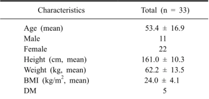
Table 1. Basic Characteristics of the Total Patients
Characteristics Total (n = 33)
Age (mean) 53.4 ± 16.9
Male 11
Female 22
Height (cm, mean) 161.0 ± 10.3 Weight (kg, mean) 62.2 ± 13.5 BMI (kg/m2, mean) 24.0 ± 4.1
DM 5
BMI: body mass index, DM: diabetes mellitus.
Fig. 1. Microcurrent therapy to a treatment site using roller type probe.
미하게 감소한 결과를 보고하였다.8 위 연구들은 통증치료 목적으로 미세전류가 사용된 연구들이나 허리 추간판 탈출 증의 통증 치료에 미세전류치료를 적용한 연구는 없었다.
이에 본 저자는 허리 추간판 탈출증 환자의 통증에 대한 고전압 미세전류치료 효과를 확인하고자 하였다.
연구 대상 및 방법 1. 연구대상
본 연구는 2016년 10월부터 2017년 11월까지 본원 재활 의학과에 입원한 환자들 중, 허리 자기공명영상(magnetic resonance imaging) 검사에서 허리 추간판 탈출이 확인 된 방사통이나 허리통증을 호소하는 33명의 환자를 대상으로 한 후향적 의무기록 분석 연구이다. 환자들의 평균 나이는 53.4세였고 남자의 수는 11명이었다. 본 연구자는 그 외 환 자들의 키, 몸무게, 신체비만지수, 당뇨병 유무 등의 정보를 조사하였다(Table 1). 통증을 유발할 수 있는 척수병증 (myelopathy)이나 척수종양(spinal cord tumor), 뇌졸중 (stroke) 등의 중추신경계 질환이나 기타 류마티스성 질환 이 있는 환자들은 배제하였다.
2. 연구방법
본 연구대상자들은 운동치료, 견인치료 및 약물치료를 받았고 일부 환자에서는 추가로 미세전류치료 및 미추차단 술을 시행받았다. 본 연구자는 미세전류치료를 위해 미세 전류 치료기기(PANACELL, Chungam Medical, Seoul, Korea)를 사용하여 연구대상자들이 통증을 주로 호소하는 부위에 시행하였고 주로 발바닥, 다리, 허리 부위로 통증 부위에 직접 적용하였다. 치료를 위한 미세전류는 롤러형 의 더듬자를 통하여 싸인파(sine wave)의 60 헤르츠(Hz)의 주파수로, 250 μA 에서 1000 uA까지의 강도로 적용되었 다(Fig. 1). 치료 시간은 치료 당 7분이며 환자가 견딜 수 있는 최대 강도의 미세전류가 적용되었고 치료는 1∼2회
또는 3회 이상으로 시행되었고 치료 1회 시행 후 환자가 느끼는 주관적인 효과 및 의료진의 판단에 따라 치료 지속 여부를 결정하였으며 3회 이상 받은 환자 중 가장 많은 치 료를 받은 환자는 최대 8회까지 시행 받았다. 또한 치료 전 과 후의 시각통증등급(VAS)의 변화를 각각 측정하였다. 치 료 전 후의 통증 변화(△VAS)를 몇 가지 기준으로 나누어 비교분석 하였는데, 그 기준은 다음과 같다. 첫째, 허리 자 기공명영상 검사에서 대상자들이 허리추간판 탈출을 보이 는 위치에 따라 각각 단일 분절의 추간판 탈출 그리고 2개 이상의 분절의 추간판 탈출로 분류하였다. 둘째, 대상자들 에게 시행한 허리 자기공명영상 검사에서 척추협착 동반 유무에 따라 분류하였다. 셋째, 대상자들의 방사통 피부분 절(dermatome) 위치에 따라 허리 5번, 천추 1번, 허리주변 부, 그리고 그 외의 다른 부위로 분류하였다. 넷째, 대상자 들이 복용하는 약물에 따라 비스테로이드소염제만 복용하 는 그룹(Medication 1), gabapentin 또는 pregabalin을 복용 하나 아편유사제(opioid) 성분의 약은 복용하지 않는 그룹 (Medication 2), gabapentin 또는 pregabalin의 복용 여부와 상관없이 아편유사제 성분의 약을 복용하는 그룹(Medicat- ion 3), 그 외의 기타 약물을 복용하는 그룹(Medication 4) 로 분류하였다. 다섯째, 대상자들의 치료부위가 허리인 경 우와 다리인 경우로 분류하였다. 여섯째, 미추차단 시행 여 부에 따라 분류하였다. 마지막 기준으로 대상자들에게 시 행한 치료횟수가 1∼2회인 경우와 3회 이상인 경우로 분류 하였다(Table 2).
Table 2. Pain Improvement after Microcurrent by Sub-Groups VAS
pre
VAS
post △VAS p Herniated level
Single level (n = 12) 5.3 3.4 1.9 0.24 Multi-level (n = 21) 6.1 4.6 1.5 Stenosis
Absent (n = 21) 6.1 4.5 1.6 0.96
Present (n = 12) 5.3 3.7 1.6 Dermatome
L5 (n = 6) 6.2 4.2 2.0 0.65
S1 (n = 13) 5.2 3.6 1.7
Paralumbar (n = 13) 6.2 4.8 1.4
Other (n = 1) 6.0 4.0 2.0
Medication
No Medication (n = 2) 5.0 2.5 2.5 0.39 Medication 1 (n = 8) 5.5 4.1 1.4 Medication 2 (n = 11) 6.0 4.2 1.8 Medication 3 (n = 9) 5.9 4.4 1.5 Medication 4 (n = 3) 6.0 4.7 1.3 Treatment site
Paralumbar (n = 13) 6.2 4.8 1.4 0.43
Limb (n = 20) 5.5 3.8 1.7
Number of treatment
< 3 (n = 21) 5.7 4.3 1.4 0.04*
≥ 3 (n = 12) 6.0 4.0 2.0
Caudal epidural block
Yes (n = 13) 6.2 4.7 1.5 0.58
No (n = 20) 5.5 3.9 1.6
VAS: visual analog scale, Medication 1: group taking only non- steroidal anti-nflammatory drug, Medication 2: group taking ga- bapentin or pregabalin but not opioid drugs, Medication 3: group taking opioid drugs with or without gabapentin or pregabalin, Medication 4: groups taking any other drugs.
*p<0.05, Kruskal Wallis test.
Table 3. Changes of Pain in Patients Treated More than 3 Times
Treatment number VAS (mean) △VAS (mean)
Pre-treatment 6.0
1st 4.0 2.0
2nd 2.9 3.1
3rd 2.7 3.3
△VAS was larger when the treatment was repeated.
VAS: visual analog scale.
3. 통계
본 저자는 치료효과를 확인하기 위하여 치료 전후의 시 각통증등급의 변화(△VAS)를 측정하였다. 통계분석을 위 하여 SPSS 23.0 프로그램을 사용하였고 치료 전후의 통증 변화에 대한 세부항목 별 비교를 위하여 크루스칼 왈리스 검정(Kruskal-Wallis test)을 이용하였으며, 유의수준은 0.05 이하인 경우를 통계적으로 유의성이 있는 것으로 정의하였다.
결 과
본 연구결과에서 치료 전 후의 통증 변화(△VAS)는 평 균 1.6점(5.8점에서 4.2점, 27.6%)으로 31명(93.9%)의 환자
에서 치료 직후 통증의 호전을 나타내었다.
앞에서 언급한 기준에 따른 미세전류치료 전후의 통증변 화는 추간판 탈출증 분절 수, 척추협착유무, 방사통 피부분 절, 약물, 치료 부위, 미추차단에 있어서는 통계적으로 유의 미하지 않았으나(p=0.24, 0.96, 0.65, 0.65, 0.39 and 0.58), 치료 횟수에 따른 통증 변화는 유의미한 결과를 보였는데, 본 연구에서 치료를 3회 이상 받은 12명의 환자들에서의
△VAS가 3회 미만으로 받았던 환자들의 △VAS에 비하여 유의미하게 큰 차이를 보였다(2.00 vs 1.38, p=0.04) (Table 2). 3회 이상 미세전류치료를 받은 환자들에서 1차에서 3차 까지 치료를 시행 함에 따라 평균 VAS는 6.0에서 4.0, 2.9, 2.7로 점차적으로 통증이 줄어들었고 시행 전에 비하여
△VAS는 각각 2.0, 3.1, 3.3으로 △VAS가 증가하였다 (Table 3).
고 찰
본 연구에서는 33명의 허리 추간판 탈출증 환자의 방사 통에 대하여 고전압 미세전류치료를 시행하였으며 치료 직 후 대부분의 환자(93.9%)에서 통증의 호전을 보였다. 본 연 구에서 미세전류치료를 받은 환자의 통증 변화(△VAS)에 영향을 미칠 수 있는 다양한 변수들인 추간판 탈출증 분절 수, 척추협착유무, 방사통 피부분절, 투여 중인 약물, 치료 부위 그리고 치료 횟수에 따른 통증 변화(△VAS)를 비교해 보았을 때, 치료 횟수의 차이가 통증 변화(△VAS)에 유의 미한 영향을 미치는 것을 알 수 있었다.
미세전류치료에 대한 과거 동물실험연구들은 미세전류 가 삼인산아데노신(adenosine triphosphate, ATP)의 생산, 아미노산의 세포막을 통한 이동 및 단백질 합성, 힘줄 및 손상된 골격근의 재생을 촉진시키는 효과가 있다는 연구결 과를 보여주었다.6,9-11 이와 관련된 가설로서 손상되어 저항 이 증가된 조직에 미세전류를 적용하면, 손상 부위의 세포 저항이 감소하게 되어 내인성 전류 흐름이 증가하고, 그렇 게 되면 생체전류가 흐르게 되어 치유의 과정에서 일어나 는 일련의 생화학적 반응들로 인하여 항상성을 회복할 수
있다는 의견이 제시되었었다.10 미세전류치료는 앞서 언급 한 근육통과 건염의 통증에 대한 효과 외에 피부와 욕창의 상처 치유 효과 그리고 당뇨신경병성 통증에도 효과가 있 다는 연구가 있다.12-16 그외에도 미세전류치료가 적용되는 예로서 두개전기자극(cranial electrotherapy stimulation)이 있는데 섬유근육통 환자에서 두개전기자극을 시행한 그룹 과 대조군을 비교하였을 때 두개전기자극을 한 그룹에서 통증, 피곤함, 수면 및 기능의 호전에 있어 이점을 보였다는 보고가 있으며 척수손상 환자의 신경병성 통증에 있어서도 유의미한 호전을 보여주었다는 연구가 있다.17,18 미세전류 치료의 기전은 기존 전기치료에서 제시하는 기전과 차이점 을 보인다. 기존 경피전기신경자극과 간섭전류치료와 같은 전기치료에서는 작용기전을 A-β 구심신경섬유의 자극으 로 척수의 뒷뿔(dorsal horn) 및 뇌로의 통증신호 전달이 억 제되는 기전(문조절이론) 그리고 내인성 아편유사제(endo- genous opioid) 및 다른 신경전달물질의 분비촉진 및 하행 성 억제 경로의 활성화로 설명하고 있다.19,20
이전 Marchand 외의 연구에서 6개월 이상의 만성허리통 증을 호소하는 환자를 대상으로 경피신경전기자극치료 (transcutaneous electrical nerve stimulation)를 10주간 주 2 회 적용하면서 16회의 통증 평가를 시행하였을 때 경피치 료군에서는 통증의 강도가 점점 줄어드는 누적효과(cumu- lative effect)가 나타났으나 플라세보군에서는 누적효과가 나타나지 않았다.21 또한 Lomarev 외의 연구에서는 16명의 보행속도저하 및 상지의 운동느림증(bradykinesia)을 보이 는 파킨슨병 환자에서 반복적 경두개자기자극술(repetitive transcranial magnetic stimulation)을 대뇌겉질(motor cor- tex) 및 배외측 전전두겉질(dorsolateral prefrontal cortex)에 4주간 총 8회 적용한 뒤 10미터 보행 시간 및 10회의 팔꿈 치 굽힘 폄 수행 시간을 측정하였을 때 실험군은 대조군과 비교하여 뚜렷한 수행 시간의 단축을 보였으며 측정에 따 라 시간이 점진적으로 단축되는 누적효과를 보여주었다.22 본 연구에서는 치료 횟수가 3회 이상이었던 군에서 유의미 하게 통증 변화(△VAS)의 효과가 큰 것으로 나타났으며 이 것은 앞서 언급된 연구와 유사하게 본 연구의 미세전류치 료에도 치료의 누적효과가 나타난 것으로 유추할 수 있다.
본 연구의 제한점은 다음과 같다. 첫번째로 본 연구의 대 상의 수가 33명으로 그 수가 적다는 점이다. 따라서 추후연 구에서는 더 많은 환자들을 대상으로 연구가 이루어질 필 요가 있다. 두번째로 본 연구의 결과는 대상자의 치료 직후 의 통증 변화로 장기적인 통증치료 효과 여부를 확인하지 못한 점이다. 따라서 장기적인 효과를 분석하기 위한 추후 연구가 필요할 것으로 사료된다. 마지막으로 본 연구에서 는 미세전류치료가 모든 환자군에서 시행되었기에 정확한 미세전류치료에 대한 효과를 대조군과 비교하지 못한 점이
다. 그러므로 추후연구에서는 대조군을 설정하여 명확한 효과를 비교하는 것이 필요할 것으로 사료된다.
본 연구는 허리 추간판 탈출증 환자의 방사통 및 허리통 증에 대하여 단기통증완화 목적으로 미세전류치료를 적용 할 수 있음을 통계적으로 분석하여 제시했다는 점에서 의 의가 있다. 그러나 위에서 언급한 몇 가지 제한점들이 존재 하는 것으로 미루어 볼 때 더 많은 추가적인 연구가 이루어 져야 할 것이다.
결 론
미세전류치료는 허리 추간판 탈출증 환자의 방사통 및 허리통증에 있어 시술 직후 통증 경감에 효과가 있었고 미 세전류치료를 3회 이상 시행 한 경우 치료가 지속됨에 따라 치료효과(△VAS)가 더 큰 것으로 보아 누적효과가 영향을 미쳤을 것으로 생각된다.
REFERENCES
1. Younes M, Bejia I, Aguir Z, Letaief M, Hassen-Zrour S, Touzi M, et al. Prevalence and risk factors of disk-related sciatica in an urban population in Tunisia. Joint Bone Spine 2006; 73: 538-542
2. Bush K, Cowan N, Katz DE, Gishen P. The natural history of sciatica associated with disc pathology. A prospective study with clinical and independent radiologic follow-up.
Spine 1992; 17: 1205-1212
3. Weber H, Holme I, Amlie E. The natural course of acute sciatica with nerve root symptoms in a double-blind place- bo-controlled trial evaluating the effect of piroxicam. Spine 1993; 18: 1433-1438
4. Chou R, Qaseem A, Snow V, Casey D, Cross JT, Shekelle P, et al. Diagnosis and treatment of low back pain: a joint clinical practice guideline from the American College of Physicians and the American Pain Society. Ann Intern Med 2007; 147: 478-491
5. Stanish WD, Rubinovich M, Kozey J, MacGillvary G. The Use of Electricity in Ligament and Tendon Repair. Phys Sportsmed 1985; 13: 108-116
6. Nessler JP, Mass DP. Direct-current electrical stimulation of tendon healing in vitro. Clin Orthop Relat Res 1987;
217: 303-312
7. Chapman-Jones D, Hill D. Novel microcurrent treatment is more effective than conventional therapy for chronic Achilles tendinopathy: randomised comparative trial. Phy- siotherapy 2002; 88: 471-480
8. Curtis D, Fallows S, Morris M, McMakin C. The efficacy of frequency specific microcurrent therapy on delayed on-
set muscle soreness. J Bodyw Mov Ther 2010; 14: 272-279 9. Ohno Y, Fujiya H, Goto A, Nakamura A, Nishiura Y,
Sugiura T, et al. Microcurrent electrical nerve stimulation facilitates regrowth of mouse soleus muscle. Int J Med Sci 2013; 10: 1286-1294
10. Cheng N, Van Hoof H, Bockx E, Hoogmarte MJ, Mulier JC, De Dijcker FJ, et al. The effects of electric currents on ATP generation, protein synthesis, and membrane transport of rat skin. Clin Orthop Relat Res 1982; 171: 264-272 11. Fujiya H, Ogura Y, Ohno Y, Goto A, Nakamura A, Ohashi
K, et al. Microcurrent electrical neuromuscular stimulation facilitates regeneration of injured skeletal muscle in mice.
J Sports Sci Med 2015; 14: 297
12. Lambert MI, Marcus P, Burgess T, Noakes TD. Electro- membrane microcurrent therapy reduces signs and symp- toms of muscle damage. Med Sci Sports Exerc 2002; 34:
602-607
13. Park RJ, Son H, Kim K, Kim S, Oh T. The effect of micro- current electrical stimulation on the foot blood circulation and pain of diabetic neuropathy. J Phys Ther Sci 2011; 23:
515-518
14. Reich JD, Tarjan PP. Electrical stimulation of skin. Int J Dermatol 1990; 29: 395-400
15. Vodovnik L, Karba R. Treatment of chronic wounds by means of electric and electromagnetic fields. Part 1. Litera- ture review. Med Biol Eng Comput 1992; 30: 257-266 16. Carley PJ, Wainapel SF. Electrotherapy for acceleration of
wound healing: low intensity direct current. Arch Phys Med Rehabil 1985; 66: 443-446
17. Taylor AG, Anderson JG, Riedel SL, Lewis JE, Kinser PA, Bourguignon C. Cranial electrical stimulation improves symptoms and functional status in individuals with fibromyalgia. Pain Manag Nurs 2013; 14: 327-35
18. Tan G, Rintala DH, Jensen MP, Richards JS, Holmes SA, Parachuri R, et al. Efficacy of cranial electrotherapy stim- ulation for neuropathic pain following spinal cord injury:
a multi-site randomized controlled trial with a secondary 6-month open-label phase. J Spinal Cord Med 2011; 34:
285-96
19. Melzack R, Wall PD. Pain mechanisms: a new theory.
Science 1965; 150: 971-9
20. Heidland A, Fazeli G, Klassen A, Sebekova K, Hennemann H, Bahner U, et al. Neuromuscular electrostimulation tech- niques: historical aspects and current possibilities in treat- ment of pain and muscle waisting. Clin Nephrol 2013; 79:
S12-S23
21. Marchand S, Charest J, Li J, Chenard JR, Lavignolle B, Laurencelle L. Is TENS purely a placebo effect? A con- trolled study on chronic low back pain. Pain 1993; 54:
99-106
22. Lomarev MP, Kanchana S, Bara-Jimenez W, Iyer M, Wassermann EM, Hallett M. Placebo‐controlled study of rTMS for the treatment of Parkinson's disease. Mov Disord 2006; 21: 325-331