ISSN 2234-3806 • eISSN 2234-3814
http://dx.doi.org/10.3343/alm.2014.34.1.71
Necrotizing Pneumonia and Empyema in an Immunocompetent Patient Caused by Nocardia cyriacigeorgica and Identified by 16S rRNA and secA1 Sequencing
Changmin Yi, M.D.1, Min-Jung Kwon, M.D.1, Chang-Seok Ki, M.D.2, Nam Yong Lee, M.D.2, Eun-Jeong Joo, M.D.3, Joon-Sup Yeom, M.D.3, Hee-Yeon Woo, M.D.1, and Hyosoon Park, M.D.1
Department of Laboratory Medicine1, Kangbuk Samsung Hospital, Sungkyunkwan University School of Medicine; Department of Laboratory Medicine and Genetics2, Samsung Medical Center, Sungkyunkwan University School of Medicine; Division of Infectious Diseases3, Kangbuk Samsung Hospital, Sungkyunkwan University School of Medicine, Seoul, Korea
Nocardiosis is a rare and potentially life-threatening infection.
Nocardia species are a group of aerobic actinomycetes, which are filamentous, branching, gram-positive, and acid-fast bacilli [1]. Worldwide, respiratory and disseminated infections are most often due to N. asteroids complex, which is increasingly recog- nized as an opportunistic infection in immunocompromised hosts with underlying HIV infection, neoplastic disease, or long- term high-dose corticosteroid therapy [2]. Recently, infection of N. cyriacigeorgica was reported in an immunocompromised pa- tient in Korea, which was identified by 16S rRNA sequencing analysis and additional biochemical tests combined to draw a conclusion [3]. Here, we describe a case of necrotizing pneu- monia and empyema caused by N. cyriacigeorgica in a 77-yr- old Korean male patient who was immunocompetent. This case was diagnosed by DNA amplification and sequence analyses of the 16S rRNA and secA1 genes.
The patient was referred to our hospital from a local clinic be- cause of dyspnea and right-side chest pain after he had a fall from a bed 2 days before presentation. He had no underlying medical problems except controlled hypertension. On initial clini-
cal examination in the emergency room, oxygen saturation was 89.7%, but it improved to 97% after initiation of 5 L oxygen. The patient had normal vital sign and electrocardiogram. Chest radi- ography revealed a pleural effusion on the right side (Fig. 1A).
The pleural effusion was drained by chest tube placement, and it was reddish and had many red blood cells (RBCs) (>10,000/
L) and 850 leukocytes/L (96% of polymorphonuclear cells). Lab- oratory investigation showed a leukocyte count of 6,900/L with 97.2% segmented neutrophils and elevation of serum C-reactive protein level of 27.8 mg/dL, erythrocyte sedimentation rate of 53 mm/hr, and serum lactate dehydrogenase of 575 IU/L. Myco- bacterium tuberculosis PCR performed using the pleural fluid isolate tested negative. Chest computed tomography (CT) scan with contrast showed pleural effusion of both lungs with pleural thickening (Fig. 1B). Pleural fluid cytology was negative for ma- lignant or severe inflammatory changes. Whole body fusion posi- tron emission tomography (PET)/CT showed diffuse hyperme- tabolism of both pleural surfaces, but it was more suggestive of an inflammatory lesion than malignancy. The patient was clini- cally diagnosed as having both necrotizing pneumonia and he-
Received: May 9, 2013 Revision received: July 17, 2013 Accepted: September 4, 2013 Corresponding author: Min-Jung Kwon
Department of Laboratory Medicine, Kangbuk Samsung Hospital, Sungkyunkwan University School of Medicine, 29 Saemunan-ro, Jongno-gu, Seoul 110-746, Korea
Tel: +82-2-2001-5211, Fax: +82-2-757-0711, E-mail: [email protected]
© The Korean Society for Laboratory Medicine.
This is an Open Access article distributed under the terms of the Creative Commons Attribution Non-Commercial License (http://creativecommons.org/licenses/by-nc/3.0) which permits unrestricted non-commercial use, distribution, and reproduction in any medium, provided the original work is properly cited.
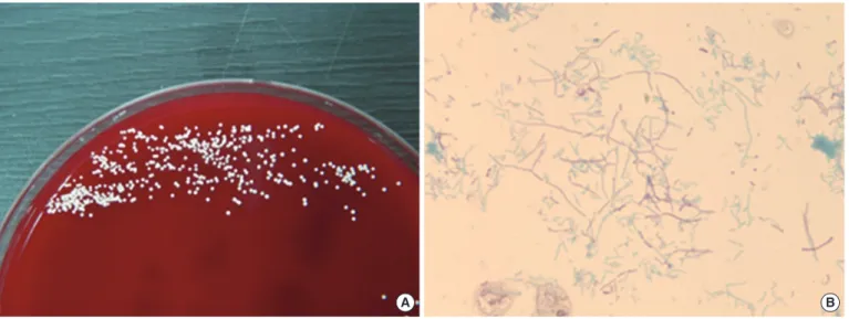
mothorax and was empirically treated intravenously with a third- generation cephalosporin and aminoglycoside. Although fever was observed on admission day 7 and aggravation of pneumonia was noted on a follow-up CT scan taken on day 15, no organism was obtained from subsequent sputum, pleural fluid, or blood cultures. On day 17, the patient was transferred to the intensive care unit and received ventilator support because of altered mental status and respiratory distress. On day 23, pleural fluid cultures on sheep blood agar plates (BAP) grew rough, chalky- white colonies with an earthy odor; the presence of abundant thin, modified acid-fast, and gram-positive branching filaments was observed on microscopic examination (Fig. 2). The same organism was isolated once again from pleural fluid; thus, the
patient was suggested to have a nocardial infection. We decided to obtain further information using molecular methods for 16S rRNA and secA1 sequence analysis. However, multidrug-resis- tant Acinetobacter baumannii was identified from the patient’s sputum obtained on day 36, and the patient died from progres- sion of hospital/ventilator-associated pneumonia and respiratory failure. The etiologic diagnosis was established post-mortem by means of molecular analysis.
To identify the bacterium, molecular identification was per- formed by DNA amplification and sequencing analysis of the 16S rRNA and secA1 genes from the chest tube drainage iso- late. 16S rRNA and secA1 gene fragments were amplified by standard methods according to CLSI guidelines [4]. The secA1
A B
Fig. 1. (A) Both chest radiograph (A) and computed tomography scan of the thorax (B) demonstrate bilateral pleural effusions.
A B
Fig. 2. Colony morphology on sub-inoculation and microscopic morphology. (A) Chalky-white colonies on blood agar plates at 36°C in 5%
CO2. (B) Beaded, branching, gram-positive rods of N. cyriacigeorgica (modified acid-fast bacilli stain, ×400).
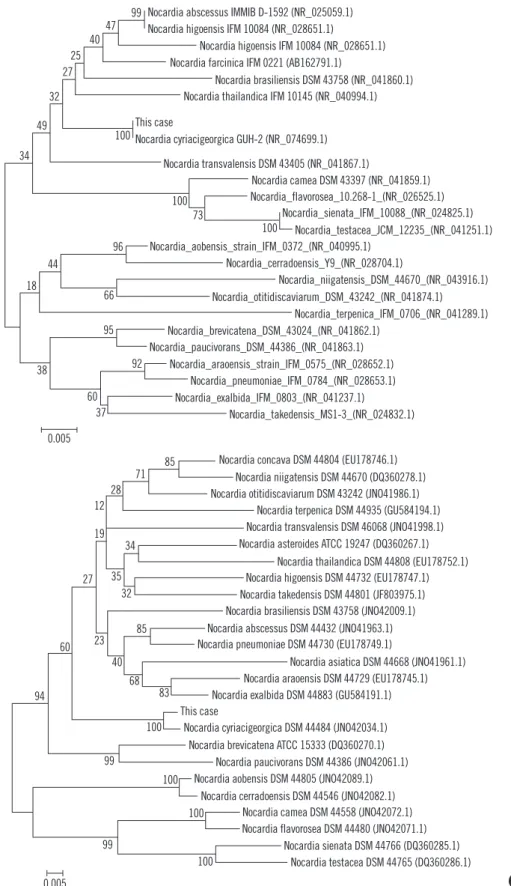
protein is an essential component of the preprotein translocase ATPase, which provides the driving force for the export of pro- teins across the bacterial cytoplasmic membrane [5]. The secA1 locus has potential for nocardia species identification as an ad- junct to 16S rRNA gene sequencing [6]. Two sub-regions of the 16S rRNA and secA1 genes were amplified and sequencing analysis using the previously reported primer pairs [4, 6]. Puri- fied PCR products were sequenced directly using the BigDye Terminator Cycle Sequencing Kit 3.1 (Applied Biosystems; Fos- ter City, CA, USA) on an ABI Prism 3100 genetic analyzer (Ap- plied Biosystems). A GenBank Basic Local Alignment Search Tool (BLAST) search revealed that the 16S rRNA gene sequence of the isolate showed 100% and 98% homology of 695 base pairs to corresponding sequences of the previously sequenced N. cyriacigeorgica and N. farcinica (GenBank accession num- bers NR_074699.1 and AB162791.1). Moreover, for the secA1 gene, we confirmed the corresponding sequence from N. cyri- acigeorgica (JQ773455.1) with 100% homology of 505 base pairs, whereas less than 94% homology was noted for other No- cardia species. For further phylogenetic analysis, 16S rRNA and secA1 gene sequences isolated from the chest tube drainage fluid were aligned using ClustalW [7]. Phylogenetic trees were constructed with the neighbor-joining method using the MEGA 5 software package (http://www.megasoftware.net) and Kimura 2 parameters as the substitution model [8]. Statistical significance of the phylogenies was assessed by bootstrap analysis with 1,000 pseudoreplicate datasets. The tree is drawn to scale, with branch lengths in the same units as those of the evolutionary distances used to infer the phylogenetic tree (Fig. 3).
Prior to the advent of molecular biological techniques, tradi- tional diagnostic methods for identifying these organisms were often ambiguous. Rapid and accurate identification of aerobic actinomycetes is becoming increasingly important in clinical mi- crobiology laboratories. Pulmonary nocardiosis may clinically be acute, subacute, or chronic. Acute nocardiosis often occurs in severely immunocompromised patients and correlates with a poor prognosis, with a high mortality of more than 40% in cases of disseminated disease. Subacute lung nocardiosis, the most common clinically encountered form, often mimics tuberculosis, pneumocystosis, invasive fungal infection, and/or malignancy [9]. Currently, the Nocardia genus has approximately 100 re- ported species of different taxonomic statuses (http://www.bacte- rio.cict.fr/n/nocardia.html). According to Wallace et al. [10], the heterogeneity of the Nocardia group in terms of drug susceptibil- ity patterns requires frequent new taxon formation to account for new species. Of the known Nocardia species, the species most
frequently involved in human infection are members of the N.
asteroides complex. The members of this complex have been sub-classified into 6 separate groups according to drug suscepti- bility patterns with numerous new species described [11]. N.
cyriacigeorgica was first characterized in 2001 by Yassin et al.
[12] from the bronchial secretions of a patient with chronic bron- chitis. At that time, this organism was identified as a novel spe- cies that was both molecularly and biochemically different from previously described members of the genus Nocardia. It corre- sponds to strains of the drug pattern type VI of the N. asteroides complex. They reported that this new species had different DNA- DNA hybridization with N. abscessus (45% reassociation) and N.
asteroides complex type strains (61% reassociation).
Nocardia species do not typically affect immunocompetent hosts. Clinically oriented criteria for nocardiosis may be useful for evaluating the significance of a positive culture from isolated specimens. Criteria include direct visualization of a rod-form mi- croorganism with gram-positive and acid-fast microscopic find- ings and repeated isolation from serial clinical samples in im- munocompromised hosts. However, their presence should sug- gest a careful evaluation of clinical conditions. In a retrospective study, 20% of Nocardia species isolates were found not to be associated with the clinical disease [13]. Most nocardiosis cases are not rapidly recognized because of lack of pathognomonic clinical signs, limitations of conventional biochemical tests for slow-growing bacteria, and the presence of commensal flora on culture. In the present case, when gram-positive bacilli were iso- lated for the first time from the pleural fluid of our patient, we considered that this strain was present owing to the transient colonization in response to chest tube replacement. However, subsequent pleural fluid cultures obtained the same results, along with aggravation of the patient’s clinical status and the lack of any other documented organism. The strain was presumed to be a member of the genus Nocardia, and further molecular analysis was planned. For therapy, Nocardia, and especially N.
cyriacigeorgica, is typically susceptible to sulfonamide, amikacin, linezolid, imipenem, and broad-spectrum cephalosporins, which are often used empirically in the absence of antibiotic suscepti- bility information [14]. In this study, the strain was tested for susceptibility using the Sensititre RAPMYCOI plate (Trek Diag- nostic Systems, Cleveland, OH, USA) according to CLSI guide- lines [15]. The isolate was susceptible to trimethoprim/sulfa- methoxazole (minimum inhibitory concentration [MIC]: 0.5/9.5 µg/mL), amikacin (≤1 µg/mL), linezolid (≤1 µg/mL), ceftriaxone (≤4 µg/mL), minocycline (≤1 µg/mL), and tobramycin (≤1 µg/
mL), and it was resistant to ciprofloxacin (>4 µg/mL), moxifloxa-
A
B Nocardia abscessus IMMIB D-1592 (NR_025059.1)
99 40 27
34
100
100 73 100 96
95
92 60
37 0.005 38
66 44
18
47 25
49 32
Nocardia higoensis IFM 10084 (NR_028651.1)
Nocardia higoensis IFM 10084 (NR_028651.1) Nocardia farcinica IFM 0221 (AB162791.1)
Nocardia brasiliensis DSM 43758 (NR_041860.1) Nocardia thailandica IFM 10145 (NR_040994.1) This case
Nocardia cyriacigeorgica GUH-2 (NR_074699.1) Nocardia transvalensis DSM 43405 (NR_041867.1)
Nocardia camea DSM 43397 (NR_041859.1) Nocardia_flavorosea_10.268-1_(NR_026525.1)
Nocardia_sienata_IFM_10088_(NR_024825.1) Nocardia_testacea_JCM_12235_(NR_041251.1) Nocardia_aobensis_strain_IFM_0372_(NR_040995.1)
Nocardia_cerradoensis_Y9_(NR_028704.1)
Nocardia_niigatensis_DSM_44670_(NR_043916.1) Nocardia_otitidiscaviarum_DSM_43242_(NR_041874.1)
Nocardia_terpenica_IFM_0706_(NR_041289.1) Nocardia_brevicatena_DSM_43024_(NR_041862.1)
Nocardia_paucivorans_DSM_44386_(NR_041863.1) Nocardia_araoensis_strain_IFM_0575_(NR_028652.1)
Nocardia_pneumoniae_IFM_0784_(NR_028653.1) Nocardia_exalbida_IFM_0803_(NR_041237.1)
Nocardia_takedensis_MS1-3_(NR_024832.1)
Nocardia concava DSM 44804 (EU178746.1) Nocardia niigatensis DSM 44670 (DQ360278.1) Nocardia otitidiscaviarum DSM 43242 (JNO41986.1)
Nocardia terpenica DSM 44935 (GU584194.1) Nocardia transvalensis DSM 46068 (JNO41998.1) Nocardia asteroides ATCC 19247 (DQ360267.1)
Nocardia thailandica DSM 44808 (EU178752.1) Nocardia higoensis DSM 44732 (EU178747.1) Nocardia takedensis DSM 44801 (JF803975.1) Nocardia brasiliensis DSM 43758 (JNO42009.1) Nocardia abscessus DSM 44432 (JNO41963.1) Nocardia pneumoniae DSM 44730 (EU178749.1)
Nocardia asiatica DSM 44668 (JNO41961.1) Nocardia araoensis DSM 44729 (EU178745.1) Nocardia exalbida DSM 44883 (GU584191.1) This case
Nocardia cyriacigeorgica DSM 44484 (JNO42034.1) Nocardia brevicatena ATCC 15333 (DQ360270.1)
Nocardia paucivorans DSM 44386 (JNO42061.1) Nocardia aobensis DSM 44805 (JNO42089.1)
Nocardia cerradoensis DSM 44546 (JNO42082.1) Nocardia camea DSM 44558 (JNO42072.1) Nocardia flavorosea DSM 44480 (JNO42071.1)
Nocardia sienata DSM 44766 (DQ360285.1) Nocardia testacea DSM 44765 (DQ360286.1) 71 85
28 12
19 34
27 35
85 32
23 40
83
100
100
100
100 99
99 94
68 60
0.005
Fig. 3. Molecular phylogenetic tree constructed by the neighbor-joining method by using (A) 16s rRNA and (B) secA1 gene sequences from the N. cyriacigeorgica isolate and various Nocardia species. Reference sequences indicating the species strain and accession numbers are given in parentheses. All names and accession numbers are as cited in GenBank. Scale bar represents 5 nucleotide substitutions per 1,000 nucleotides.
cin (4 µg/mL), imipenem (64 µg/mL), cefepime (32 µg/mL), and amoxicillin/clavulanic acid (64/32 µg/mL). Although the patient received empirical treatment with third-generation cephalosporin and aminoglycoside, he died from progression of hospital/venti- lator-associated pneumonia and respiratory failure.
Application of molecular methods for sequence analysis of the 16S rRNA and secA1 genes can assist in a quicker diagno- sis of most Nocardia species. For certain groups of aerobic acti- nomycetes, the use of alternative gene targets such as the secA1 gene can serve as adjuncts to 16S rRNA sequencing of Nocardia species. The use of targeted genes is useful because some species may share 99.6-100% 16S rRNA sequence ho- mology, despite being different species [16, 17]. Thus, definitive identification was obtained by 16S rRNA analysis and alternate DNA target analysis with the secA1 gene.
Although this is the second case of pulmonary nocardiosis caused by N. cyriacigeorgica in Korea [3], to the best of our knowledge, this is the first report on an immunocompetent pa- tient with N. cyriacigeorgica infection, which was identified us- ing molecular analysis for 16S rRNA and secA1 sequences.
This case shows that necrotizing pneumonia and empyema can occur in an immunocompetent patient and should be consid- ered in the differential diagnosis of suspected cases.
Authors’ Disclosures of Potential Conflicts of Interest
No potential conflicts of interest relevant to this article were re- ported.
REFERENCES
1. Conville PS and Witebsky FG. Nocardia, Rhodococcus, Gordonia, Acti- nomadura, Streptomyces, and other aerobic actinomycetes. In: Versa- lovic J, et al. ed. Manual of clinical microbiology. 10th ed. Washinton DC: American Society for Microbiology, 2011:443-71.
2. Uttamchandani RB, Daikos GL, Reyes RR, Fischl MA, Dickinson GM, Yamaguchi E, et al. Nocardiosis in 30 patients with advanced human immunodeficiency virus infection: clinical features and outcome. Clin Infect Dis 1994;18:348-53.
3. Oh H, Kim YS, Jang SJ, Li XM, Kim WY, Park G, et al. A strain of Nocar- dia cyriacigeorgica isolated from a patient with pulmonary infection. Ko- rean J Clin Microbiol 2008;11:136-40.
4. Clinical and Laboratory Standards Institute. Interpretive criteria for iden- tification of bacteria and fungi by DNA target sequencing; approved guideline. Document MM18-A. Wayne, PA: Clinical and Laboratory Standards Institute, 2008.
5. Economou A and Wickner W. SecA promotes preprotein translocation by undergoing ATP-driven cycles of membrane insertion and deinser- tion. Cell 1994;78:835-43.
6. Conville PS, Zelazny AM, Witebsky FG. Analysis of secA1 gene se- quences for identification of Nocardia species. J Clin Microbiol 2006;
44:2760-6.
7. Aiyar RS, Gagneur J, Steinmetz LM. Identification of mitochondrial dis- ease genes through integrative analysis of multiple datasets. Methods 2008;46:248-55.
8. Tamura K, Peterson D, Peterson N, Stecher G, Nei M, Kumar S. MEGA5:
molecular evolutionary genetics analysis using maximum likelihood, evo- lutionary distance, and maximum parsimony methods. Mol Biol Evol 2011;28:2731-9.
9. Matulionyte R, Rohner P, Uçkay I, Lew D, Garbino J. Secular trends of nocardia infection over 15 yr in a tertiary care hospital. J Clin Pathol 2004;
57:807-12.
10. Wallace RJ Jr, Brown BA, Blacklock Z, Ulrich R, Jost K, Brown JM, et al. New Nocardia taxon among isolates of Nocardia brasiliensis associ- ated with invasive disease. J Clin Microbiol 1995;33:1528-33.
11. Conville PS, Fischer SH, Cartwright CP, Witebsky FG. Identification of nocardia species by restriction endonuclease analysis of an amplified portion of the 16S rRNA gene. J Clin Microbiol 2000;38:158-64.
12. Yassin AF, Rainey FA, Steiner U. Nocardia cyriacigeorgici sp. nov. Int J Syst Evol Microbiol 2001;51:1419-23.
13. Georghiou PR and Blacklock ZM. Infection with Nocardia species in Queensland. A review of 102 clinical isolates. Med J Aust 1992;156:
692-7.
14. Schlaberg R, Huard RC, Della-Latta P. Nocardia cyriacigeorgica, an emerging pathogen in the United States. J Clin Microbiol 2008;46:265- 73.
15. Clinical and Laboratory Standards Institute. Susceptibility testing of My- cobacteria, Nocardiae, and other actinomycetes; approved Standard- Second edition, Document M24-A2. Wayne, PA: Clinical and Laboratory Standards Institute, 2011.
16. Roth A, Andrees S, Kroppenstedt RM, Harmsen D, Mauch H. Phylogeny of the genus Nocardia based on reassessed 16S rRNA gene sequences reveals underspeciation and division of strains classified as Nocardia as- teroides into three established species and two unnamed taxons. J Clin Microbiol 2003;41:851-6.
17. Kong F, Wang H, Zhang E, Sintchenko V, Xiao M, Sorrell TC, et al.
secA1 gene sequence polymorphisms for species identification of No- cardia species and recognition of intraspecies genetic diversity. J Clin Microbiol 2010;48:3928-34.